. 2014 Mar;306(6):H789-96.
doi: 10.1152/ajpheart.00559.2013. Epub 2014 Jan 24.

Vascular endothelial growth factor-C: its unrevealed role in fibrogenesis

Affiliations

Vascular endothelial growth factor-C: its unrevealed role in fibrogenesis

Tieqiang Zhao et al. Am J Physiol Heart Circ Physiol. 2014 Mar.

Abstract

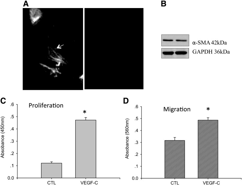
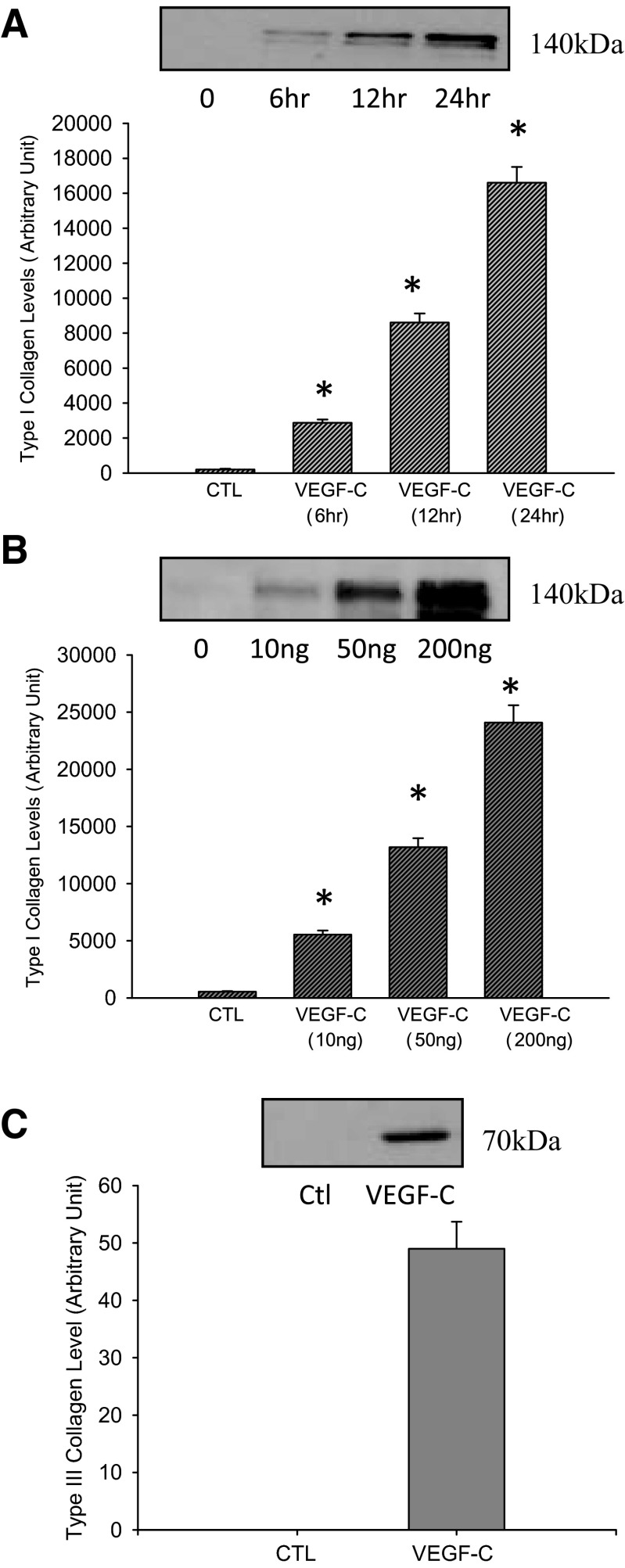
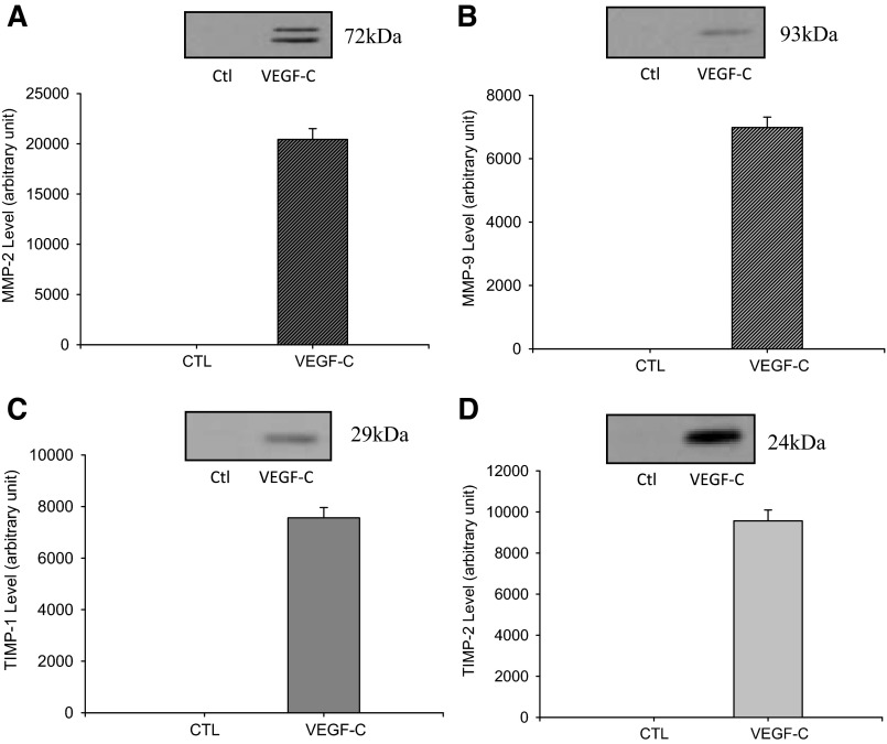
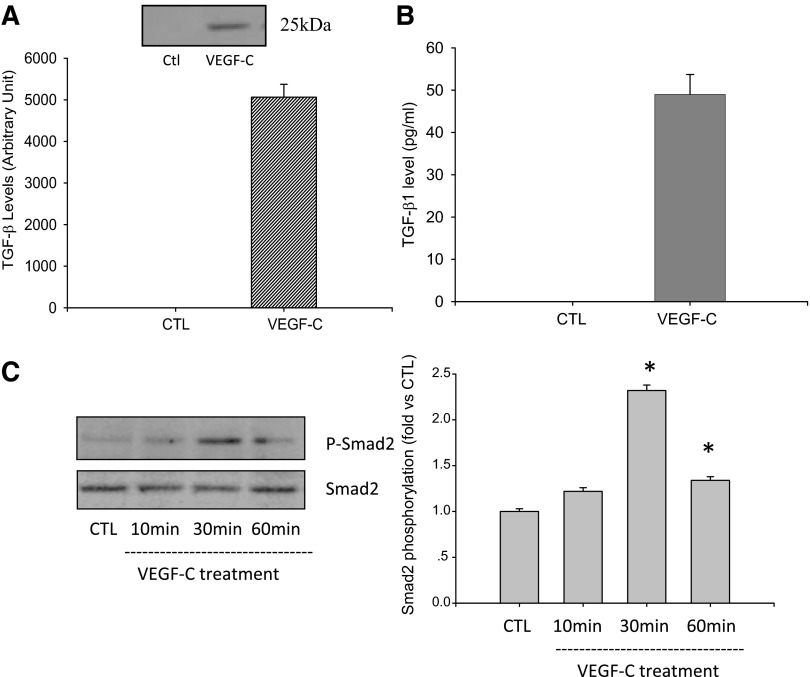
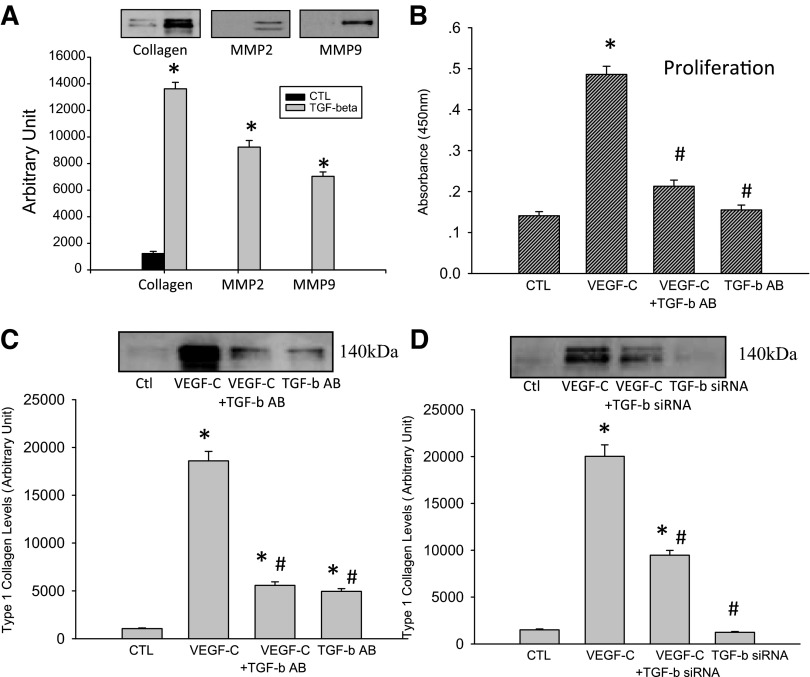
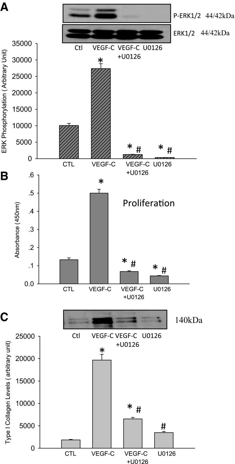
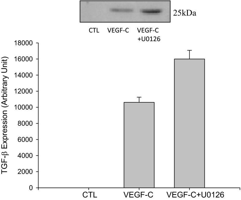
Vascular endothelial growth factor (VEGF)-C is a key mediator of lymphangiogenesis. Our recent study shows that VEGF-C/VEGF receptors (VEGFR)-3 are significantly increased in the infarcted rat myocardium, where VEGFR-3 is expressed not only in lymph ducts but also in myofibroblasts, indicating that VEGF-C has an unrevealed role in fibrogenesis during cardiac repair. The current study is to explore the regulation and molecular mechanisms of VEGF-C in fibrogenesis. The potential regulation of VEGF-C on myofibroblast differentiation/growth/migration, collagen degradation/synthesis, and transforming growth factor (TGF)-β and ERK pathways was detected in cultured cardiac myofibroblasts. Our results showed that VEGF-C significantly increased myofibroblast proliferation, migration, and type I/III collagen production. Matrix metalloproteinase (MMP)-2 and -9 were significantly elevated in the medium of VEGF-C-treated cells, coincident with increased tissue inhibitor of metalloproteinase (TIMP)-1 and TIMP-2. Furthermore, VEGF-C activated the TGF-β1 pathway and ERK phosphorylation, which was significantly suppressed by TGF-β or ERK blockade. This is the first study indicating that in addition to lymphangiogenesis, VEGF-C is also involved in fibrogenesis through stimulation of myofibroblast proliferation, migration, and collagen synthesis, via activation of the TGF-β1 and ERK pathways.

Keywords: ERK; TGF-β1; VEGF-C; collagen degradation; collagen synthesis; myofibroblasts.


Figures

Fig. 1.
Fig. 1.
Effect of VEGF-C on myofibroblast differentiation, proliferation, and migration. Cultured cardiac fibroblasts automatically differentiated into myofibroblasts at P1 and expressed α-smooth muscle actin (α-SMA; A, left, arrows). Compared with vehicle-treated myofibroblasts, VEGF-C did not affect myofibroblast differentiation (B), significantly increased myofibroblast proliferation (C) and migration (D). A, right: negative control of α-SMA labeling. *P < 0.05 vs. controls (CTL); n = 8/group.
Fig. 2.
Fig. 2.
VEGF-C promotes type I and III collagen synthesis in myofibroblasts. Compared with controls, VEGF-C treatment significantly stimulated type I collagen secretion in a time (A)- and dose-dependent (B) manner. VEGF-C also promoted type III collagen production (C). *P < 0.05 vs. controls (CTL); n = 6/group.
Fig. 3.
Fig. 3.
VEGF-C increases matrix metalloproteinase (MMP)-2 (A) and -9 (B) and tissue inhibitor of metalloproteinase (TIMP)-1 (C) and -2 (D) synthesis in myofibroblasts. MMP-2, MMP-9, TIMP-1 and TIMP-2 were not detectable in the medium of vehicle-treated myofibroblasts. VEGF-C treatment induced MMP-2, MMP-9, TIMP-1, and TIMP-2 release; n = 6/group.
Fig. 4.
Fig. 4.
VEGF-C upregulates TGF-β1 production and Smad2 phosphorylation. Detected by Western blot (A) and ELISA (B), TGF-β1 was not detectable in the medium of vehicle-treated cells, while VEGF-C treatment significantly enhanced TGF-β1 levels in the medium. C: VEGF-C treatment significantly enhanced Smad2 phosphorylation in myofibroblasts. *P < 0.05 vs. controls (CTL); n = 6/group.
Fig. 5.
Fig. 5.
TGF-β blockade abolishes the role of VEGF-C on fibrogenesis. Compared with control cells, TGF-β1 neutralizing antibody significantly suppressed VEGF-C-induced myofibroblast proliferation (B) and type I collagen synthesis (A). TGF-β1 siRNA treatment significantly suppressed VEGF-C-induced type I collagen production (C and D). *P < 0.05 vs. controls; #P < 0.05 vs. VEGFC treatment; n = 6/group.
Fig. 6.
Fig. 6.
VEGF-C stimulates ERK phosphorylation in myofibroblasts. Compared with controls, VEGF-C elevated ERK phosphorylation (p-ERK1/2), which was blocked by the ERK inhibitor U0126 (A). VEGF-C-induced myofibroblast proliferation (B) and type I collagen synthesis (C) were significantly reduced by U0126; n = 6/group. *P < 0.05 vs. controls; #P < 0.05 vs. VEGFC treatment.
Fig. 7.
Fig. 7.
Effect of the ERK inhibitor on VEGF-C-induced TGF-β1 production in myofibroblasts. VEGF-C enhanced TGF-β1 production by myofibroblasts, which was not diminished by U0126; n = 6/group.

References

    1. Bujak M, Frangogiannis NG. The role of TGF-beta signaling in myocardial infarction and cardiac remodeling. Cardiovasc Res 74: 184–195, 2007 - PMC - PubMed
    1. Chaudary N, Milosevic M, Hill RP. Suppression of vascular endothelial growth factor receptor 3 (VEGFR3) and vascular endothelial growth factor C (VEGFC) inhibits hypoxia-induced lymph node metastases in cervix cancer. Gynecol Oncol 123: 393–400, 2011 - PubMed
    1. Chen W, Frangogiannis NG. Fibroblasts in post-infarction inflammation and cardiac repair. Biochim Biophys Acta 1833: 945–953, 2013 - PMC - PubMed
    1. Cohen S, Efraim AN, Levi-Schaffer F, Eliashar R. The effect of hypoxia and cycloxygenase inhibitors on nasal polyp derived fibroblasts. Am J Otolaryngol 32: 564–573, 2011 - PubMed
    1. Daskalopoulos EP, Hermans KC, Blankesteijn WM. Cardiac (myo)fibroblast: novel strategies for its targeting following myocardial infarction. Curr Pharm Des 2013. June 18 [Epub ahead of print] - PubMed

Publication types

MeSH terms

Substances

LinkOut - more resources