. 2014 Mar;306(6):H807-15.
doi: 10.1152/ajpheart.00913.2013. Epub 2014 Jan 24.

Contractile dysfunction in a mouse model expressing a heterozygous MYBPC3 mutation associated with hypertrophic cardiomyopathy

Affiliations

Contractile dysfunction in a mouse model expressing a heterozygous MYBPC3 mutation associated with hypertrophic cardiomyopathy

David Barefield et al. Am J Physiol Heart Circ Physiol. 2014 Mar.

Abstract

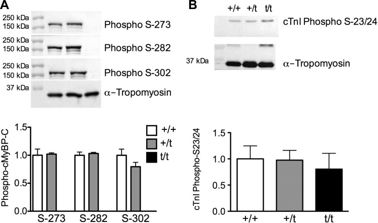
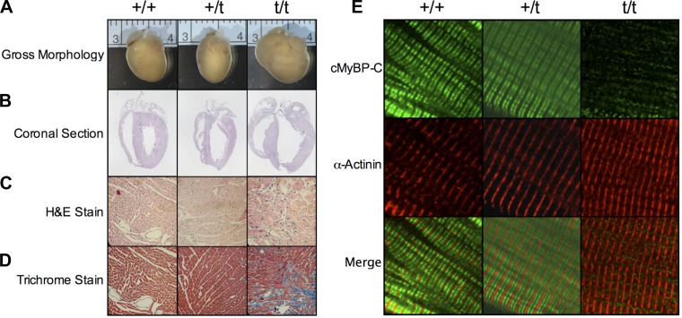
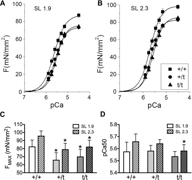
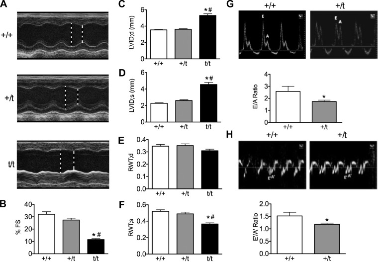
The etiology of hypertrophic cardiomyopathy (HCM) has been ascribed to mutations in genes encoding sarcomere proteins. In particular, mutations in MYBPC3, a gene which encodes cardiac myosin binding protein-C (cMyBP-C), have been implicated in over one third of HCM cases. Of these mutations, 70% are predicted to result in C'-truncated protein products, which are undetectable in tissue samples. Heterozygous carriers of these truncation mutations exhibit varying penetrance of HCM, with symptoms often occurring later in life. We hypothesize that heterozygous carriers of MYBPC3 mutations, while seemingly asymptomatic, have subtle functional impairments that precede the development of overt HCM. This study compared heterozygous (+/t) knock-in MYBPC3 truncation mutation mice with wild-type (+/+) littermates to determine whether functional alterations occur at the whole-heart or single-cell level before the onset of hypertrophy. The +/t mice show ∼40% reduction in MYBPC3 transcription, but no changes in cMyBP-C level, phosphorylation status, or cardiac morphology. Nonetheless, +/t mice show significantly decreased maximal force development at sarcomere lengths of 1.9 μm (+/t 68.5 ± 4.1 mN/mm(2) vs. +/+ 82.2 ± 3.2) and 2.3 μm (+/t 79.2 ± 3.1 mN/mm(2) vs. +/+ 95.5 ± 2.4). In addition, heterozygous mice show significant reductions in vivo in the early/after (E/A) (+/t 1.74 ± 0.12 vs. +/+ 2.58 ± 0.43) and E'/A' (+/t 1.18 ± 0.05 vs. +/+ 1.52 ± 0.15) ratios, indicating diastolic dysfunction. These results suggest that seemingly asymptomatic heterozygous MYBPC3 carriers do suffer impairments that may presage the onset of HCM.

Keywords: cardiac myosin binding protein-C; haploinsufficiency.


Figures

Fig. 1.
Fig. 1.
Expression of cardiac myosin binding protein-C gene (MYBPC3) and hypertrophic markers. A: myofilament protein fraction resolved with SDS-PAGE and stained with Coomassie blue. B: Western blot showing levels of cMyBP-C and actin. Western blot quantification shows no cMyBP-C present in the homozygous MYBPC3 truncation mutant mouse hearts (t/t) samples and no significant change in cMyBP-C levels between wild-type MYBPC3 mouse hearts (+/+) and heterozygous MYBPC3 truncation mutant mouse hearts (+/t) (n = 5). C: total heart homogenate used for analysis of myosin heavy chain isoforms (α-MHC and β-MHC) resolved on a Hoefer format SDS-PAGE and stained with Sypro-Ruby. The +/+ and +/t hearts show no changes in α-MHC isoform levels, with t/t hearts showing a significant increase in the hypertrophic marker β-MHC (N = 11, 8, 9). D: MYBPC3 transcription is reduced in +/t hearts compared with +/+ hearts, and t/t hearts show an even further reduction in MYBPC3 expression. E and F: transcript levels of hypertrophic markers cardiac β-myosin heavy chain gene (MYH7) and atrial natriuretic factor gene (NPPA) are unchanged in +/+ and +/t hearts, but they show a significant increase in the t/t hearts (N = 6,7, 3). *P < 0.05 vs. +/+; #P < 0.05 vs. +/t. ELC, essential light chain; RLC, regulatory light chain; cTnI, cardiac troponin I protein; α-TM, α-tropomyosin.
Fig. 2.
Fig. 2.
Phosphorylation of cMyBP-C and cTnI. A: Western blot analysis detecting cMyBP-C phosphorylated at serine-273, -282, and -302. No significant change in phosphorylation of cMyBP-C was observed at any of the phosphorylation sites between the +/+ and +/t groups (n = 5). B: phosphorylation status of cTnI at serine-23 and -24, as determined by Western blot, shows no significant changes in any of the groups (n = 5).
Fig. 3.
Fig. 3.
Gross morphology and protein incorporation. Representative whole heart images (A) and coronal sections (B) show no overt hypertrophy in the +/t hearts, whereas t/t hearts display a hypertrophic phenotype. Cross-sectional hematoxylin and eosin (H&E; C) and Trichrome staining (D) reveal deranged muscle arrangement and fibrosis, respectively, in the t/t heart, with no notable differences in +/+ and +/t. E: confocal microcopy immunofluorescence of isolated cardiomyocytes stained for cMyBP-C (green) and α-actinin (red). The cMyBP-C signal shows the characteristic C-zone doublet pattern between each Z-disk, stained for α-actinin in both +/+ and +/t cardiomyocytes. Staining of the t/t cells reveals only minimal cMyBP-C signaling and no structural localization.
Fig. 4.
Fig. 4.
Alterations of force development and Ca2+ sensitivity in skinned cardiomyocytes. AC: maximum development of force (Fmax) is significantly decreased in +/t and t/t skinned cardiomyocytes compared with +/+ at both short [sarcomere length (SL) 1.9 μm] and long (SL 2.3 μm) sarcomere lengths. D: relative force comparisons show a significant decrease in Ca2+ sensitivity of force development in t/t hearts compared with +/+ at SL 2.3 μm. No significant changes in Ca2+ sensitivity were seen between +/t and +/+ (N = 6, 7, and 7 hearts per group, with 3 to 4 cells averaged per heart). *P < 0.05 vs. +/+.
Fig. 5.
Fig. 5.
In vivo measurements of cardiac morphology and function. A: representative parasternal long-axis M-mode echocardiography tracings from 10- to 12-wk-old +/+, +/t, and t/t mouse hearts. B: percentage of left ventricular fractional shortening (FS) shows that +/t hearts trend toward reduced pump function compared with +/+, whereas t/t hearts show a significant deficit compared with both +/+ and +/t groups. C and D: left ventricular internal diameter (LVID) at peak systole (s) and diastole (d) shows significant dilation in the t/t hearts with no changes between +/+ and +/t. E and F: relative wall thickness (RWT; anterior + posterior wall/total heart thickness) at peak systole and diastole shows no change between +/t and +/+, whereas the wall-to-heart ratio is reduced in t/t. G and H: representative power and tissue Doppler images depicting the early (E) and late (A) blood filling of the left ventricle and mitral valve motion. E/A and E′/A′ ratios were significantly reduced in +/t hearts compared with +/+, with t/t hearts showing a significant deficit compared with both groups (N = 7, 10, 9). *P < 0.05 vs. +/+; #P < 0.05 vs. +/t.

References

    1. Alcalai R, Seidman J, Seidman C. Genetic basis of hypertrophic cardiomyopathy: from bench to the clinics. J Cardiovasc Electrophysiol 19: 104–110, 2008 - PubMed
    1. Barefield D, Sadayappan S. Phosphorylation and function of cardiac myosin binding protein-C in health and disease. J Mol Cell Cardiol 48: 866–875, 2010 - PMC - PubMed
    1. Bonne G, Carrier L, Bercovici J, Cruaud C, Richard P, Hainque B, Gautel M, Labeit S, James M, Beckmann J, Weissenbach J, Vosberg HP, Fiszman M, Komajda M, Schwartz K. Cardiac myosin binding protein-C gene splice acceptor site mutation is associated with familial hypertrophic cardiomyopathy. Nat Genet 11: 438–440, 1995 - PubMed
    1. Carrier L, Bonne G, Bahrend E, Yu B, Richard P, Niel F, Hainque B, Cruaud C, Gary F, Labeit S, Bouhour J, Dubourg O, Desnos M, Hagege A, Trent R, Komajda M, Fiszman M, Schwartz K. Organization and sequence of human cardiac myosin binding protein C gene (MYBPC3) and identification of mutations predicted to produce truncated proteins in familial hypertrophic cardiomyopathy. Circ Res 80: 427–434, 1997 - PubMed
    1. Carrier L, Knoll R, Vignier N, Keller D, Bausero P, Prudhon B, Isnard R, Ambroisine M, Fiszman M, Ross J, Schwartz K, Chien K. Asymmetric septal hypertrophy in heterozygous cMyBP-C null mice. Cardiovasc Res 63: 293–304, 2004 - PubMed

Publication types

LinkOut - more resources