. 2014 Apr;42(7):4640-51.
doi: 10.1093/nar/gkt1413. Epub 2014 Jan 24.

Interplay between pre-mRNA splicing and microRNA biogenesis within the supraspliceosome

Affiliations

Interplay between pre-mRNA splicing and microRNA biogenesis within the supraspliceosome

Lily Agranat-Tamir et al. Nucleic Acids Res. 2014 Apr.

Abstract

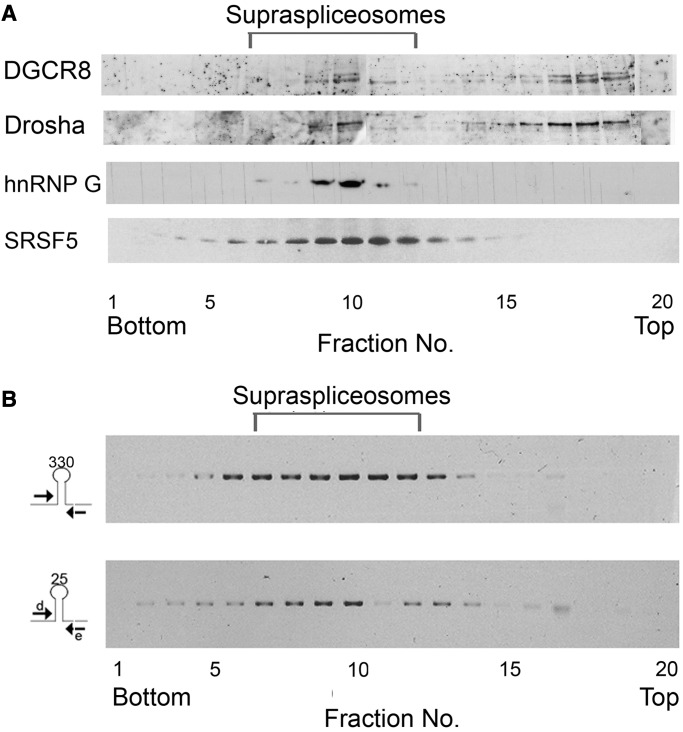
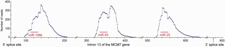
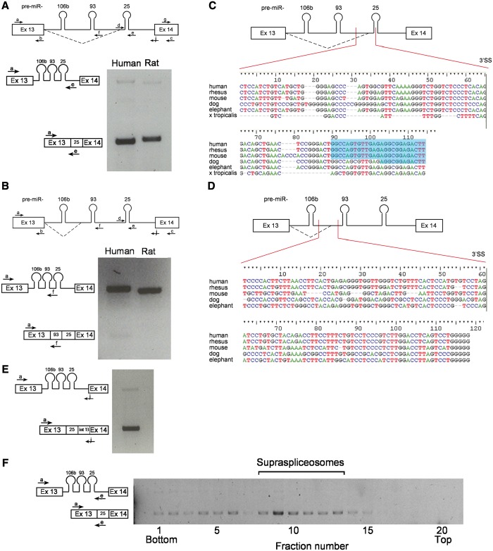
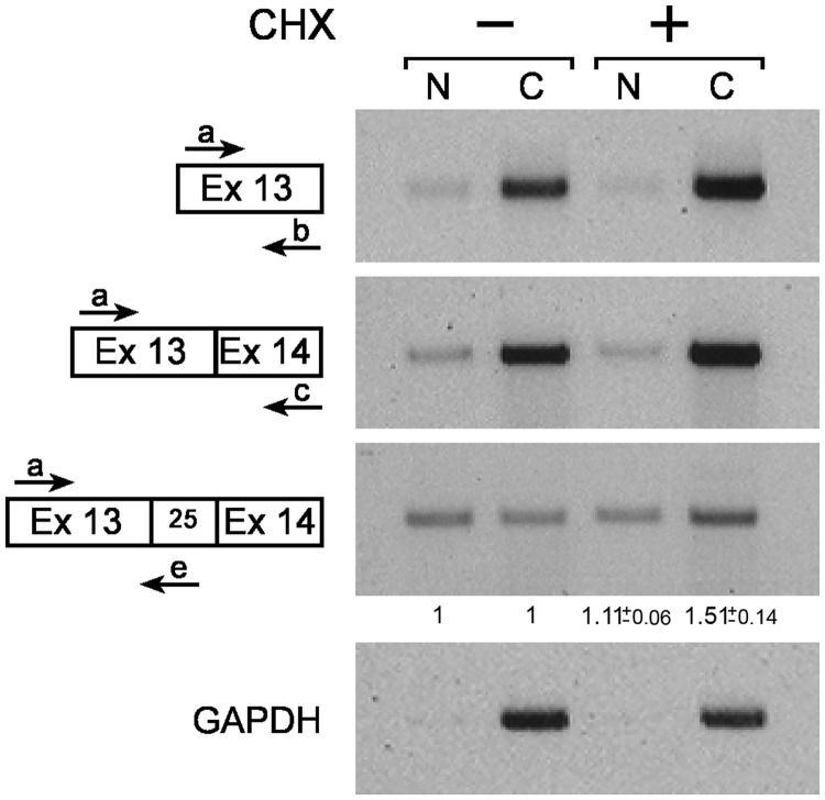
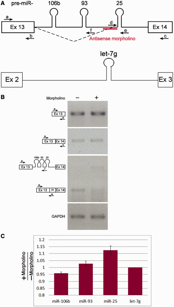
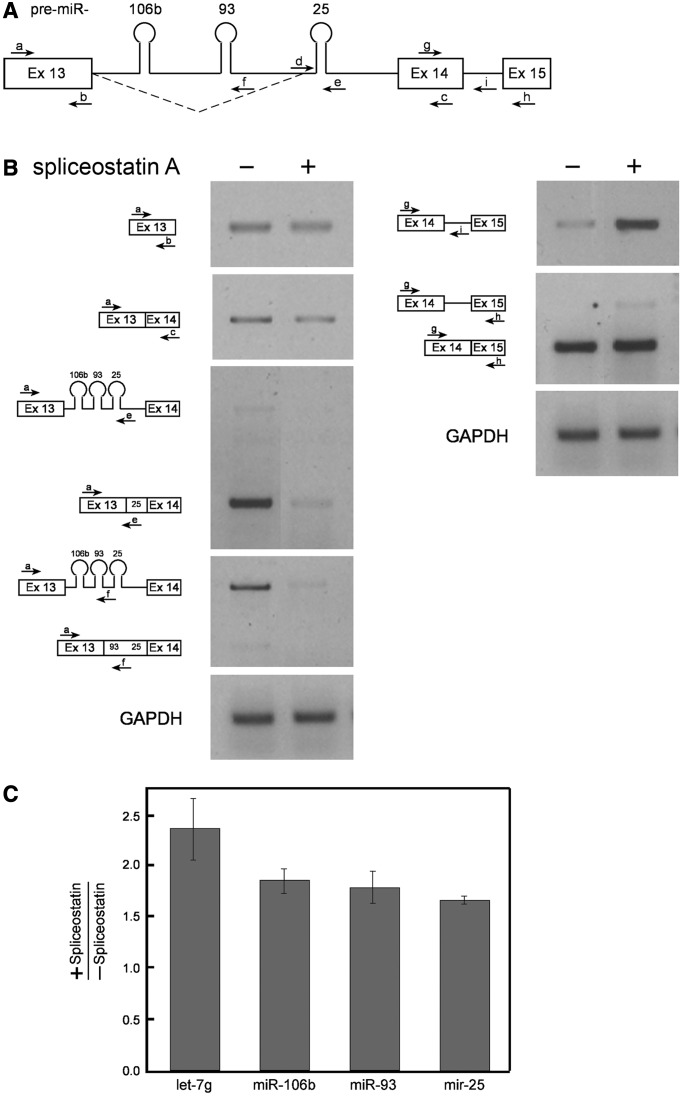
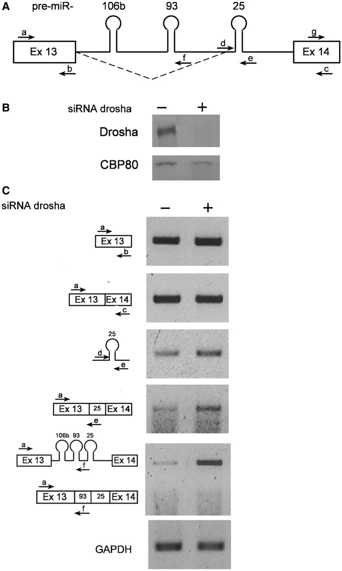
MicroRNAs (miRNAs) are central regulators of gene expression, and a large fraction of them are encoded in introns of RNA polymerase II transcripts. Thus, the biogenesis of intronic miRNAs by the microprocessor and the splicing of their host introns by the spliceosome require coordination between these processing events. This cross-talk is addressed here. We show that key microprocessor proteins Drosha and DGCR8 as well as pre-miRNAs cosediment with supraspliceosomes, where nuclear posttranscriptional processing is executed. We further show that inhibition of splicing increases miRNAs expression, whereas knock-down of Drosha increases splicing. We identified a novel splicing event in intron 13 of MCM7, where the miR-106b-25 cluster is located. The unique splice isoform includes a hosted pre-miRNA in the extended exon and excludes its processing. This indicates a possible mechanism of altering the levels of different miRNAs originating from the same transcript. Altogether, our study indicates interplay between the splicing and microprocessor machineries within a supraspliceosome context.


Figures

Figure 1.
Figure 1.
Microprocessor components and pri-miRNA sequences are found in supraspliceosomes. (A) Nuclear supernatants enriched for supraspliceosomes were prepared from HeLa cells and were fractionated in 10–45% glycerol gradients (28). Supraspliceosome peak fractions (8–10) were pooled and refractionated on a second glycerol gradient. Aliquots from gradient fractions were analyzed by WB using anti-Drosha and anti-DGCR8 antibodies. For comparison, distribution across glycerol gradients of regulatory splicing factors associated with supraspliceosomes, hnRNP G and SRSF5 SR proteins is shown (B) Nuclear supernatants enriched for supraspliceosomes were prepared from HeLa cells and were fractionated in 10–45% glycerol gradients (28). RT-PCR analyses of the distribution of pri-miR-330 and pri-miR-25 across the gradient using the indicated primer pairs that flank the respective pri-miRs.
Figure 2.
Figure 2.
Processing of pri-miRNA in supraspliceosomes. RNA was extracted from supraspliceosomes prepared from frozen HeLa cells, as previously described (28). Searching for sequences of pre-miRNAs of the 106b-25 cluster within the deep sequencing data of small RNAs (<200 nt), from this supraspliceosomal RNA (fractions 8–10), revealed that sequences of the pre-miRNA 106 b, 93 and 25 were found in supraspliceosomes.
Figure 3.
Figure 3.
Conserved novel 3′SS in intron 13 of MCM7. (A) A novel alternative 3′SS between pre-miR-93 and 25. A diagram of MCM7 exon 13 through exon 14 with splicing at the novel 93-25 3′SS (broken line) and the primers used for RT-PCR (upper panel) indicated. RT-PCR on RNA extracted from HeLa cells using primer pair a/e (lower panel). The amplified RT-PCR products are depicted on the left. (B) A second, yet minor, novel 3′SS between pre-miR-106 b and 93. A diagram of MCM7 exon 13 through exon 14 with splicing at the novel 106 b-93 3′SS (broken line) and the primers used for RT-PCR (upper panel) are indicated. RT-PCR using primer pair a/f (lower panel). The amplified RT-PCR products are depicted on the left. (C) Conservation of the sequences flanking the novel 93-25 3′SS. Sequence alignment of the region surrounding the novel 3′SS between pre-miRNAs 93 and 25. The different organisms are marked on the left. The splice point is marked by a vertical line. Above is a diagram of exon 13 through exon 14 of MCM7 with the aligned region marked by two vertical lines. The 5′-end of sequences of pre-miR-25 are highlighted in blue. (D) Conservation of the sequences flanking the novel 106b-93 3′SS. The same as in (C), with sequence alignment of the region surrounding the novel 3′SS between pre-miRNAs 106 b and 93. (E) The novel splice isoform, the product of splicing at the 93-25 3′SS, incorporates the sequence of pre-miR-25 into exon 14 of MCM7. RT-PCR analysis using primer pair a/j. (F) The novel splice isoform at the 93-25 3'SS is found in supraspliceosomes. RT-PCR analysis of aliquots of RNA is extracted from gradient fractions, using the indicated primer pairs.
Figure 4.
Figure 4.
The novel alternative 93-25 3′ splice isoform is found in the cytoplasm. RNA was extracted from nuclear (N) and cytoplasmic (C) fractions of HeLa cells, either treated (+) or untreated (−) with 50 µg/ml CHX for 2 h, and analyzed by RT-PCR. Fold change of the novel alternative 93-25 3′ splice isoform in nucleus and cytoplasm after CHX treatment, compared with fold change in the MCM7 constitutive mRNA, is indicated below the specific lanes with standard error (n = 2).
Figure 5.
Figure 5.
An antisense morpholino oligonucleotide abolishes splicing at the alternative 3'SS. Morpholino molecules antisense to the 93-25 3'SS were transfected into HeLa cells. RNA was then extracted and analyzed. (A) Schematic representation of exon 13– exon 14 of the MCM7 gene and the antisense morpholino is indicated; and schematic representation of exon 2–exon 3 of the WDR82 gene. (B) RT-PCR analyses of RNA extracted from the cells treated with antisense morpholino and from control untreated cells were performed using the indicated primers. (C) Graph showing the fold change of the indicated miRNAs after morpholino treatment measured using the miRNA TaqMan assay, normalized to the change in let-7 g miRNA. Standard errors are shown. P < 0.05 for miR-106 b and miR-25 (t-test). The results represent triplicates of each of two independent experiments.
Figure 6.
Figure 6.
SSA upregulates the levels of intronic miRNAs. SSA was added to HeLa cells (100 ng/ml for 5 h), and RNA was then extracted. (A) Schematic representation of exon 13– exon 15 of the MCM7 gene. The novel 93-25 3′ splicing pattern is indicated (broken line), and so are the primers used. (B) RT-PCR analyses of RNA extracted from cells treated (+) or untreated (−) with SSA, using the indicated primer pairs from MCM7 and with primers from GAPDH. (C) Graph showing the fold change of the indicated miRNAs after SSA treatment measured using the miRNA TaqMan assay. Standard errors are shown. P < 0.05 for let-7 g and miR-93; <0.01 for miR-106 b; and <0.005 for miR-25 (t test). The results represent triplicates of each of two independent experiments.
Figure 7.
Figure 7.
Knock-down of Drosha is accompanied by increase in the level of the novel 3′ splice isoform of MCM7 mRNA. HeLa cells were treated with siRNA molecules against Drosha. Proteins and RNAs were extracted from siRNA treated (+), and untreated (−) cells, and were analyzed. (A) Schematic representation of exon 13–exon 14 of the MCM7 gene. The novel 93-25 3′ splicing pattern is indicated (broken line), and so are the primers used. (B) WB with anti-Drosha and anti-CBP80 antibodies as a reference. Drosha was knocked-down by siRNA to 18%. (C) RT-PCR analyses with the indicated primers.

Similar articles

Cited by

References

    1. Eulalio A, Huntzinger E, Izaurralde E. Getting to the root of miRNA-mediated gene silencing. Cell. 2008;132:9–14. - PubMed
    1. Bartel DP. MicroRNAs: target recognition and regulatory functions. Cell. 2009;136:215–233. - PMC - PubMed
    1. Fabian MR, Sonenberg N. The mechanics of miRNA-mediated gene silencing: a look under the hood of miRISC. Nat. Struct. Mol. Biol. 2012;19:586–593. - PubMed
    1. Rodriguez A, Griffiths-Jones S, Ashurst JL, Bradley A. Identification of mammalian microRNA host genes and transcription units. Genome Res. 2004;14:1902–1910. - PMC - PubMed
    1. Kim YK, Kim VN. Processing of intronic microRNAs. EMBO J. 2007;26:775–783. - PMC - PubMed

Publication types