. 2014 Apr;42(7):4712-22.
doi: 10.1093/nar/gkt1407. Epub 2014 Jan 23.

The crystal structure of TDP-43 RRM1-DNA complex reveals the specific recognition for UG- and TG-rich nucleic acids

Affiliations

The crystal structure of TDP-43 RRM1-DNA complex reveals the specific recognition for UG- and TG-rich nucleic acids

Pan-Hsien Kuo et al. Nucleic Acids Res. 2014 Apr.

Abstract

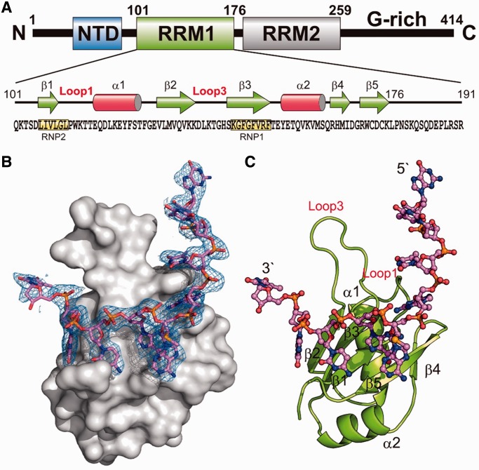
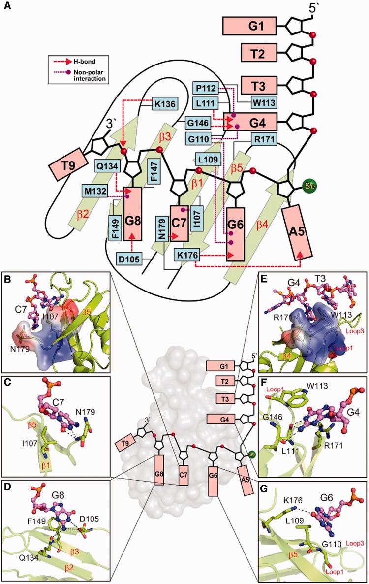
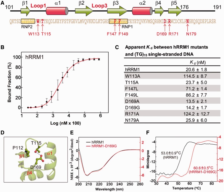
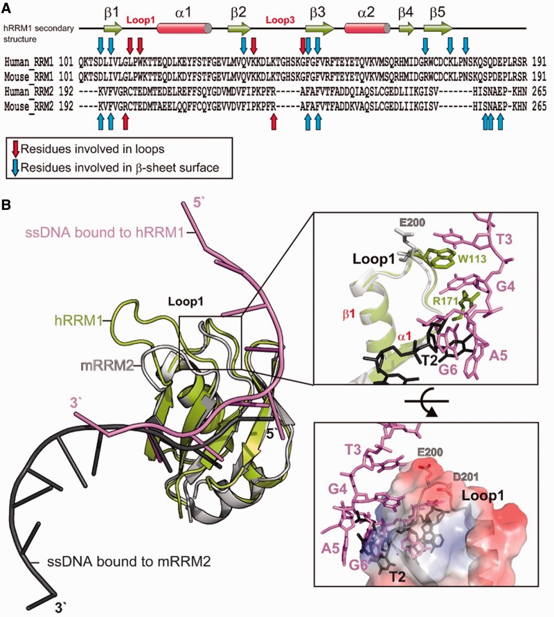
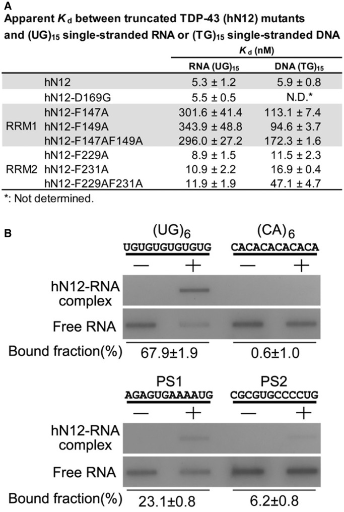
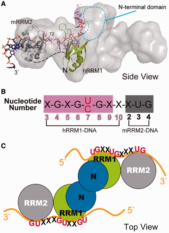
TDP-43 is an important pathological protein that aggregates in the diseased neuronal cells and is linked to various neurodegenerative disorders. In normal cells, TDP-43 is primarily an RNA-binding protein; however, how the dimeric TDP-43 binds RNA via its two RNA recognition motifs, RRM1 and RRM2, is not clear. Here we report the crystal structure of human TDP-43 RRM1 in complex with a single-stranded DNA showing that RRM1 binds the nucleic acid extensively not only by the conserved β-sheet residues but also by the loop residues. Mutational and biochemical assays further reveal that both RRMs in TDP-43 dimers participate in binding of UG-rich RNA or TG-rich DNA with RRM1 playing a dominant role and RRM2 playing a supporting role. Moreover, RRM1 of the amyotrophic lateral sclerosis-linked mutant D169G binds DNA as efficiently as the wild type; nevertheless, it is more resistant to thermal denaturation, suggesting that the resistance to degradation is likely linked to TDP-43 proteinopathies. Taken together all the data, we suggest a model showing that the two RRMs in each protomer of TDP-43 homodimer work together in RNA binding and thus the dimeric TDP-43 recognizes long clusters of UG-rich RNA to achieve high affinity and specificity.


Figures

Figure 1.
Figure 1.
Overall crystal structure of hRRM1-DNA complex. (A) Domain structure of TDP-43. (B) The molecular surface of hRRM1 bound with a ssDNA. The difference Fourier (Fo-Fc) electron density map was superimposed on the structural model of DNA. (C) The overall crystal structure of hRRM1 in complex with DNA.
Figure 2.
Figure 2.
The interactions between hRRM1 and DNA. (A) Schematic diagram of the interactions between hRRM1 and DNA. I107, F147 and F149 are the conserved aromatic/hydrophobic residues in RNP2 and RNP1 segments that interact with DNA bases and sugar rings of C7-G8. (B) The cytosine of C7 stacks with the side chains of I107 and N179. (C) C7 forms hydrogen bonds to N179. (D) G8 forms hydrogen bonds with D105 and Q134. (E) G4 stacks between R179 and W113, whereas T3 stacks with W113. (F) G4 hydrogen bonds with L111, W113 and G146. (G) G6 hydrogen bonds with K176.
Figure 3.
Figure 3.
Mutation of the critical residues for nucleic acid binding and TDP-43 pathogenesis in hRRM1. (A) Mutation of a number of residues (indicated by arrows) in hRRM1. (B) The binding affinity between the wild-type hRRM1 and (TG)15 was measured by the nitrocellulose filter binding assay. The 5′-end 32P-labeled DNA (10 pmol) was incubated with hRRM1 (0.0002–300 µM) and the hRRM1-DNA complexes trapped in the nitrocellulose filters were quantified. (C) The apparent Kd between hRRM1 mutants and (TG)15. (D) D169 located in Loop6 between β4 and β5 forms a hydrogen bond to the side chain of T115 located in Loop1. (E) The CD spectra of hRRM1 and hRRM1-D169G mutant. (F) The thermal melting points of hRRM1 and hRRM1-D169G mutant were estimated by CD at a wavelength of 208 nm.
Figure 4.
Figure 4.
hRRM1 interacts more extensively with DNA, as revealed by the comparison between the crystal structures of hRRM1-DNA and mRRM2-DNA. (A) Sequence alignment of human and mouse RRM domains. Blue and red arrows mark the residues that interact with ssDNA via nonbonded interactions or hydrogen bonding. Red arrows are located in the loops and blue arrows are located within or close to β-strands. (B) The crystal structure of hRRM1-DNA (green and purple) is superimposed on mRRM2-DNA (gray and black) complex. A close look in the right top panel shows that only Loop1 of hRRM1 interacts with DNA: W113/R171 stacking with T3/G4. On the other hand, the Loop1 of RRM2 does not interact with DNA because the molecular surface of Loop1 is acidic and not suitable for DNA binding as shown in the right bottom panel.
Figure 5.
Figure 5.
Mutations in either RRM1 or RRM2 reduce the TDP-43 high-affinity binding for UG-repeated RNA or TG-repeated DNA. (A) The C-terminal truncated TDP-43 (hN12, residues 1–259) was incubated with 5′-end 32P-labeled single-stranded (UG)15 RNA or (TG)15 DNA (10 pmol) for the measurement of binding affinity by the nitrocellulose filter binding assay. The estimated apparent Kd between the wild-type and mutated hN12 show that mutations in both RRM1and RRM2 generated defective mutants in RNA and DNA binding. (B) The nitrocellulose filter binding assays reveal sequence specificity of TDP-43 for UG repeats and UG-rich sequences. The hN12 (2 µM) bound to (UG)6 with the highest affinity (estimated bound fraction: 67.9 ± 1.9%), the UG-rich PS1 and PS2 with moderate affinity (23.1 ± 0.8 and 6.2 ± 0.8%) and (CA)6 with the lowest affinity (0.6 ± 1.0%).
Figure 6.
Figure 6.
The structural model of TDP-43 bound to single-stranded nucleic acids. (A) The SAXS envelope of TDP-43 dimer fitted with the crystal structure of hRRM1-DNA and mRRM2-DNA in the orientation that the DNA forms a continuous 5′–3′ strand, as it is bound from hRRM1 to mRRM2 in TDP-43. (B) The putative RNA binding sequence of TDP-43 is derived from the sequence in hRRM1-DNA and mRRM2-DNA complexes. (C) TDP-43 homodimer likely binds to a long UG-rich RNA via its RRM1 and RRM2 domains.

Similar articles

Cited by

References

    1. Wang I-F, Wu L-S, Shen C-KJ. TDP-43: an emerging new player in neurodegenerative diseases. Trends Mol. Med. 2008;14:479–485. - PubMed
    1. Neumann M, Sampathu DM, Kwong LK, Truax AC, Micsenyi MC, Chou TT, Bruce J, Schuck T, Grossman M, Clark CM, et al. Ubiquitinated TDP-43 in frontotemporal lobar degeneration and amyotrophic lateral sclerosis. Science. 2006;314:130–133. - PubMed
    1. Arai T, Hasegawa M, Akiyama H, Ikeda K, Nonaka T, Mori H, Mann D, Tsuchiya K, Yoshida M, Hashizume Y, et al. TDP-43 is a component of ubiquitin-positive tau-negative inclusions in frontotemporal lobar degeneration and amyotrophic lateral sclerosis. Biochem. Biophys. Res. Comm. 2006;351:602–611. - PubMed
    1. Gendron TF, Josephs KA, Petrucelli L. Review: transactive response DNA-binding protein 43 (TDP-43): mechanisms of neurodegeneration. Neuropathol. Appl. Neurobiol. 2010;36:97–112. - PMC - PubMed
    1. Geser F, Lee VM, Trojanowski JQ. Amyotrophic lateral sclerosis and frontotemporal lobar degeneration: a spectrum of TDP-43 proteinopathies. Neuropathology. 2010;30:103–112. - PMC - PubMed

Publication types

Associated data