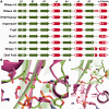
The RNase H-like superfamily: new members, comparative structural analysis and evolutionary classification
- PMID: 24464998
- PMCID: PMC3985635
- DOI: 10.1093/nar/gkt1414
The RNase H-like superfamily: new members, comparative structural analysis and evolutionary classification
Abstract
Ribonuclease H-like (RNHL) superfamily, also called the retroviral integrase superfamily, groups together numerous enzymes involved in nucleic acid metabolism and implicated in many biological processes, including replication, homologous recombination, DNA repair, transposition and RNA interference. The RNHL superfamily proteins show extensive divergence of sequences and structures. We conducted database searches to identify members of the RNHL superfamily (including those previously unknown), yielding >60 000 unique domain sequences. Our analysis led to the identification of new RNHL superfamily members, such as RRXRR (PF14239), DUF460 (PF04312, COG2433), DUF3010 (PF11215), DUF429 (PF04250 and COG2410, COG4328, COG4923), DUF1092 (PF06485), COG5558, OrfB_IS605 (PF01385, COG0675) and Peptidase_A17 (PF05380). Based on the clustering analysis we grouped all identified RNHL domain sequences into 152 families. Phylogenetic studies revealed relationships between these families, and suggested a possible history of the evolution of RNHL fold and its active site. Our results revealed clear division of the RNHL superfamily into exonucleases and endonucleases. Structural analyses of features characteristic for particular groups revealed a correlation between the orientation of the C-terminal helix with the exonuclease/endonuclease function and the architecture of the active site. Our analysis provides a comprehensive picture of sequence-structure-function relationships in the RNHL superfamily that may guide functional studies of the previously uncharacterized protein families.
Figures


Similar articles
-
Structural and evolutionary bioinformatics of the SPOUT superfamily of methyltransferases.BMC Bioinformatics. 2007 Mar 5;8:73. doi: 10.1186/1471-2105-8-73. BMC Bioinformatics. 2007. PMID: 17338813 Free PMC article.
-
Functional insight into Maelstrom in the germline piRNA pathway: a unique domain homologous to the DnaQ-H 3'-5' exonuclease, its lineage-specific expansion/loss and evolutionarily active site switch.Biol Direct. 2008 Nov 25;3:48. doi: 10.1186/1745-6150-3-48. Biol Direct. 2008. PMID: 19032786 Free PMC article.
-
Sequence and comparative structural analysis of the murine leukaemia virus amphotropic strain 4070A RNase H domain.Arch Virol. 1999;144(11):2185-99. doi: 10.1007/s007050050632. Arch Virol. 1999. PMID: 10603172
-
Retroviral integrase superfamily: the structural perspective.EMBO Rep. 2009 Feb;10(2):144-51. doi: 10.1038/embor.2008.256. Epub 2009 Jan 23. EMBO Rep. 2009. PMID: 19165139 Free PMC article. Review.
-
RNases H: Structure and mechanism.DNA Repair (Amst). 2019 Dec;84:102672. doi: 10.1016/j.dnarep.2019.102672. Epub 2019 Jul 20. DNA Repair (Amst). 2019. PMID: 31371183 Review.
Cited by
-
Oxylipin biosynthetic gene families of Cannabis sativa.PLoS One. 2023 Apr 26;18(4):e0272893. doi: 10.1371/journal.pone.0272893. eCollection 2023. PLoS One. 2023. PMID: 37099560 Free PMC article.
-
Comprehensive classification of the PIN domain-like superfamily.Nucleic Acids Res. 2017 Jul 7;45(12):6995-7020. doi: 10.1093/nar/gkx494. Nucleic Acids Res. 2017. PMID: 28575517 Free PMC article.
-
Regulatory RNAs: A Universal Language for Inter-Domain Communication.Int J Mol Sci. 2020 Nov 24;21(23):8919. doi: 10.3390/ijms21238919. Int J Mol Sci. 2020. PMID: 33255483 Free PMC article. Review.
-
Biology and applications of CRISPR-Cas12 and transposon-associated homologs.Nat Biotechnol. 2024 Dec;42(12):1807-1821. doi: 10.1038/s41587-024-02485-9. Epub 2024 Dec 4. Nat Biotechnol. 2024. PMID: 39633151 Review.
-
Resilience of biochemical activity in protein domains in the face of structural divergence.Curr Opin Struct Biol. 2014 Jun;26:92-103. doi: 10.1016/j.sbi.2014.05.008. Epub 2014 Jun 19. Curr Opin Struct Biol. 2014. PMID: 24952217 Free PMC article. Review.
References
-
- Katayanagi K, Miyagawa M, Matsushima M, Ishikawa M, Kanaya S, Ikehara M, Matsuzaki T, Morikawa K. Three- dimensional structure of ribonuclease H from E. coli. Nature. 1990;347:306–309. - PubMed
-
- Yang W, Hendrickson WA, Crouch RJ, Satow Y. Structure of ribonuclease H phased at 2 A resolution by MAD analysis of the selenomethionyl protein. Science. 1990;249:1398–1405. - PubMed
-
- Rice PA, Baker TA. Comparative architecture of transposase and integrase complexes. Nat. Struct. Biol. 2001;8:302–307. - PubMed
-
- Ariyoshi M, Vassylyev DG, Iwasaki H, Nakamura H, Shinagawa H, Morikawa K. Atomic structure of the RuvC resolvase: a holliday junction-specific endonuclease from E. coli. Cell. 1994;78:1063–1072. - PubMed
Publication types
MeSH terms
Substances
Grants and funding
LinkOut - more resources
Full Text Sources
Other Literature Sources
Molecular Biology Databases

