

Absence of Delayed Neuronal Death in ATP-Injected Brain: Possible Roles of Astrogliosis
- PMID: 24465146
- PMCID: PMC3897692
- DOI: 10.5607/en.2013.22.4.308
Absence of Delayed Neuronal Death in ATP-Injected Brain: Possible Roles of Astrogliosis
Abstract
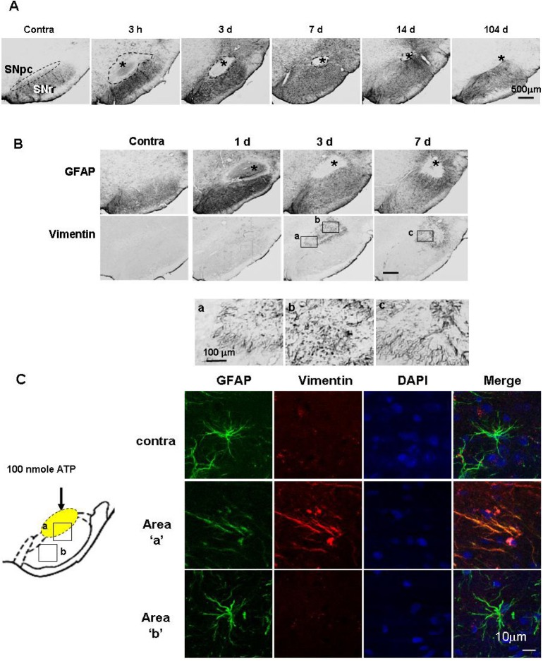
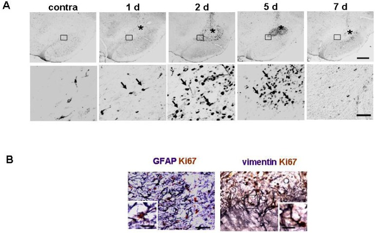
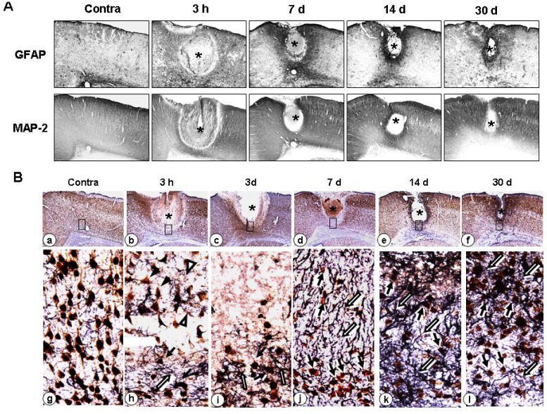
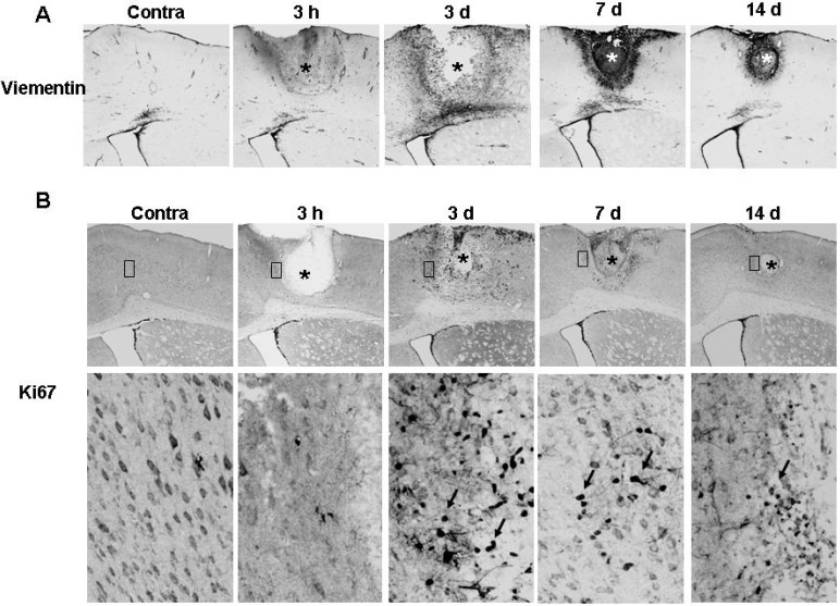
Although secondary delayed neuronal death has been considered as a therapeutic target to minimize brain damage induced by several injuries, delayed neuronal death does not occur always. In this study, we investigated possible mechanisms that prevent delayed neuronal death in the ATP-injected substantia nigra (SN) and cortex, where delayed neuronal death does not occur. In both the SN and cortex, ATP rapidly induced death of the neurons and astrocytes in the injection core area within 3 h, and the astrocytes in the penumbra region became hypertropic and rapidly surrounded the damaged areas. It was observed that the neurons survived for up to 1-3 months in the area where the astrocytes became hypertropic. The damaged areas of astrocytes gradually reduced at 3 days, 7 days, and 1-3 months. Astrocyte proliferation was detectable at 3-7 days, and vimentin was expressed in astrocytes that surrounded and/or protruded into the damaged sites. The NeuN-positive cells also reappeared in the injury sites where astrocytes reappeared. Taken together, these results suggest that astroycte survival and/or gliosis in the injured brain may be critical for neuronal survival and may prevent delayed neuronal death in the injured brain.
Keywords: astrogliosis; brain injury; delayed neuronal death.
Figures
Similar articles
-
Astrogliosis is a possible player in preventing delayed neuronal death.Mol Cells. 2014 Apr;37(4):345-55. doi: 10.14348/molcells.2014.0046. Epub 2014 Apr 22. Mol Cells. 2014. PMID: 24802057 Free PMC article.
-
Inflammatory responses are not sufficient to cause delayed neuronal death in ATP-induced acute brain injury.PLoS One. 2010 Oct 29;5(10):e13756. doi: 10.1371/journal.pone.0013756. PLoS One. 2010. PMID: 21060796 Free PMC article.
-
Adenosine kinase facilitated astrogliosis-induced cortical neuronal death in traumatic brain injury.J Mol Histol. 2016 Jun;47(3):259-71. doi: 10.1007/s10735-016-9670-7. Epub 2016 Mar 16. J Mol Histol. 2016. PMID: 26983602
-
Nucleotide signaling in astrogliosis.Neurosci Lett. 2014 Apr 17;565:14-22. doi: 10.1016/j.neulet.2013.09.056. Epub 2013 Oct 5. Neurosci Lett. 2014. PMID: 24103370 Review.
-
Reactive astrogliosis in stroke: Contributions of astrocytes to recovery of neurological function.Neurochem Int. 2017 Jul;107:88-103. doi: 10.1016/j.neuint.2016.12.016. Epub 2017 Jan 3. Neurochem Int. 2017. PMID: 28057555 Review.
Cited by
-
Neuroprotective effects of Ilexonin A following transient focal cerebral ischemia in rats.Mol Med Rep. 2016 Apr;13(4):2957-66. doi: 10.3892/mmr.2016.4921. Epub 2016 Feb 22. Mol Med Rep. 2016. PMID: 26936330 Free PMC article.
-
Systemic inflammation attenuates the repair of damaged brains through reduced phagocytic activity of monocytes infiltrating the brain.Mol Brain. 2024 Jul 29;17(1):47. doi: 10.1186/s13041-024-01116-3. Mol Brain. 2024. PMID: 39075534 Free PMC article.
-
Astrogliosis is a possible player in preventing delayed neuronal death.Mol Cells. 2014 Apr;37(4):345-55. doi: 10.14348/molcells.2014.0046. Epub 2014 Apr 22. Mol Cells. 2014. PMID: 24802057 Free PMC article.
-
PINK1 expression increases during brain development and stem cell differentiation, and affects the development of GFAP-positive astrocytes.Mol Brain. 2016 Jan 8;9:5. doi: 10.1186/s13041-016-0186-6. Mol Brain. 2016. PMID: 26746235 Free PMC article.
-
Astrocytes, Microglia, and Parkinson's Disease.Exp Neurobiol. 2018 Apr;27(2):77-87. doi: 10.5607/en.2018.27.2.77. Epub 2018 Apr 24. Exp Neurobiol. 2018. PMID: 29731673 Free PMC article. Review.
References
-
- Lee DY, Oh YJ, Jin BK. Thrombin-activated microglia contribute to death of dopaminergic neurons in rat mesencephalic cultures: dual roles of mitogen-activated protein kinase signaling pathways. Glia. 2005;51:98–110. - PubMed
-
- Meda L, Cassatella MA, Szendrei GI, Otvos L, Jr, Baron P, Villalba M, Ferrari D, Rossi F. Activation of microglial cells by beta-amyloid protein and interferon-gamma. Nature. 1995;374:647–650. - PubMed
-
- Chao CC, Hu S, Molitor TW, Shaskan EG, Peterson PK. Activated microglia mediate neuronal cell injury via a nitric oxide mechanism. J Immunol. 1992;149:2736–2741. - PubMed
LinkOut - more resources
Full Text Sources
Other Literature Sources
