

Developmental Role of Anoctamin-1/TMEM16A in Ca(2+)-Dependent Volume Change in Supporting Cells of the Mouse Cochlea
- PMID: 24465148
- PMCID: PMC3897694
- DOI: 10.5607/en.2013.22.4.322
Developmental Role of Anoctamin-1/TMEM16A in Ca(2+)-Dependent Volume Change in Supporting Cells of the Mouse Cochlea
Abstract
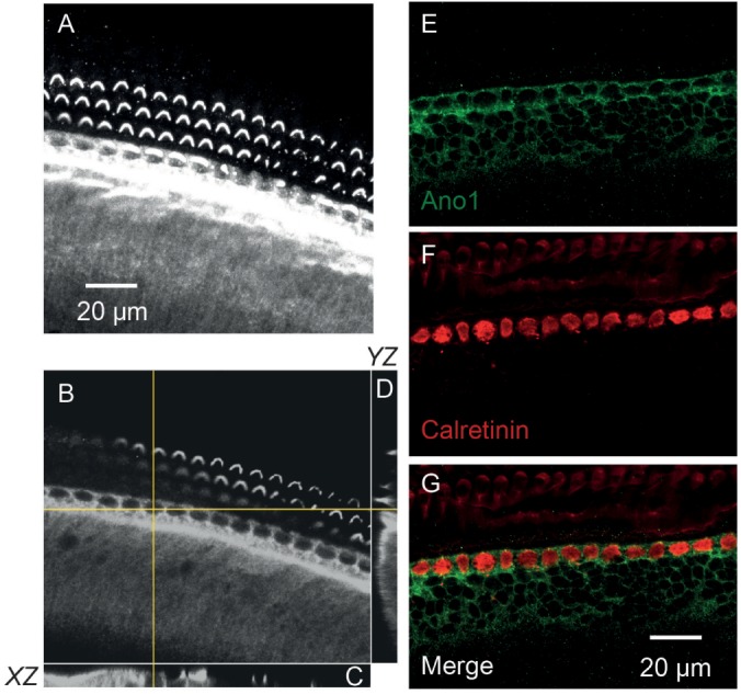
Mammalian cochlea undergoes morphological and functional changes during the postnatal period, around the hearing onset. Major changes during the initial 2 postnatal weeks of mouse include maturation of sensory hair cells and supporting cells, and acquisition of afferent and efferent innervations. During this period, supporting cells in the greater epithelial ridge (GER) of the cochlea exhibit spontaneous and periodic activities which involves ATP, increase in intracellular Ca(2+), and cell volume change. This Ca(2+)-dependent volume change has been proposed to involve chloride channels or transporters. We found that the spontaneous volume changes were eliminated by anion channel blocker, 100 µM NPPB. Among candidates, expression of Anoctamin-1 (Ano1 or TMEM16A), bestriphin-1 and NKCC1 were investigated in whole-mount cochlea of P9-10 mice. Immunolabeling indicated high level of Ano1 expression in the GER, but not of betrophin-1 or NKCC1. Double-labeling with calretinin and confocal image analysis further elucidated the cellular localization of Ano1 immunoreactivity in supporting cells. It was tested if the Ano1 expression exhibits similar time course to the spontaneous activities in postnatal cochlear supporting cells. Cochlear preparations from P2-3, P5-6, P9-10, P15-16 mice were subjected to immunolabeling. High level of Ano1 immunoreactivity was observed in the GER of P2-3, P5-6, P9-10 cochleae, but not of P15-17 cochleae. Taken together, the localization and time course in Ano1 expression pattern correlates with the spontaneous, periodic volume changes recorded in postnatal cochlear supporting cells. From these results we propose that Ano1 is the pacemaker of spontaneous activities in postnatal cochlea.
Keywords: Anoctamin-1 (Ano1)/TMEM16A; Ca2+-activated chloride channel; cell volume change; cochlea; greater epithelial ridge; supporting cell.
Figures


Similar articles
-
Expression and immunohistochemical localization of TMEM16A/anoctamin 1, a calcium-activated chloride channel in the mouse cochlea.Cell Tissue Res. 2011 Aug;345(2):223-30. doi: 10.1007/s00441-011-1206-6. Epub 2011 Jul 21. Cell Tissue Res. 2011. PMID: 21779783
-
Relationship between TMEM16A/anoctamin 1 and LRRC8A.Pflugers Arch. 2016 Oct;468(10):1751-63. doi: 10.1007/s00424-016-1862-1. Epub 2016 Aug 12. Pflugers Arch. 2016. PMID: 27514381
-
The Cl--channel TMEM16A is involved in the generation of cochlear Ca2+ waves and promotes the refinement of auditory brainstem networks in mice.Elife. 2022 Feb 7;11:e72251. doi: 10.7554/eLife.72251. Elife. 2022. PMID: 35129434 Free PMC article.
-
The Ca2+-activated chloride channel ANO1/TMEM16A: An emerging therapeutic target for epithelium-originated diseases?Acta Pharm Sin B. 2021 Jun;11(6):1412-1433. doi: 10.1016/j.apsb.2020.12.003. Epub 2020 Dec 9. Acta Pharm Sin B. 2021. PMID: 34221860 Free PMC article. Review.
-
Calcium-Activated Chloride Channel ANO1/TMEM16A: Regulation of Expression and Signaling.Front Physiol. 2020 Nov 5;11:590262. doi: 10.3389/fphys.2020.590262. eCollection 2020. Front Physiol. 2020. PMID: 33250781 Free PMC article. Review.
Cited by
-
A nonsense TMEM43 variant leads to disruption of connexin-linked function and autosomal dominant auditory neuropathy spectrum disorder.Proc Natl Acad Sci U S A. 2021 Jun 1;118(22):e2019681118. doi: 10.1073/pnas.2019681118. Proc Natl Acad Sci U S A. 2021. PMID: 34050020 Free PMC article.
-
Suppression of 14-3-3γ-mediated surface expression of ANO1 inhibits cancer progression of glioblastoma cells.Sci Rep. 2016 May 23;6:26413. doi: 10.1038/srep26413. Sci Rep. 2016. PMID: 27212225 Free PMC article.
-
Spontaneous Activity of Cochlear Hair Cells Triggered by Fluid Secretion Mechanism in Adjacent Support Cells.Cell. 2015 Dec 3;163(6):1348-59. doi: 10.1016/j.cell.2015.10.070. Epub 2015 Nov 25. Cell. 2015. PMID: 26627734 Free PMC article.
-
Kölliker's organ and the development of spontaneous activity in the auditory system: implications for hearing dysfunction.Biomed Res Int. 2014;2014:367939. doi: 10.1155/2014/367939. Epub 2014 Aug 20. Biomed Res Int. 2014. PMID: 25210710 Free PMC article. Review.
-
Modulating Ca²⁺ signals: a common theme for TMEM16, Ist2, and TMC.Pflugers Arch. 2016 Mar;468(3):475-90. doi: 10.1007/s00424-015-1767-4. Epub 2015 Dec 23. Pflugers Arch. 2016. PMID: 26700940 Review.
References
-
- Kros CJ, Ruppersberg JP, Rüsch A. Expression of a potassium current in inner hair cells during development of hearing in mice. Nature. 1998;394:281–284. - PubMed
-
- Sobkowicz HM, Slapnick SM, August BK. Differentiation of spinous synapses in the mouse organ of corti. Synapse. 2002;45:10–24. - PubMed
-
- Simmons DD, Mansdorf NB, Kim JH. Olivocochlear innervation of inner and outer hair cells during postnatal maturation: evidence for a waiting period. J Comp Neurol. 1996;370:551–562. - PubMed
LinkOut - more resources
Full Text Sources
Other Literature Sources
Research Materials
Miscellaneous
