

Sex-steroid regulation of relaxin receptor isoforms (RXFP1 & RXFP2) expression in the patellar tendon and lateral collateral ligament of female WKY rats
- PMID: 24465164
- PMCID: PMC3894403
- DOI: 10.7150/ijms.6283
Sex-steroid regulation of relaxin receptor isoforms (RXFP1 & RXFP2) expression in the patellar tendon and lateral collateral ligament of female WKY rats
Abstract
The incidence of non-contact knee injury was found higher in female than in male and is related to the phases of the menstrual cycle. This raised the possibility that female sex-steroids are involved in the mechanism underlying this injury via affecting the expression of the receptors for relaxin, a peptide hormone known to modulate ligament laxity. Therefore, this study aims to investigate the effect of sex-steroids on relaxin receptor isoforms (RXFP1 & RXFP2) expression in the ligaments and tendons of the knee.
Methods: Ovariectomized adult female WKY rats were treated with different doses of estrogen (0.2, 2, 20 μg/kg), progesterone (4mg) and testosterone (125 & 250μg/kg) for three consecutive days. At the end of the treatment, the animals were sacrificed and the patellar tendon and lateral collateral ligament were harvested for mRNA and protein expression analyses by Real Time PCR and Western blotting respectively.
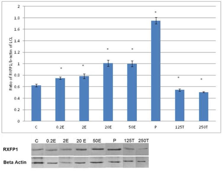
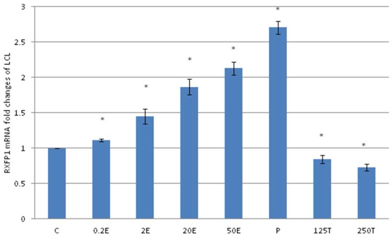
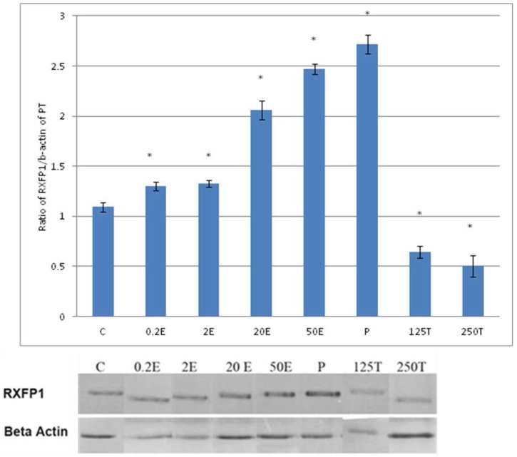
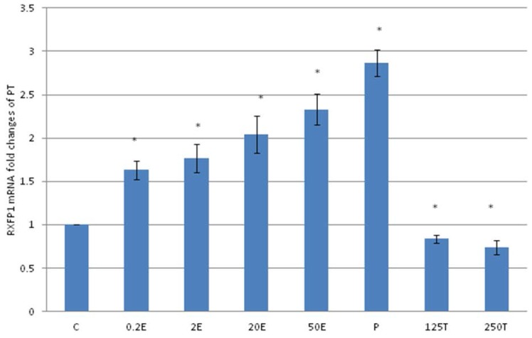
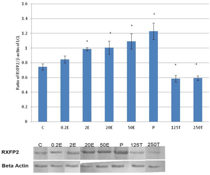
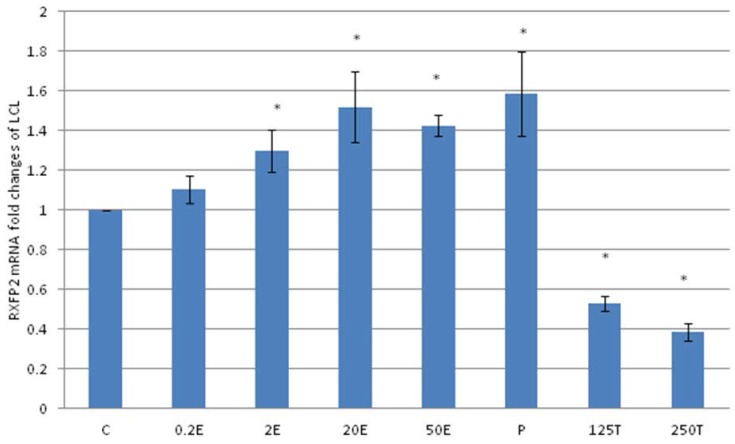
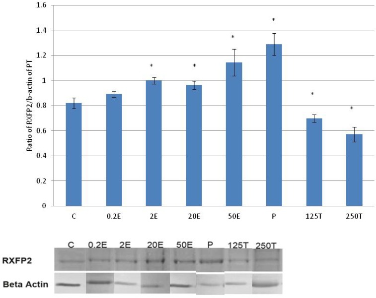
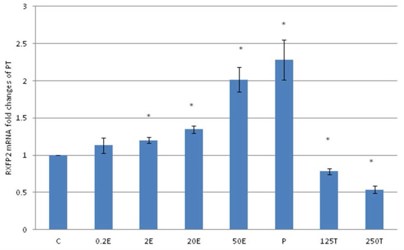
Results: RXFP1, the main isoform expressed in these knee structures and RXFP2 showed a dose-dependent increase in expression with estrogen. Progesterone treatment resulted in an increase while testosterone caused a dose-dependent decrease in the mRNA and protein expression of both relaxin receptor isoforms.
Discussion: Progesterone and high dose estrogen up-regulate while testosterone down-regulates RXFP1 and RXFP2 expression in the patellar tendon and lateral collateral ligament of rat's knee.
Conclusion: Relaxin receptor isoforms up-regulation by progesterone and high dose estrogen could provide the basis for the reported increase in knee laxity while down-regulation of these receptor isoforms by testosterone could explain low incidence of non-contact knee injury in male.
Keywords: RXFP1; RXFP2; collateral ligaments.; patellar tendon; sex-steroids.
Conflict of interest statement
Conflict of interest: There is no conflict of interest in this study.
Figures
Similar articles
-
Changes in Knee Laxity and Relaxin Receptor Isoforms Expression (RXFP1/RXFP2) in the Knee throughout Estrous Cycle Phases in Rodents.PLoS One. 2016 Aug 11;11(8):e0160984. doi: 10.1371/journal.pone.0160984. eCollection 2016. PLoS One. 2016. PMID: 27513858 Free PMC article.
-
Estrogen receptor (ER)-α, β and progesterone receptor (PR) mediates changes in relaxin receptor (RXFP1 and RXFP2) expression and passive range of motion of rats' knee.Environ Toxicol Pharmacol. 2015 Nov;40(3):785-91. doi: 10.1016/j.etap.2015.09.004. Epub 2015 Sep 8. Environ Toxicol Pharmacol. 2015. PMID: 26447688
-
Testosterone reduces knee passive range of motion and expression of relaxin receptor isoforms via 5α-dihydrotestosterone and androgen receptor binding.Int J Mol Sci. 2014 Mar 17;15(3):4619-34. doi: 10.3390/ijms15034619. Int J Mol Sci. 2014. PMID: 24642882 Free PMC article.
-
Relaxin family peptide receptors--former orphans reunite with their parent ligands to activate multiple signalling pathways.Br J Pharmacol. 2007 Mar;150(6):677-91. doi: 10.1038/sj.bjp.0707140. Epub 2007 Feb 12. Br J Pharmacol. 2007. PMID: 17293890 Free PMC article. Review.
-
Sex Hormones and Tendon.Adv Exp Med Biol. 2016;920:139-49. doi: 10.1007/978-3-319-33943-6_13. Adv Exp Med Biol. 2016. PMID: 27535256 Review.
Cited by
-
Changes in Knee Laxity and Relaxin Receptor Isoforms Expression (RXFP1/RXFP2) in the Knee throughout Estrous Cycle Phases in Rodents.PLoS One. 2016 Aug 11;11(8):e0160984. doi: 10.1371/journal.pone.0160984. eCollection 2016. PLoS One. 2016. PMID: 27513858 Free PMC article.
-
Testosterone decreases the expression of endometrial pinopode and L-selectin ligand (MECA-79) in adult female rats during uterine receptivity period.Int J Clin Exp Pathol. 2014 Apr 15;7(5):1967-76. eCollection 2014. Int J Clin Exp Pathol. 2014. PMID: 24966906 Free PMC article.
-
ACL Research Retreat VII: An Update on Anterior Cruciate Ligament Injury Risk Factor Identification, Screening, and Prevention.J Athl Train. 2015 Oct;50(10):1076-93. doi: 10.4085/1062-6050-50.10.06. Epub 2015 Sep 4. J Athl Train. 2015. PMID: 26340613 Free PMC article. No abstract available.
-
MicroRNA-144-3p targets relaxin/insulin-like family peptide receptor 1 (RXFP1) expression in lung fibroblasts from patients with idiopathic pulmonary fibrosis.J Biol Chem. 2019 Mar 29;294(13):5008-5022. doi: 10.1074/jbc.RA118.004910. Epub 2019 Feb 1. J Biol Chem. 2019. PMID: 30709904 Free PMC article.
-
Relaxin as a treatment for musculoskeletal fibrosis: What we know and future directions.Biochem Pharmacol. 2024 Jul;225:116273. doi: 10.1016/j.bcp.2024.116273. Epub 2024 May 8. Biochem Pharmacol. 2024. PMID: 38729446 Free PMC article. Review.
References
-
- Park JI, Chang C, Hsu S. New Insights into Biological Roles of Relaxin and Relaxin-related Peptides. Rev Endocr Metab Disord. 2005;6(4):291–296. - PubMed
-
- Nair VB Samuel, C S, Separovic F, Hossain M A, Wade JD. Human relaxin-2: historical perspectives and role in cancer biology. Amino acids. 2012;43(3):1131–1140. - PubMed
-
- Scott DJ, Layfield S, Riesewijk A, Morita H, Tregear GW, Bathgate RA. Characterization of the mouse and rat relaxin receptors. Ann N Y Acad Sci. 2005;1041:8–12. - PubMed
Publication types
MeSH terms
Substances
LinkOut - more resources
Full Text Sources
Other Literature Sources
Medical
Molecular Biology Databases
