. 2014 Jan;10(1):e1004005.
doi: 10.1371/journal.pgen.1004005. Epub 2014 Jan 23.

Down-regulation of Rad51 activity during meiosis in yeast prevents competition with Dmc1 for repair of double-strand breaks

Affiliations

Down-regulation of Rad51 activity during meiosis in yeast prevents competition with Dmc1 for repair of double-strand breaks

Yan Liu et al. PLoS Genet. 2014 Jan.

Abstract

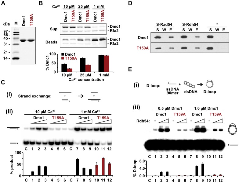
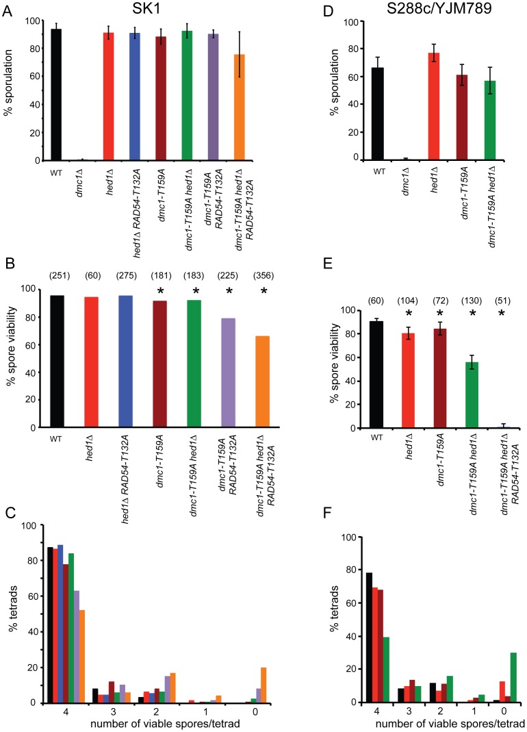
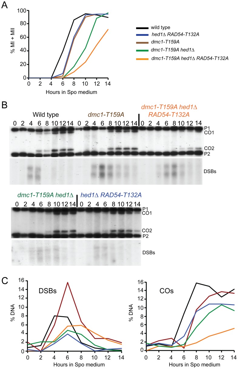
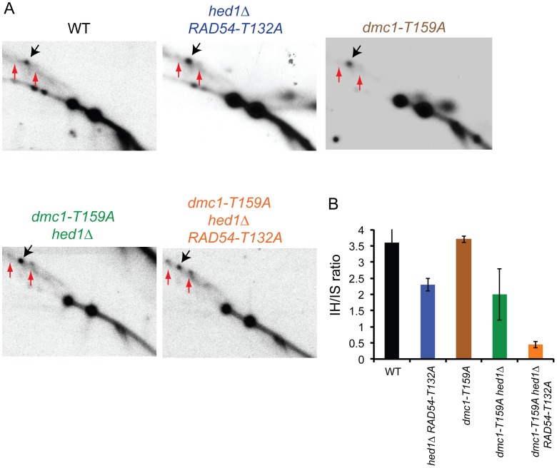
Interhomolog recombination plays a critical role in promoting proper meiotic chromosome segregation but a mechanistic understanding of this process is far from complete. In vegetative cells, Rad51 is a highly conserved recombinase that exhibits a preference for repairing double strand breaks (DSBs) using sister chromatids, in contrast to the conserved, meiosis-specific recombinase, Dmc1, which preferentially repairs programmed DSBs using homologs. Despite the different preferences for repair templates, both Rad51 and Dmc1 are required for interhomolog recombination during meiosis. This paradox has recently been explained by the finding that Rad51 protein, but not its strand exchange activity, promotes Dmc1 function in budding yeast. Rad51 activity is inhibited in dmc1Δ mutants, where the failure to repair meiotic DSBs triggers the meiotic recombination checkpoint, resulting in prophase arrest. The question remains whether inhibition of Rad51 activity is important during wild-type meiosis, or whether inactivation of Rad51 occurs only as a result of the absence of DMC1 or checkpoint activation. This work shows that strains in which mechanisms that down-regulate Rad51 activity are removed exhibit reduced numbers of interhomolog crossovers and noncrossovers. A hypomorphic mutant, dmc1-T159A, makes less stable presynaptic filaments but is still able to mediate strand exchange and interact with accessory factors. Combining dmc1-T159A with up-regulated Rad51 activity reduces interhomolog recombination and spore viability, while increasing intersister joint molecule formation. These results support the idea that down-regulation of Rad51 activity is important during meiosis to prevent Rad51 from competing with Dmc1 for repair of meiotic DSBs.


Conflict of interest statement

The authors have declared that no competing interests exist.

Figures

Figure 1
Figure 1. Biochemical characterization of recombinant Dmc1-T159A protein.
A. For Dmc1 and Dmc1-T159A, 2.5 µg of each protein were analyzed by SDS-polyacrylimide gel electrophoresis (PAGE) and Coomassie staining. B. The stability of Dmc1 filaments on bead-immobilized ssDNA was assessed by exposing the filaments to RPA and measuring the amount of Dmc1 retained on the beads (n = 3, +/− standard error). Proteins were monitored by SDS-PAGE and Coomassie staining. The experiments were performed with different Ca2+ concentrations as indicated. Rfa2, the 30 kDa subunit of RPA is indicated. The histogram shows the percent of Dmc1 protein that remained associated with the beads after challenge by RPA as determined by band desitometry. C. (i) Schematic of the strand exchange recombination assay. (ii) Strand exchange activity of Dmc1 and Dmc1-T159A was monitored using 2, 4, or 8 µM protein in the presence of either 10 µM or 1 mM Ca2+. The histogram indicates the percent of radioactively labeled oligonucleotide that was incorporated into the slower migrating product by strand exchange (n = 3, +/− standard error). At 1 mM Ca2+, the inhibition of strand exchange seen at elevated concentrations of wild-type Dmc1 is likely due to coating of the dsDNA substrate by the recombinase, thereby blocking access of the ssDNA filament. D. Dmc1 and Dmc1-T159A interactions with Rad54 and Rdh54 were assayed by pull-down experiments. S-tagged Rad54 or Rdh54 (2 µg each) was incubated with 1.2 µg Dmc1 or Dmc1-T159A. The S-tagged protein was captured on S-protein agarose resin, which was washed and the protein eluted with SDS. S = supernatant after collecting the beads, W = supernatant obtained from washing the beads, E = eluate from beads. The “-” indicates that no tagged protein was added to the reaction. E. (i) Schematic of the D-loop recombination assay. (ii) Homologous DNA pairing activity of Dmc1 and Dmc1-T159A was assessed by a D-loop formation assay (n = 3, +/− standard error). 0.5 or 1.0 µM of Dmc1 or Dmc1-T159A was combined with 0, 150, or 250 nM of Rdh54. The histogram shows the percent of radioactive ssDNA that is incorporated into the slower migrating D-loop product via homologous pairing.
Figure 2
Figure 2. Characterization of various meiotic phenotypes in diploids containing dmc1-T159A.
A. Sporulation in SK1 diploids: Wild-type (NH716), dmc1Δ (NH792), hed1Δ (NH1065), hed1Δ RAD54-T132A (NH1065::pHN104(S/N)2, dmc1-T159A (NH792::pNH301-T159A2), dmc1-T159A hed1Δ (NH942::pNH301-T159A2), dmc1-T159A RAD54-T132A (NH2231) and dmc1-T159A hed1Δ RAD54-T132A (NH2184) cells were transferred to Spo medium on plates for two days at 30°C and the percent sporulation was determined by phase contrast microscopy. 200 cells from at least four independent colonies were examined. Error bars represent the standard error. B. Spore viability in SK1 strains was assayed by tetrad dissection from at least four independent colonies. Numbers in parentheses indicate the number of tetrads dissected. * indicates that the spore viability is statistically significantly different from wild type by χ2 analysis. The p values are dmc1-T159A (<0.001); dmc1-T159A hed1Δ (<0.006); dmc1-T159A RAD54-T132A (<0.0001); dmc1-T159A hed1Δ RAD54-T132A (<0.0001) C. The distribution of viable spores in tetrads from the asci dissected for Panel B. D. Sporulation in S288c/YJM789 diploids: Wild-type (NH1053), dmc1Δ (NH2030), hed1Δ (NH2038), dmc1-T159A (NH2142), dmc1-T159A hed1Δ (NH2145), and dmc1-T159A hed1Δ RAD54-T132A (NH2146) cells were assayed for sporulation after four days on Spo plates at 30°C. E. Spore viability was assayed by dissection of at least 3 independent colonies. * indicates that the spore viability is statistically significantly different from wild type by χ2 analysis. The p values are hed1Δ (<0.001); dmc1-T159A (<0.02); dmc1-T159A hed1Δ (<0.0001); dmc1-T159A hed1Δ RAD54-T132A (<0.0001). F. The distribution of viable spores in tetrads from the asci dissected for Panel E.
Figure 3
Figure 3. Meiotic progression and crossover formation in various dmc1-T159A SK1 strains.
Wild-type, hed1Δ RAD54-T132A, dmc1-T159A, dmc1-T159A hed1Δ and dmc1-T159A hed1Δ RAD54-T132A diploids were transferred to Spo medium at 30°C at 0 hr and samples were taken at two hour intervals. Color coding is the same as in Figure 2. A. Meiotic progression was measured by staining the nuclei with DAPI and counting the fraction of bi-nucleate (MI) and tetranucleate (MII) cells. B. Crossovers and DSBs at the HIS4/LEU2 hotspot. The DNA was digested with XhoI and probed as described in . P1 and P2 represent the parental fragments and CO1 and CO2 represent the two products of reciprocal recombination. Numbers above each lane indicate the hours after transfer to Spo medium. C. Quantitation of the crossover and DSBs bands shown in Panel B. A replicate of this experiment is shown in Figure S1.
Figure 4
Figure 4. Meiotic joint molecule analysis in various SK1 dmc1-T159A ndt80Δ strains.
ndt80Δ (NH2188), hed1Δ ndt80Δ RAD54-T132A (NH2223::pHN104(S/N)2, dmc1-T159A ndt80Δ (NH2235), dmc1-T159A hed1Δ ndt80Δ (NH2190) and dmc1-T159A hed1Δ ndt80Δ RAD54-T132A (NH2193) diploids were transferred to Spo medium for nine hours to arrest the cells in pachytene and the DNA was crosslinked with psoralen, extracted and digested with XhoI. Color coding is the same as in Figure 2. A. Southern blots of two-dimensional gels probed to detect interhomolog JMs (indicated by black arrows) and intersister JMs (indicated by red arrows) as described in . B. Quantitation of the ratio of interhomolog:intersister joint molecules in the gels shown in A averaged with a second replicate. Error bars indicate the standard error.

Comment in

Similar articles

Cited by

References

    1. Petronczki M, Siomos MF, Nasmyth K (2003) Un menage a quatre: the molecular biology of chromosome segregation in meiosis. Cell 112: 423–440. - PubMed
    1. Hassold T, Hunt P (2001) To err (meiotically) is human: the genesis of human aneuploidy. Nature reviews 2: 280–291. - PubMed
    1. Keeney S (2008) Spo11 and the formation of DNA double-strand breaks in meiosis. Genome dynamics and stability 2: 81–123. - PMC - PubMed
    1. Neale MJ, Keeney S (2006) Clarifying the mechanics of DNA strand exchange in meiotic recombination. Nature 442: 153–158. - PMC - PubMed
    1. Sheridan S, Bishop DK (2006) Red-Hed regulation: recombinase Rad51, though capable of playing the leading role, may be relegated to supporting Dmc1 in budding yeast meiosis. Genes Dev 20: 1685–1691. - PubMed

Publication types

MeSH terms