. 2014 Jan;10(1):e1004091.
doi: 10.1371/journal.pgen.1004091. Epub 2014 Jan 23.

Arabidopsis AL PHD-PRC1 complexes promote seed germination through H3K4me3-to-H3K27me3 chromatin state switch in repression of seed developmental genes

Affiliations

Arabidopsis AL PHD-PRC1 complexes promote seed germination through H3K4me3-to-H3K27me3 chromatin state switch in repression of seed developmental genes

Anne Marie Molitor et al. PLoS Genet. 2014 Jan.

Abstract

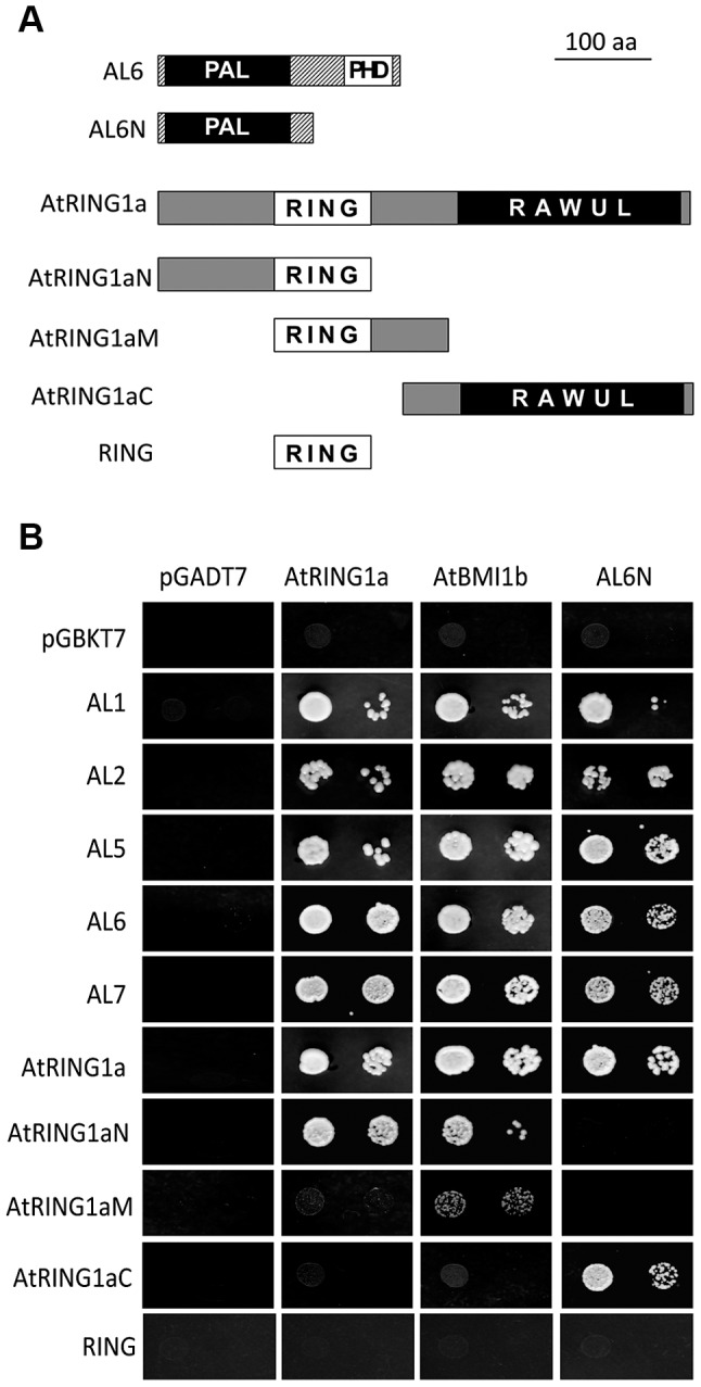
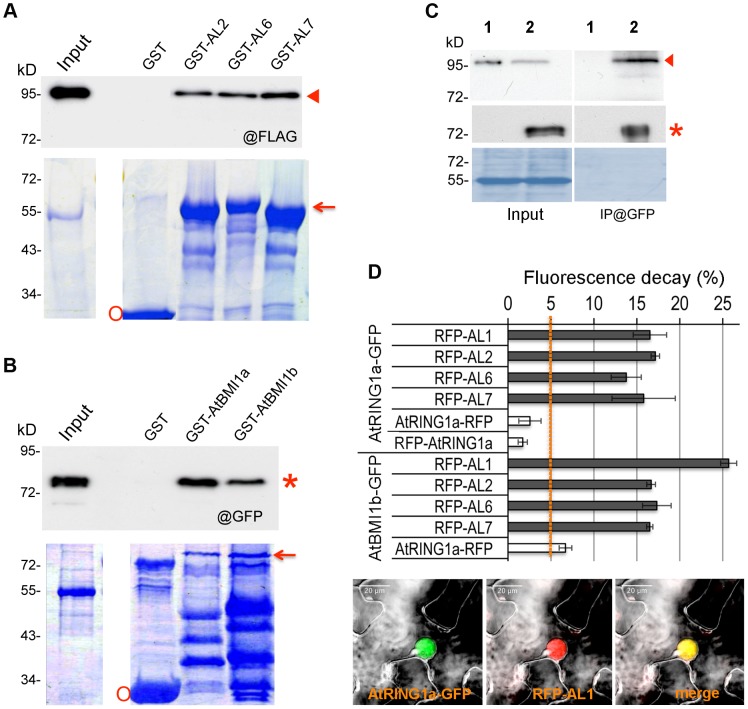
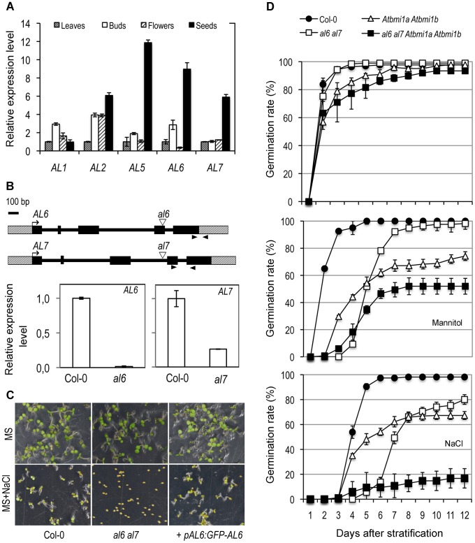
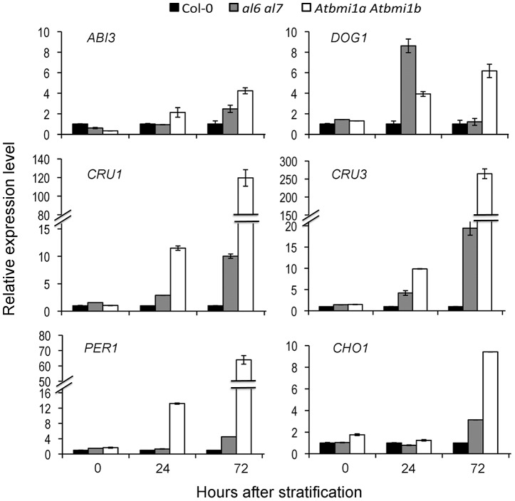
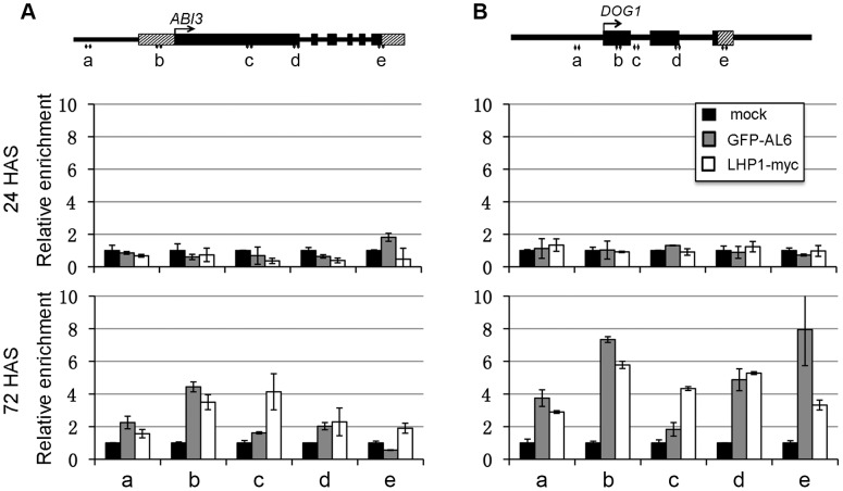
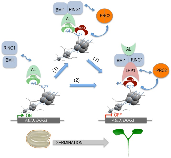
Seed germination and subsequent seedling growth define crucial steps for entry into the plant life cycle. For those events to take place properly, seed developmental genes need to be silenced whereas vegetative growth genes are activated. Chromatin structure is generally known to play crucial roles in gene transcription control. However, the transition between active and repressive chromatin states during seed germination is still poorly characterized and the underlying molecular mechanisms remain largely unknown. Here we identified the Arabidopsis PHD-domain H3K4me3-binding ALFIN1-like proteins (ALs) as novel interactors of the Polycomb Repressive Complex 1 (PRC1) core components AtBMI1b and AtRING1a. The interactions were confirmed by diverse in vitro and in vivo assays and were shown to require the AL6 N-terminus containing PAL domain conserved in the AL family proteins and the AtRING1a C-terminus containing RAWUL domain conserved in animal and plant PRC1 ring-finger proteins (including AtRNIG1a/b and AtBMI1a/b). By T-DNA insertion mutant analysis, we found that simultaneous loss of AL6 and AL7 as well as loss of AtBMI1a and AtBMI1b retards seed germination and causes transcriptional derepression and a delayed chromatin state switch from H3K4me3 to H3K27me3 enrichment of several seed developmental genes (e.g. ABI3, DOG1, CRU3, CHO1). We found that AL6 and the PRC1 H3K27me3-reader component LHP1 directly bind at ABI3 and DOG1 loci. In light of these data, we propose that AL PHD-PRC1 complexes, built around H3K4me3, lead to a switch from the H3K4me3-associated active to the H3K27me3-associated repressive transcription state of seed developmental genes during seed germination. Our finding of physical interactions between PHD-domain proteins and PRC1 is striking and has important implications for understanding the connection between the two functionally opposite chromatin marks: H3K4me3 in activation and H3K27me3 in repression of gene transcription.


Conflict of interest statement

The authors have declared that no competing interests exist.

Figures

Figure 1
Figure 1. Interactions of ALs and PRC1 ring-finger proteins in yeast two-hybrid assay.
(A) Schematic representation of full-length and truncated AtRING1a and AL6 proteins. The conserved domains PAL, PHD, RING and RAWUL are indicated. (B) Yeast two-hybrid assays. Yeast cultures co-expressing the indicated protein combinations from pGADT7 and pGBKT7 were plated as a 1∶10 dilution from left to right onto SD-LTA selective media. Growth of yeast cells indicates positive protein-protein interaction.
Figure 2
Figure 2. ALs physically interact with PRC1 ring-finger proteins in both in vitro and in vivo assays.
(A) Pulldown assay. Agarose beads coated with GST, GST-AL2, GST-AL6 or GST-AL7 were incubated with an equal aliquot of total protein extracts of Arabidopsis plants expressing FLAG-AtRING1a. The pulldown fractions and inputs were analyzed by Western blot using antibodies against FLAG (@FLAG, top panel). Coomassie staining is shown as loading control (bottom panel). The positions of FLAG-AtRING1a, GST-AL and GST are indicated by arrowhead, arrow and circle, respectively. (B) Pulldown assay. Agarose beads coated with GST, GST-AtBMI1a or GST-AtBMI1b were incubated with an equal aliquot of total protein extracts of Arabidopsis plants expressing GFP-AL6. The pulldown fractions and inputs were analyzed by Western blot using antibodies against GFP (@GFP, top panel). Coomassie staining is shown as loading control (bottom panel). The positions of GFP-AL6, GST-AtBMI1 and GST are indicated by star, arrow and circle, respectively. (C) Co-IP detection of AtRING1a and AL6 interaction in planta. Total protein extracts from plants expressing FLAG-AtRING1a alone (lane 1) or both FLAG-AtRING1a and GFP-AL6 (lane 2) were immunoprecipitated with a polyclonal anti-GFP antibody, and the resulting fractions were analyzed by Western blot using anti-FLAG (top panel) or HRP-conjugated anti-GFP (middle panel) monoclonal antibodies. Coomassie staining (bottom panel) is shown as loading quality control. Arrowhead and star indicate FLAG-AtRING1a and GFP-AL6 positions, respectively. (D) FLIM detection of AtRING1a-GFP or AtBMI1b-GFP interaction with RFP-ALs in planta. GFP- and RFP-tagged proteins as indicated were transiently co-expressed in Nicotiana benthamiana leaves. The fluorescence lifetime of GFP fusion proteins was recorded two days post infiltration. Data represent average GFP fluorescence lifetime decay ± SD of three biological replicates, each recording over 30 nuclei. Values above 5% indicate positive protein-protein interactions. The bottom image panels show co-localization of AtRING1a-GFP and RFP-AL1 in the nucleus of a leaf epidermal cell.
Figure 3
Figure 3. Functional characterization of AL genes.
(A) Tissue-specificity of AL gene expression. Relative expression levels of AL1, AL2, AL5, AL6, and AL7 were determined by quantitative RT-PCR in different plant organs. Leaves: rosette leaves from 4-week-old plants; Buds: floral buds before anthesis; Flowers: flowers at anthesis; Seeds: dry seeds. Data represent means ± SD of three biological replicates. (B) AL6 and AL7 genomic structure and T-DNA insertion mutants. Genes are schematically represented by black boxes for exons, black lines for introns and dashed boxes for untranslated regions. Triangles indicate T-DNA insertion sites and arrowheads indicate RT-PCR primer positions. Relative expression levels of AL6 and AL7 in Col-0 and in al6 and al7 mutants are shown as means ± SD of three biological replicates. (C) Representative seed germination images of Col-0, al6 al7 double mutant, and the double mutant complemented by the AL6 promoter driving GFP-AL6 fusion gene (+pAL6:GFP-AL6). Images were taken five days after stratification from plates containing MS media or MS supplemented with 100 mM NaCl (MS+NaCl). (D) Germination rate of Col-0, double mutants al6 al7 and Atbmi1a Atbmi1b, and the quadruple mutant al6 al7 Atbmi1a Atbmi1b plated on MS (top graph), MS supplemented with 200 mM mannitol (middle graph) or with 100 mM NaCl (bottom graph). Data represent average germination percentages ± SD of three biological replicates, each >60 seeds, observed daily for 12 days after stratification.
Figure 4
Figure 4. Relative expression levels of seed developmental genes in Col-0, al6 al7 and Atbmi1a Atbmi1b.
Relative expression levels of ABI3, DOG1, CRU1, CRU3, PER1 and CHO1 were analyzed by quantitative RT-PCR using seeds/seedlings at 0, 24 and 72 hours after stratification. Data represent means ± SD of three biological replicates.
Figure 5
Figure 5. Relative enrichments of H3K4me3 and H3K27me3 at seed developmental genes in Col-0, al6 al7 and Atbmi1a Atbmi1b.
H3K4me3 and H3K27me3 levels were analyzed by ChIP at five regions (a to e) of ABI3 (A) and DOG1 (B). Gene structures are schematically represented by black boxes for exons, black lines for introns and promoters, and dashed boxes for untranslated regions. Seeds/seedlings at 0, 24 and 72 hours after stratification (HAS) were analyzed. Values were normalized to internal controls (relative to input and to TUB2). Data represent means ± SD of three biological replicates.
Figure 6
Figure 6. LHP1 and AL6 binding at ABI3 and DOG1 chromatin.
Relative enrichments of LHP1-myc and GFP-AL6 proteins were analyzed at the five regions (a to e) of ABI3 (A) and DOG1 (B) loci. Transgenic seeds/seedlings expressing LHP1-myc or GFP-AL6 were analyzed at 24 or 72 hours after stratification (HAS) by ChIP using anti-myc or anti-GFP antibodies. Samples in the absence of antibodies serve as negative controls (mock). Values were normalized to internal controls (relative to input and to TUB2). Data represent means ± SD of three biological replicates.
Figure 7
Figure 7. A proposed model for AL PHD-PRC1 complexes in silencing seed developmental genes during seed germination.
ALs, via their highly conserved PHD domains, bind H3K4me3 of chromatin, triggering the recruitment of PRC1 components BMI1 and RING1 via AL-AtBMI1, AL-AtRING1, and AtBMI1-AtRING1 physical interactions. Next, two possible pathways (1 and 2) can lead to stable repressive chromatin state formation. In the first case (1), PRC2 is recruited via its subunit CLF interaction with AtRING1 and deposits H3K27me3, favoring further LHP1 recruitment via H3K27me3-LHP1 binding. In the second case (2), LHP1 is first recruited via its interaction with AtRING1 or AtBMI1, and then PRC2 is recruited via its subunit MSI1 interaction with LHP1 and deposits H3K27me3. In both cases, H3K27me3-LHP1 and PRC2 MSI1-LHP1 interactions form a positive loop in H3K27me3 enrichment. This hypothetic model can explain how seed developmental genes (e.g. ABI3, DOG1) are switched from active transcription to a stably repressed state, which is necessary for timely seed germination and proper seedling growth and development.

Similar articles

Cited by

References

    1. Bentsink L, Koornneef M (2008) Seed dormancy and germination. Arabidopsis Book 6: e0119. - PMC - PubMed
    1. Koornneef M, Hanhart CJ, Hilhorst HW, Karssen CM (1989) In vivo inhibition of seed development and reserve protein accumulation in recombinants of abscisic acid biosynthesis and responsiveness mutants in Arabidopsis thaliana . Plant Physiol 90: 463–469. - PMC - PubMed
    1. Giraudat J, Hauge BM, Valon C, Smalle J, Parcy F, et al. (1992) Isolation of the Arabidopsis ABI3 gene by positional cloning. Plant Cell 4: 1251–1261. - PMC - PubMed
    1. Parcy F, Valon C, Raynal M, Gaubier-Comella P, Delseny M, et al. (1994) Regulation of gene expression programs during Arabidopsis seed development: roles of the ABI3 locus and of endogenous abscisic acid. Plant Cell 6: 1567–1582. - PMC - PubMed
    1. Mönke G, Seifert M, Keilwagen J, Mohr M, Grosse I, et al. (2012) Toward the identification and regulation of the Arabidopsis thaliana ABI3 regulon. Nucleic Acids Res 40: 8240–8254. - PMC - PubMed

Publication types

MeSH terms

LinkOut - more resources