

Focused fluorescence excitation with time-reversed ultrasonically encoded light and imaging in thick scattering media
- PMID: 24465244
- PMCID: PMC3900304
- DOI: 10.1088/1612-2011/10/7/075604
Focused fluorescence excitation with time-reversed ultrasonically encoded light and imaging in thick scattering media
Abstract
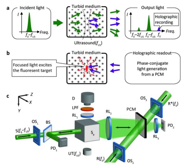
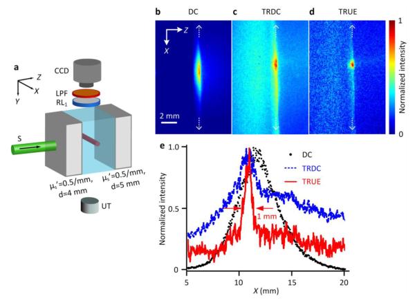
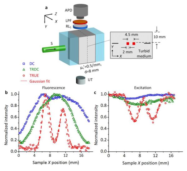
Scattering dominates light propagation in biological tissue, and therefore restricts both resolution and penetration depth in optical imaging within thick tissue. As photons travel into the diffusive regime-typically 1 mm beneath human skin, their trajectories transition from ballistic to diffusive due to increased number of scattering events, which makes it impossible to focus, much less track, photon paths. Consequently, imaging methods that rely on controlled light illumination are ineffective in deep tissue. This problem has recently been addressed by a novel method capable of dynamically focusing light in thick scattering media via time reversal of ultrasonically encoded (TRUE) diffused light. Here, using photorefractive materials as phase conjugate mirrors, we show a direct visualization and dynamic control of optical focusing with this light delivery method, and demonstrate its application for focused fluorescence excitation and imaging in thick turbid media. These abilities are increasingly critical to understanding the dynamic interactions of light with biological matter and processes at different system levels, as well as their applications for biomedical diagnosis and therapy.
Keywords: Fluorescence imaging; Optical focusing; Optical imaging; Optical scattering; Phase conjugation; Photorefractive effect; Time-reversal; Ultrasound-modulation.
Figures
Similar articles
-
Time-reversed ultrasonically encoded optical focusing into tissue-mimicking media with thickness up to 70 mean free paths.J Biomed Opt. 2011 Aug;16(8):086009. doi: 10.1117/1.3609004. J Biomed Opt. 2011. PMID: 21895321 Free PMC article.
-
Snapshot time-reversed ultrasonically encoded optical focusing guided by time-reversed photoacoustic wave.Photoacoustics. 2022 Apr 5;26:100352. doi: 10.1016/j.pacs.2022.100352. eCollection 2022 Jun. Photoacoustics. 2022. PMID: 35433254 Free PMC article.
-
Time-reversed ultrasonically encoded optical focusing into scattering media.Nat Photonics. 2011 Mar;5(3):154. doi: 10.1038/nphoton.2010.306. Nat Photonics. 2011. PMID: 21532925 Free PMC article.
-
Wavefront shaping: A versatile tool to conquer multiple scattering in multidisciplinary fields.Innovation (Camb). 2022 Aug 2;3(5):100292. doi: 10.1016/j.xinn.2022.100292. eCollection 2022 Sep 13. Innovation (Camb). 2022. PMID: 36032195 Free PMC article. Review.
-
Scanless two-photon excitation with temporal focusing.Nat Methods. 2020 Jun;17(6):571-581. doi: 10.1038/s41592-020-0795-y. Epub 2020 Apr 13. Nat Methods. 2020. PMID: 32284609 Review.
Cited by
-
Frequency-swept time-reversed ultrasonically encoded optical focusing.Appl Phys Lett. 2014 Nov 10;105(19):191108. doi: 10.1063/1.4901955. Epub 2014 Nov 11. Appl Phys Lett. 2014. PMID: 25425744 Free PMC article.
-
Ultrasonically encoded wavefront shaping for focusing into random media.Sci Rep. 2014 Jan 29;4:3918. doi: 10.1038/srep03918. Sci Rep. 2014. PMID: 24472822 Free PMC article.
-
Photoacoustically guided wavefront shaping for enhanced optical focusing in scattering media.Nat Photonics. 2015 Feb;9(2):126-132. doi: 10.1038/nphoton.2014.322. Nat Photonics. 2015. PMID: 25914725 Free PMC article.
-
New generation ICG-based contrast agents for ultrasound-switchable fluorescence imaging.Sci Rep. 2016 Oct 24;6:35942. doi: 10.1038/srep35942. Sci Rep. 2016. PMID: 27775014 Free PMC article.
-
High-Resolution Ultrasound-Switchable Fluorescence Imaging in Centimeter-Deep Tissue Phantoms with High Signal-To-Noise Ratio and High Sensitivity via Novel Contrast Agents.PLoS One. 2016 Nov 9;11(11):e0165963. doi: 10.1371/journal.pone.0165963. eCollection 2016. PLoS One. 2016. PMID: 27829050 Free PMC article.
References
-
- Tuchin V. Tissue Optics: Light Scattering Methods and Instruments for Medical Diagnosis. Second Edition SPIE Press; Bellingham, Washington: 2007.
-
- Wang LV, Wu H. Biomedical Optics: Principles and Imaging. John Wiley and Sons; Hoboken, New Jersey: 2007.
-
- Ntziachristos V. Going deeper than microscopy: the optical imaging frontier in biology. Nat Meth. 2010;7(8):603–614. - PubMed
-
- Fisher RA. Optical phase conjugation. Academic Press; New York: 1983.
Grants and funding
LinkOut - more resources
Full Text Sources
Other Literature Sources
Molecular Biology Databases
Research Materials