. 2014 Jan 20;9(1):e84040.
doi: 10.1371/journal.pone.0084040. eCollection 2014.

Induction of endoplasmic reticulum-derived replication-competent membrane structures by West Nile virus non-structural protein 4B

Affiliations

Induction of endoplasmic reticulum-derived replication-competent membrane structures by West Nile virus non-structural protein 4B

Pakieli H Kaufusi et al. PLoS One. .

Abstract

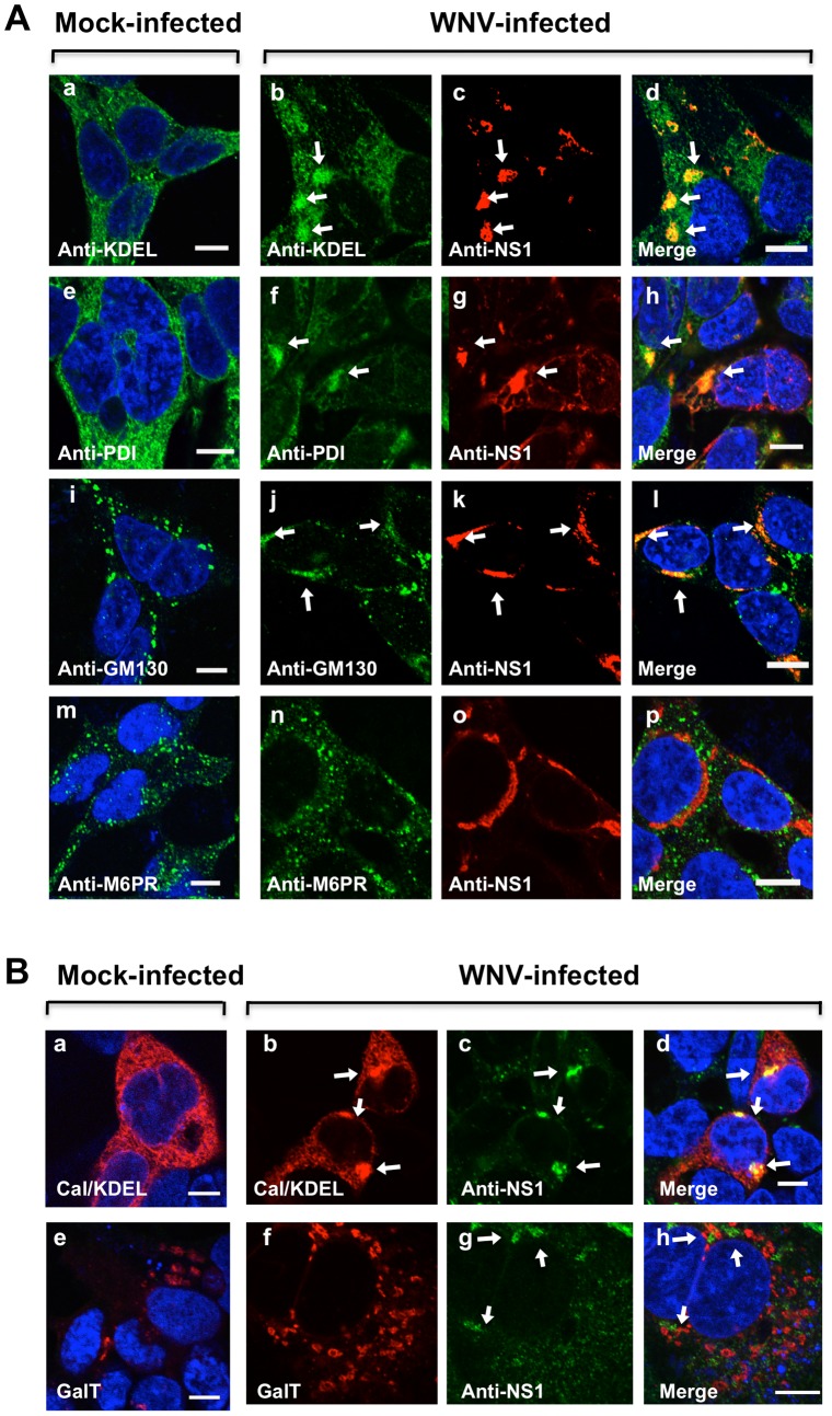
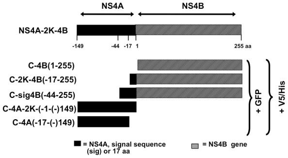
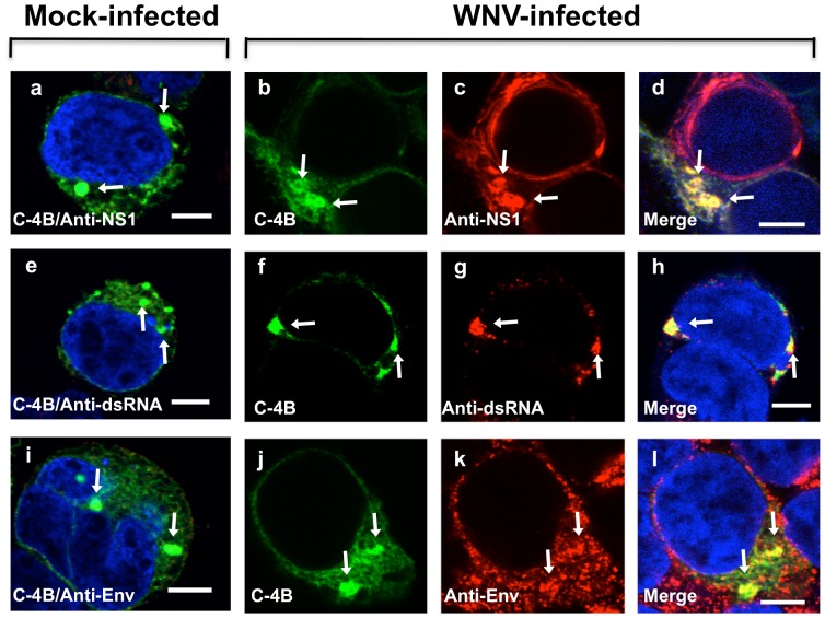
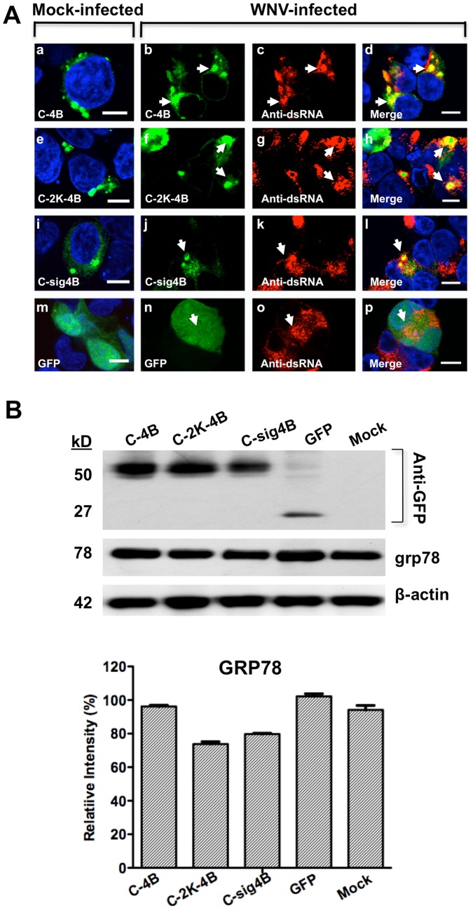
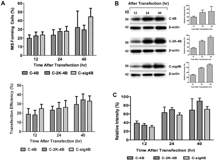
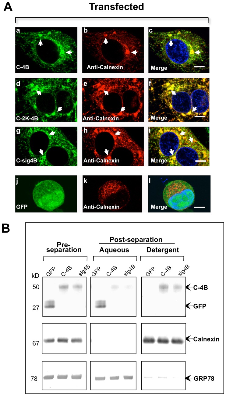
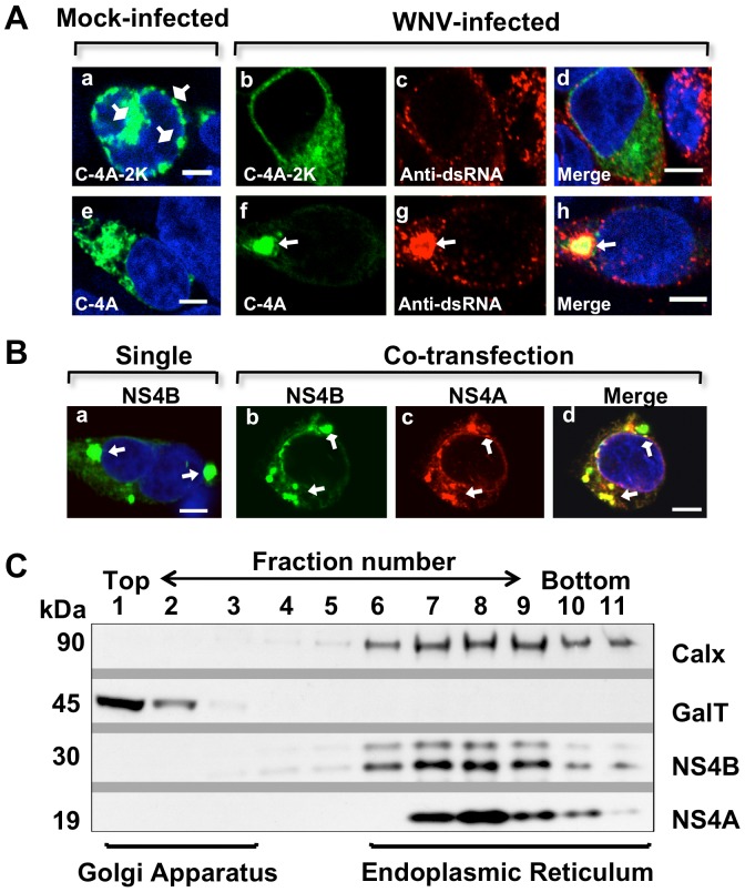
Replication of flaviviruses (family Flaviviridae) occurs in specialized virus-induced membrane structures (IMS). The cellular composition of these IMS varies for different flaviviruses implying different organelle origins for IMS biogenesis. The role of flavivirus non-structural (NS) proteins for the alteration of IMS remains controversial. In this report, we demonstrate that West Nile virus strain New York 99 (WNVNY99) remodels the endoplasmic reticulum (ER) membrane to generate specialized IMS. Within these structures, we observed an element of the cis-Golgi, viral double-stranded RNA, and viral-envelope, NS1, NS4A and NS4B proteins using confocal immunofluorescence microscopy. Biochemical analysis and microscopy revealed that NS4B lacking the 2K-signal peptide associates with the ER membrane where it initiates IMS formation in WNV-infected cells. Co-transfection studies indicated that NS4A and NS4B always remain co-localized in the IMS and are associated with the same membrane fractions, suggesting that these proteins function cooperatively in virus replication and may be an ideal target for antiviral drug discovery.


Conflict of interest statement

Competing Interests: The authors have declared that no competing interests exist.

Figures

Figure 1
Figure 1. WNV NS1 antigen and IMS are localized to the endoplasmic reticulum (ER) and cis-Golgi organelles.
(A) HEK293 cells were infected with WNV and after 24 hr, cells were fixed and immunostained with mouse anti-KDEL (a and b) and rabbit anti-PDI (e and f) polyclonal antibodies to detect the ER, mouse anti-GM130 to detect the cis-Golgi (i and j) and mouse anti-M6PR to detect early endosomes (m and n). The cells were washed and co-immunostained with WNV mouse anti-NS1 antibody (red; c, d, g, h, k, l, o and p). (B) HEK293 cells were transfected for 12 hr with pDsRed2-ER containing the ER targeting sequence of calreticulin and the ER retention sequence, KDEL (a, b, and d) or pDsRed-Monomer-Golgi encoding the N-terminal 81 amino acids of human beta 1,4-GalT, (e, f, and h). After 2 hr, cells were infected for 24 hr with WNV (b–d and f–h), fixed and co-immunostained with WNV mouse anti-NS1 antibody (green; c, d, g, and h). The nuclei were counterstained by 4,6-diamidino-2-phenylindole (DAPI). Arrows indicate the IMS. Confocal fluorescence images were of optical slice thickness ∼1 µm. Scale bar, 10 µm.
Figure 2
Figure 2. Schematic representation of plasmid constructs used in this study.
The full-length NS4A-2K-NS4B (C-4AB) is depicted with amino acid (aa) numbers, 1 representing the first aa at the N-terminus of NS4B. Also depicted are the plasmids NS4B (C-4B), NS4B retaining 17 N-terminal aa (C-2K-4B), NS4B retaining 44 N-terminal aa (C-sig4B), NS4A retaining the 17-aa to the C-terminal segment (C-4A-2K), and NS4A without the 2K-signal peptide (C-4A). NS4A and the 44- or 17-aa preceding C-4B are indicated by a black box. Plasmid fusion detection peptides, either GFP or V5-His epitope, are noted to the right.
Figure 3
Figure 3. Localization of WNV NS4B to IMS in infected cells.
HEK293 cells were infected with WNV and after 2-GFP plasmid (C-4B; b–d, f–h, and j–l). Slice confocal IF images of (a, e, and i) mock-infected but transfected, and (b–d, f–h, and j–l) WNV-infected and transfected fixed HEK293 cells at 24 hr immunostained with WNV mouse anti-NS1 antibody (red; c–d), mouse anti-dsRNA antibody (red; g–h), or WNV mouse anti-envelope antibody (red; k–l). The mock-infected cells were transfected with the NS4B-GFP plasmid and immunostained with the indicated antibodies (a, e, and i). Nuclear DNA was labeled with DAPI. The arrowheads indicate IMS. Confocal IF images were of optical slice thickness ∼1 µm. Scale bar, 10 µm.
Figure 4
Figure 4. IMS localization and expression of WNV NS4B-GFP with or without the 2K-signal peptide in infected cells.
(A) HEK293 cells were infected (b–d, f–h, j–l, and n–p) or mock-infected (a, e, i, and m) with WNV and after 2 hr transfected with C-4B (b–d), C-2K-4B (f–h), C-sig4B (j–l) or GFP (n–p). Cells were fixed at 24 hr after infection and immunostained with mouse anti-dsRNA antibody (red; c–d, g–h, k–l and o–p). The mock-infected cells were transfected and immunostained with mouse anti-dsRNA antibody (a, e, i, and m). Nuclear DNA was labeled with DAPI. The arrowheads indicate IMS. Confocal IF images were of optical slice thickness ∼1 µm. Scale bar, 10 µm. (B) Western blot analysis of WNV NS4B plasmids and stress response in transfected HEK293 cells. Fifty µg of cell lysate was loaded and electrophoresed on SDS-PAGE followed by immunoblotting with rabbit anti-GFP polyclonal antibody, an anti-GRP78 monoclonal antibody or anti-β-actin monoclonal antibody followed by a peroxidase-conjugated secondary antibody. ImageJ analysis of the western blot was conducted to determine GRP78 protein levels relative to β-actin.
Figure 5
Figure 5. Quantitation of IMS in HEK293 cells expressing NS4B with and without the 2K-signal peptide.
(A) Average number of IMS forming cells at 12, 24 and 40 hr after transfection. Each data point represents the percent of IMS forming cells in 50–100 GFP positive cells in 10 fields from two independent transfections. The average percent transfection efficiency for each data point is shown for 12, 24 and 40 hr after transfection. (B) HEK293 cells were transfected with a NS4B-GFP plasmid with and without the 2K signal peptide and harvested at 12, 24 and 40 hr after transfection. Fifty µg of harvested cell lysates were electrophoresed on SDS-PAGE followed by immunoblotting with rabbit anti-GFP antibody followed by a peroxidase-conjugated secondary antibody. The western blot images were analyzed using ImageJ to determine the relative intensity of the GFP expression level relative to β-actin, depicted as a percent relative intensity. (C) The average relative intensity of NS4B with the 2K signal peptide (C-2K-4B and C-sig4B) and without 2K (C-4B) at 12, 24 and 40 hr after transfection were determined using ImageJ.
Figure 6
Figure 6. Co-localization of WNV NS4B lacking or retaining the 2K-signal peptide with the ER marker, calnexin, to NS4B-IMS.
(A) Transfected HEK293 cells were fixed after 24 hr and processed for IF analysis. Left panels (a, d, g, j) depict different NS4B-GFP fusion constructs, and panels in the middle column (b, e, h, k) depict cells stained with calnexin. Merged images are depicted in the right panels (c, f, i, l). The arrowheads indicate IMS co-localized with calnexin; GFP alone does not co-localize with calnexin (l). (B) NS4B is associated with the cellular membrane. Triton X-114 phase separation of the total cell lysates from HEK293 cells transfected for 24 hr with GFP, NS4B (C-4B) or NS4B-retaining 2K (C-sig4B) plasmid. Total cell lysates were subjected to SDS-PAGE, immunostained against GFP (top panel), anti-calnexin (middle panel), anti-GRP78 (bottom panel) and visualized with AP-conjugated secondary antibodies. Twenty-five µL of total protein was loaded into each lane.
Figure 7
Figure 7. Association of WNV NS4A and NS4B proteins in IMS formation.
(A) HEK293 cells were infected with WNV and after 2 hr transfected with NS4A-2K-GFP (C-4A-2K, b–d) or NS4A-GFP plasmid (C-4A, f–h). Cells were fixed at 24 hr after infection and immunostained with mouse anti-dsRNA antibody (red; c–d and g–h). The mock-infected cells were transfected with (a) C-4A-2K or (e) C-4A and were immunostained with mouse anti-dsRNA antibody (a and e). Confocal IF images were of optical slice thickness ∼1 µm. The arrowheads indicate IMS whereas the square arrowhead indicates structures (potential artifacts) induced by NS4A retaining the 2K (a). Scale bar, 5 µm. (B) IF of NS4A and NS4B localization in transfected cells. HEK293 cells were co-transfected with NS4A-V5 (C-4A) and NS4B-GFP plasmids (C-4B), (b–d) or with NS4B-GFP only (a). Cells were fixed at 24 hr after transfection and immunostained with mouse anti-V5 antibody (red, b and c). Merge image (d). Confocal immunofluorescence images were of optical slice thickness ∼1 µm. The arrowheads indicate IMS. Scale bar, 5 µm. (C) Subcellular fractionation of HEK293 cells expressing NS4A and NS4B in transfected cells. HEK293 cells were co-transfected with NS4A-V5/His and NS4B-V5/His plasmids. Cell fractions were separated using sucrose gradient and subjected to SDS-PAGE before immunoblotting with a mouse monoclonal anti-V5 antibody and HRP-conjugated secondary antibody. The ER and Golgi apparatus were detected using antibodies against the markers calnexin (Calx) and 1,4-galactosyltransferase (GalT), respectively.

Similar articles

Cited by

References

    1. Brinton MA (2002) The molecular biology of West Nile Virus: a new invader of the western hemisphere. Annu Rev Microbiol 56: 371–402. - PubMed
    1. Roosendaal J, Westaway EG, Khromykh A, Mackenzie JM (2006) Regulated cleavages at the West Nile virus NS4A-2K-NS4B junctions play a major role in rearranging cytoplasmic membranes and Golgi trafficking of the NS4A protein. J Virol 80: 4623–4632. - PMC - PubMed
    1. Westaway EG, Khromykh AA, Kenney MT, Mackenzie JM, Jones MK (1997) Proteins C and NS4B of the flavivirus Kunjin translocate independently into the nucleus. Virology 234: 31–41. - PubMed
    1. Miller S, Sparacio S, Bartenschlager R (2006) Subcellular localization and membrane topology of the Dengue virus type 2 Non-structural protein 4B. J Biol Chem 281: 8854–8863. - PubMed
    1. Liu WJ, Wang XJ, Mokhonov VV, Shi PY, Randall R, et al. (2005) Inhibition of interferon signaling by the New York 99 strain and Kunjin subtype of West Nile virus involves blockage of STAT1 and STAT2 activation by nonstructural proteins. J Virol 79: 1934–1942. - PMC - PubMed

Publication types

Substances