

A glycosaminoglycan based, modular tissue scaffold system for rapid assembly of perfusable, high cell density, engineered tissues
- PMID: 24465401
- PMCID: PMC3896358
- DOI: 10.1371/journal.pone.0084287
A glycosaminoglycan based, modular tissue scaffold system for rapid assembly of perfusable, high cell density, engineered tissues
Abstract
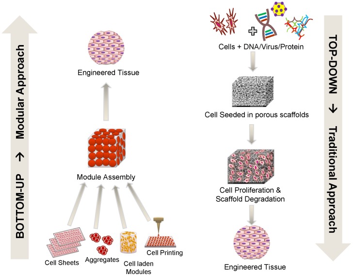
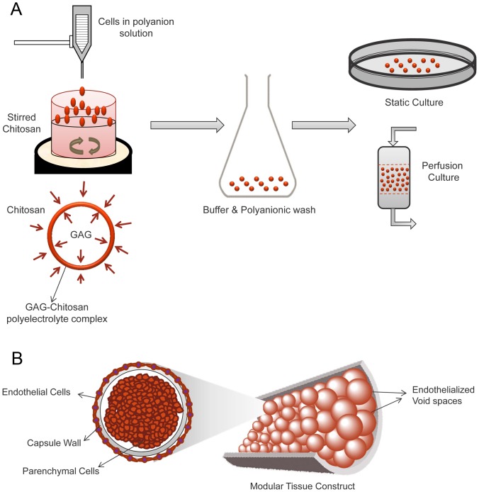
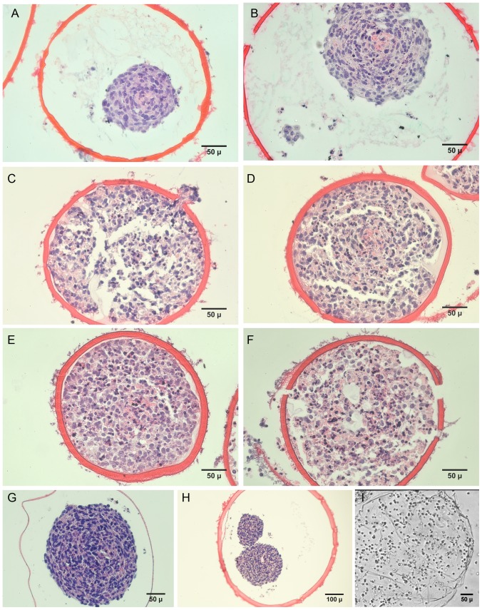
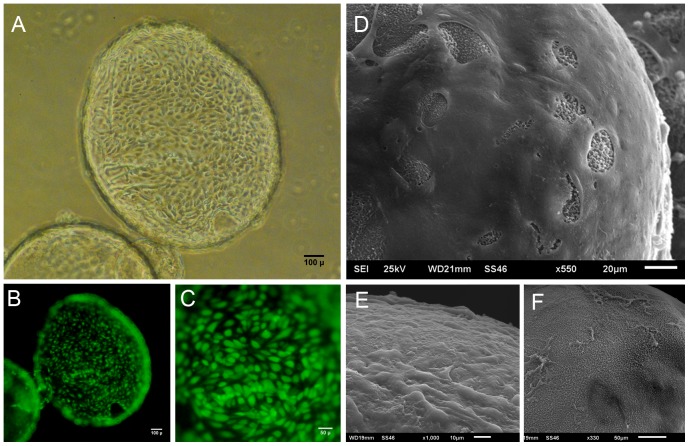
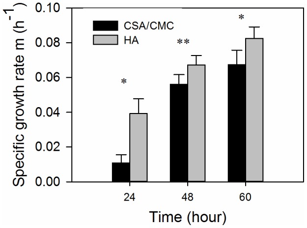
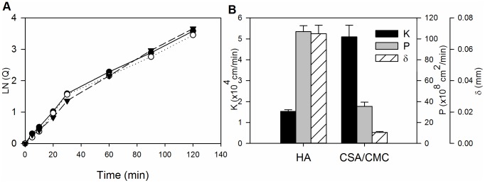
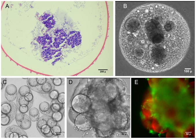
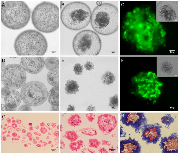
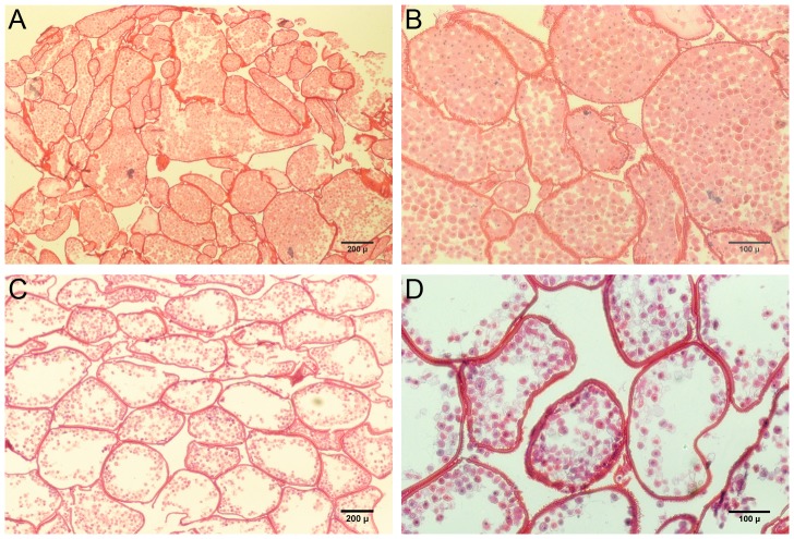
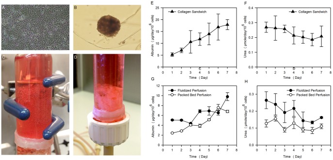
The limited ability to vascularize and perfuse thick, cell-laden tissue constructs has hindered efforts to engineer complex tissues and organs, including liver, heart and kidney. The emerging field of modular tissue engineering aims to address this limitation by fabricating constructs from the bottom up, with the objective of recreating native tissue architecture and promoting extensive vascularization. In this paper, we report the elements of a simple yet efficient method for fabricating vascularized tissue constructs by fusing biodegradable microcapsules with tunable interior environments. Parenchymal cells of various types, (i.e. trophoblasts, vascular smooth muscle cells, hepatocytes) were suspended in glycosaminoglycan (GAG) solutions (4%/1.5% chondroitin sulfate/carboxymethyl cellulose, or 1.5 wt% hyaluronan) and encapsulated by forming chitosan-GAG polyelectrolyte complex membranes around droplets of the cell suspension. The interior capsule environment could be further tuned by blending collagen with or suspending microcarriers in the GAG solution These capsule modules were seeded externally with vascular endothelial cells (VEC), and subsequently fused into tissue constructs possessing VEC-lined, inter-capsule channels. The microcapsules supported high density growth achieving clinically significant cell densities. Fusion of the endothelialized, capsules generated three dimensional constructs with an embedded network of interconnected channels that enabled long-term perfusion culture of the construct. A prototype, engineered liver tissue, formed by fusion of hepatocyte-containing capsules exhibited urea synthesis rates and albumin synthesis rates comparable to standard collagen sandwich hepatocyte cultures. The capsule based, modular approach described here has the potential to allow rapid assembly of tissue constructs with clinically significant cell densities, uniform cell distribution, and endothelialized, perfusable channels.
Conflict of interest statement
Figures
Similar articles
-
Transport Analysis of Engineered Liver Tissue Fabricated Using a Capsule-Based, Modular Approach.Ann Biomed Eng. 2019 May;47(5):1223-1236. doi: 10.1007/s10439-018-02192-y. Epub 2019 Feb 22. Ann Biomed Eng. 2019. PMID: 30796550 Free PMC article.
-
Bioengineering vascularized tissue constructs using an injectable cell-laden enzymatically crosslinked collagen hydrogel derived from dermal extracellular matrix.Acta Biomater. 2015 Nov;27:151-166. doi: 10.1016/j.actbio.2015.09.002. Epub 2015 Sep 5. Acta Biomater. 2015. PMID: 26348142 Free PMC article.
-
Fabrication of biomimetic vascular scaffolds for 3D tissue constructs using vascular corrosion casts.Acta Biomater. 2016 Mar 1;32:190-197. doi: 10.1016/j.actbio.2016.01.005. Epub 2016 Jan 6. Acta Biomater. 2016. PMID: 26772527
-
Modular Tissue Assembly Strategies for Biofabrication of Engineered Cartilage.Ann Biomed Eng. 2017 Jan;45(1):100-114. doi: 10.1007/s10439-016-1609-3. Epub 2016 Apr 12. Ann Biomed Eng. 2017. PMID: 27073109 Review.
-
Bioprinting for vascular and vascularized tissue biofabrication.Acta Biomater. 2017 Mar 15;51:1-20. doi: 10.1016/j.actbio.2017.01.035. Epub 2017 Jan 11. Acta Biomater. 2017. PMID: 28087487 Review.
Cited by
-
Encapsulation of mesenchymal stem cells in glycosaminoglycans-chitosan polyelectrolyte microcapsules using electrospraying technique: Investigating capsule morphology and cell viability.Bioeng Transl Med. 2018 Oct 1;3(3):265-274. doi: 10.1002/btm2.10111. eCollection 2018 Sep. Bioeng Transl Med. 2018. PMID: 30377665 Free PMC article.
-
Preparation and Drug Release Profile of Chitosan-Siloxane Hybrid Capsules Coated with Hydroxyapatite.Pharmaceutics. 2022 May 23;14(5):1111. doi: 10.3390/pharmaceutics14051111. Pharmaceutics. 2022. PMID: 35631697 Free PMC article.
-
3D Printing for Soft Tissue Regeneration and Applications in Medicine.Biomedicines. 2021 Mar 26;9(4):336. doi: 10.3390/biomedicines9040336. Biomedicines. 2021. PMID: 33810541 Free PMC article. Review.
-
Perspectives on scaling production of adipose tissue for food applications.Biomaterials. 2022 Jan;280:121273. doi: 10.1016/j.biomaterials.2021.121273. Epub 2021 Nov 29. Biomaterials. 2022. PMID: 34933254 Free PMC article.
-
Vasculogenesis and Angiogenesis in Modular Collagen-Fibrin Microtissues.Biomater Sci. 2014 Oct 1;2(10):1497-1508. doi: 10.1039/C4BM00141A. Biomater Sci. 2014. PMID: 25177487 Free PMC article.
References
-
- Langer R, Vacanti JP (1993) Tissue engineering. Science 260: 920–926. - PubMed
-
- MacNeil S (2007) Progress and opportunities for tissue-engineered skin. Nature 445: 874–880. - PubMed
-
- Jones E, Yang X (2011) Mesenchymal stem cells and bone regeneration: Current status. Injury 42: 562–568. - PubMed
-
- Hammouche S, Hammouche D, McNicholas M (2012) Biodegradable bone regeneration synthetic scaffolds: in tissue engineering. Curr Stem Cell Res Ther 7: 134–142. - PubMed
-
- Zhang Y, Venugopal JR, El-Turki A, Ramakrishna S, Su B, et al. (2008) Electrospun biomimetic nanocomposite nanofibers of hydroxyapatite/chitosan for bone tissue engineering. Biomaterials 29: 4314–4322. - PubMed
Publication types
MeSH terms
Substances
Grants and funding
LinkOut - more resources
Full Text Sources
Other Literature Sources
