

Patterning the cone mosaic array in zebrafish retina requires specification of ultraviolet-sensitive cones
- PMID: 24465536
- PMCID: PMC3897441
- DOI: 10.1371/journal.pone.0085325
Patterning the cone mosaic array in zebrafish retina requires specification of ultraviolet-sensitive cones
Abstract
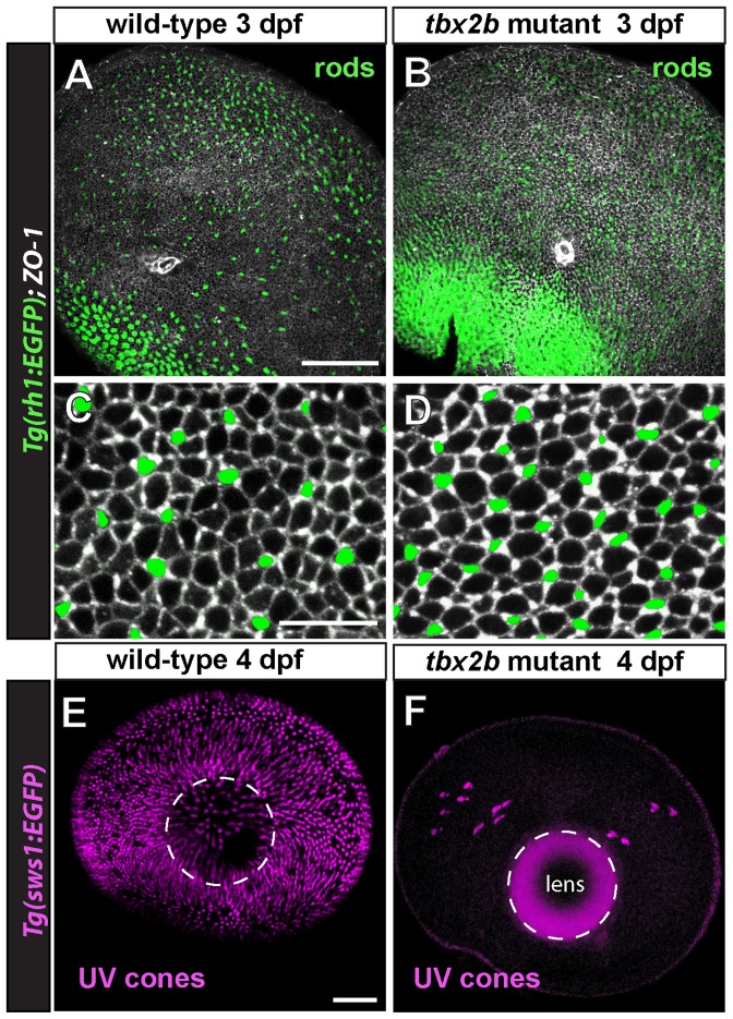
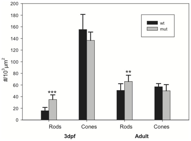
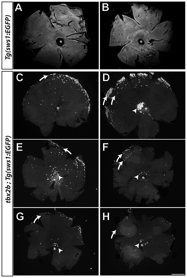
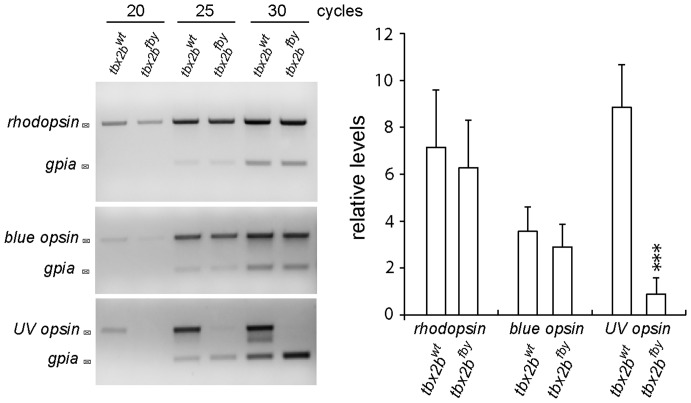
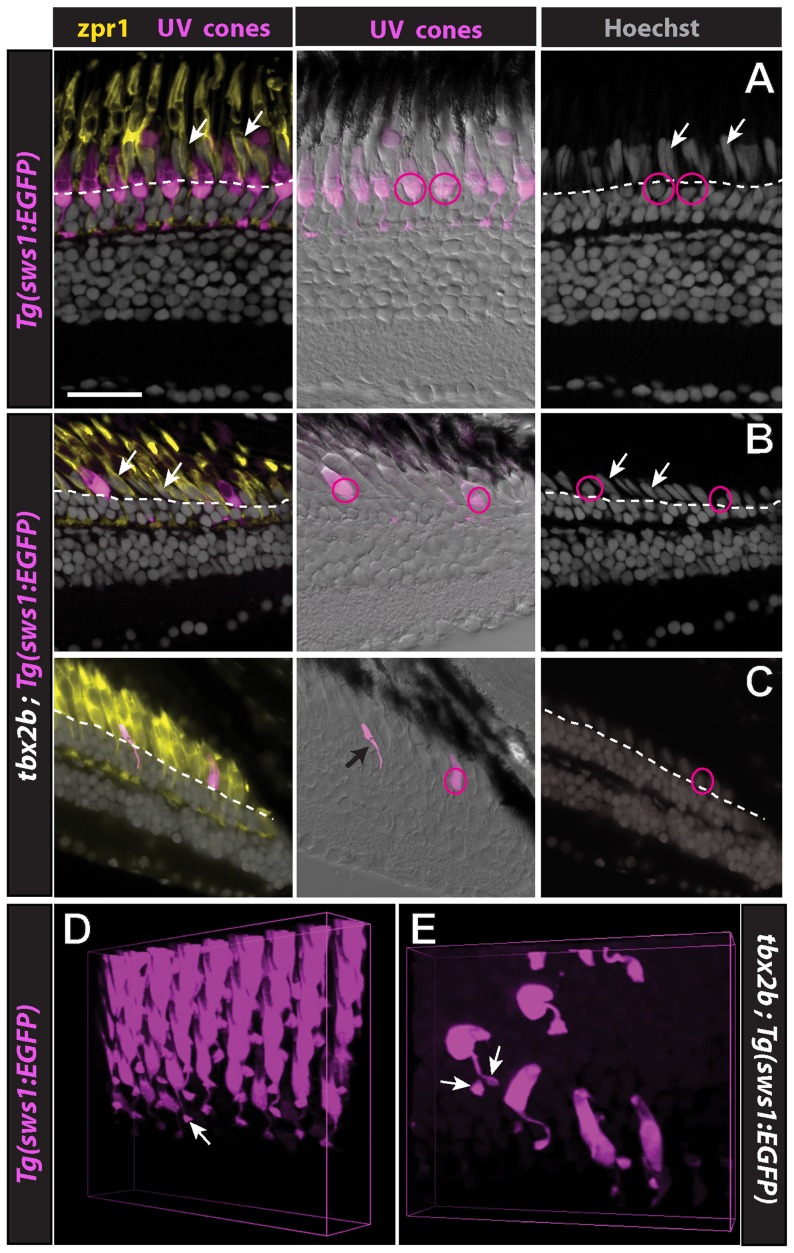
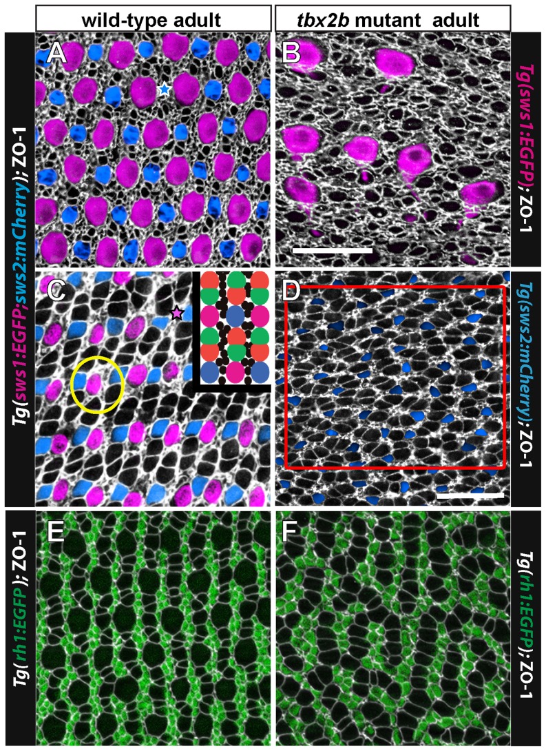
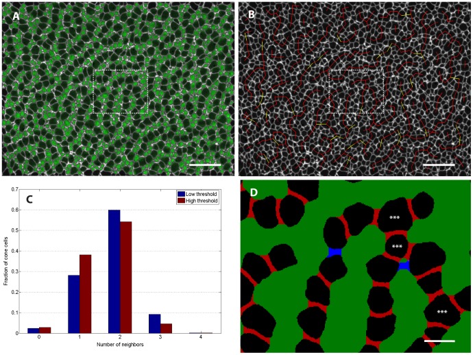
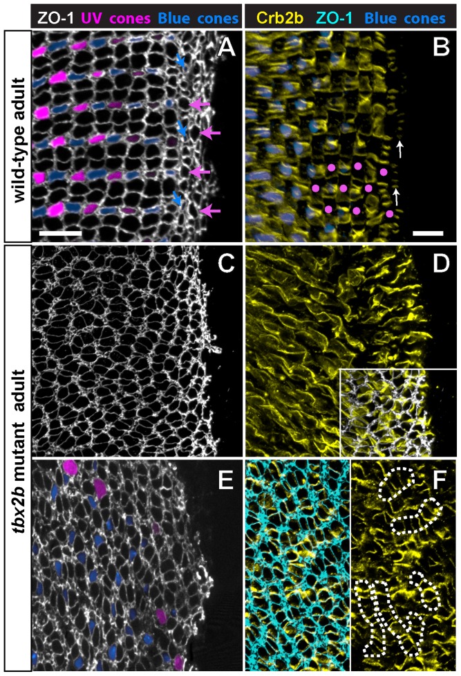
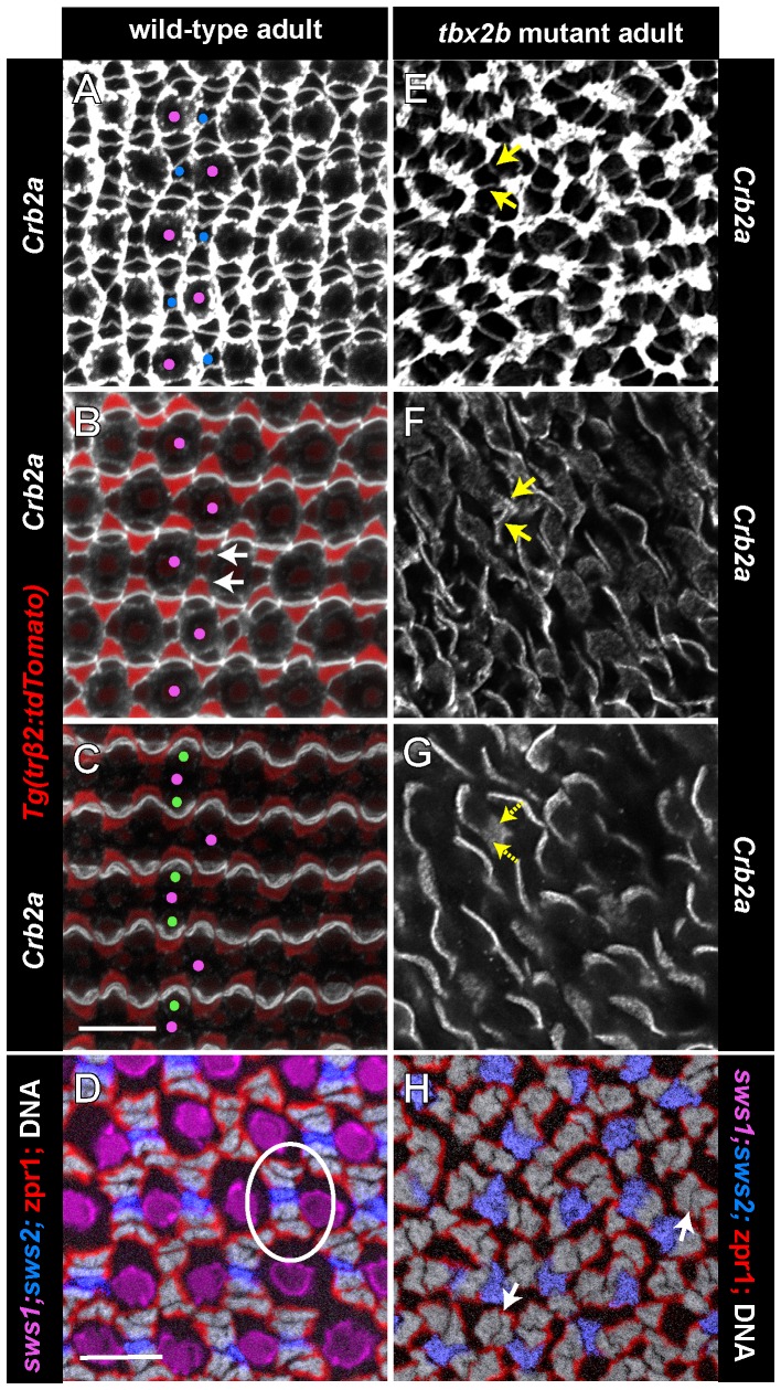
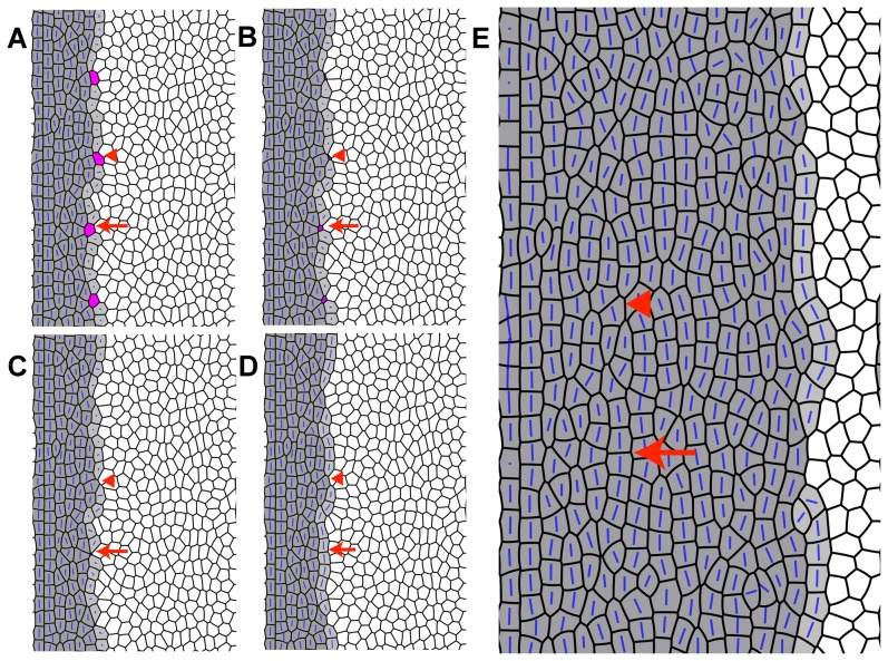
Cone photoreceptors in teleost fish are organized in precise, crystalline arrays in the epithelial plane of the retina. In zebrafish, four distinct morphological/spectral cone types occupy specific, invariant positions within a regular lattice. The cone lattice is aligned orthogonal and parallel to circumference of the retinal hemisphere: it emerges as cones generated in a germinal zone at the retinal periphery are incorporated as single-cell columns into the cone lattice. Genetic disruption of the transcription factor Tbx2b eliminates most of the cone subtype maximally sensitive to ultraviolet (UV) wavelengths and also perturbs the long-range organization of the cone lattice. In the tbx2b mutant, the other three cone types (red, green, and blue cones) are specified in the correct proportion, differentiate normally, and acquire normal, planar polarized adhesive interactions mediated by Crumbs 2a and Crumbs 2b. Quantitative image analysis of cell adjacency revealed that the cones in the tbx2b mutant primarily have two nearest neighbors and align in single-cell-wide column fragments that are separated by rod photoreceptors. Some UV cones differentiate at the dorsal retinal margin in the tbx2b mutant, although they are severely dysmorphic and are eventually eliminated. Incorporating loss of UV cones during formation of cone columns at the margin into our previously published mathematical model of zebrafish cone mosaic formation (which uses bidirectional interactions between planar cell polarity proteins and anisotropic mechanical stresses in the plane of the retinal epithelium to generate regular columns of cones parallel to the margin) reproduces many features of the pattern disruptions seen in the tbx2b mutant.
Conflict of interest statement
Figures
References
-
- Dowling JE (1987) The Retina: An Approachable Part of the Brain. Cambridge, Mass.: Belknap Press of Harvard University Press. 282 pp.
-
- Williams DS, Arikawa K, Paallysaho T (1990) Cytoskeletal components of the adherens junctions between the photoreceptors and the supportive Müller cells. J Comp Neurol 295: 155–164. - PubMed
-
- Gosens I, den Hollander AI, Cremers FP, Roepman R (2008) Composition and function of the Crumbs protein complex in the mammalian retina. Exp Eye Res 86: 713–726. - PubMed
-
- Lyall AH (1957) Cone arrangements in teleost retinae. Quart J Microsc Sci 98: 189–201.
-
- Engström K (1963) Cone types and cone arrangements in teleost retinae. Acta Zool 44: 179–243.
Publication types
MeSH terms
Substances
Grants and funding
LinkOut - more resources
Full Text Sources
Other Literature Sources
Molecular Biology Databases
Research Materials
