. 2014 Jan 22;9(1):e85671.
doi: 10.1371/journal.pone.0085671. eCollection 2014.

Antenatal glucocorticoid treatment affects hippocampal development in mice

Affiliations

Antenatal glucocorticoid treatment affects hippocampal development in mice

Cornelle W Noorlander et al. PLoS One. .

Abstract

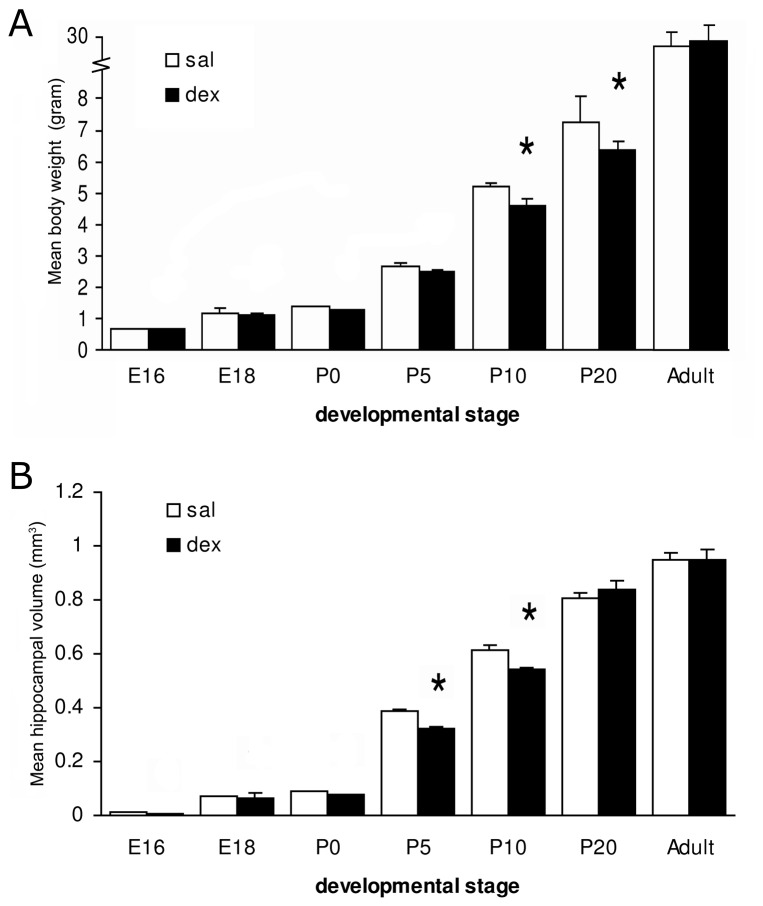
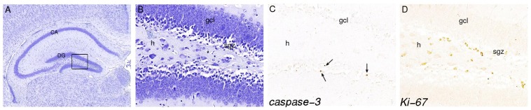
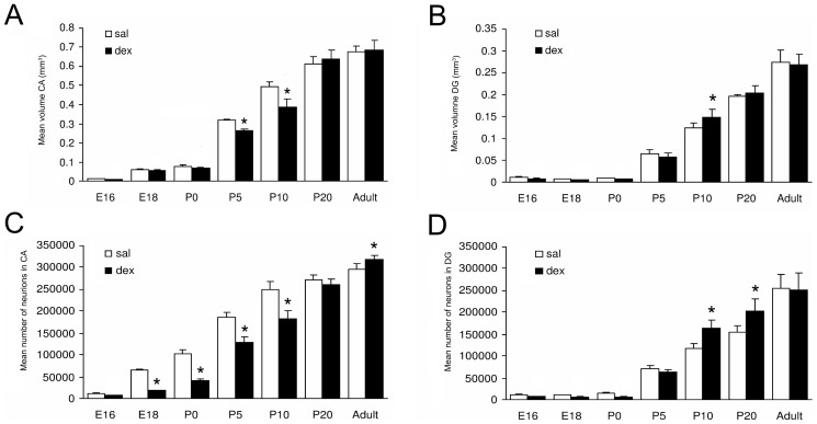
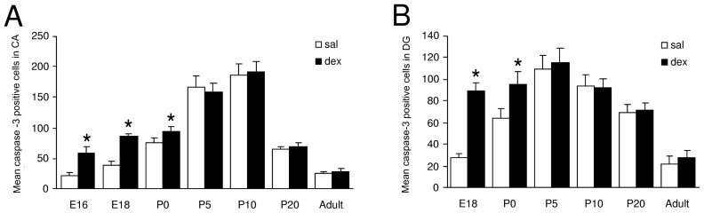
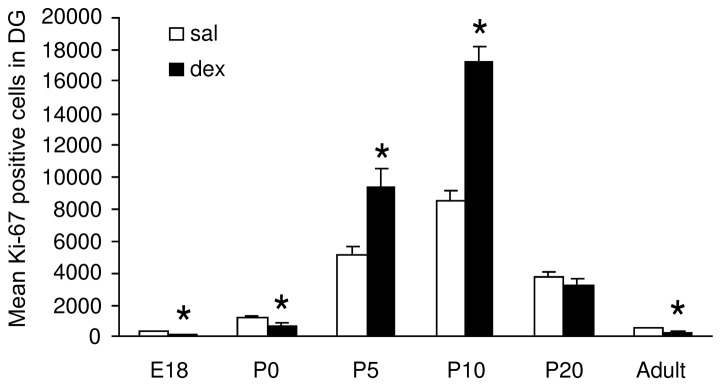
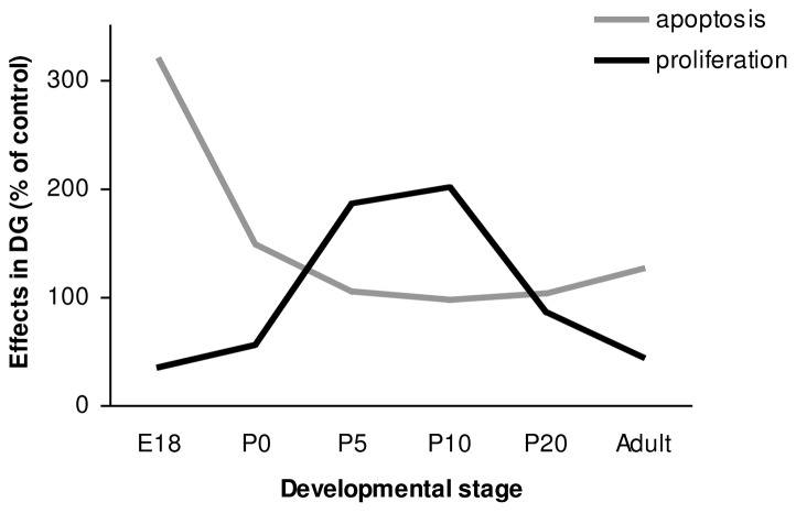
Synthetic glucocorticoids are administered to pregnant women at risk for preterm delivery, to enhance fetal lung maturation. The benefit of this treatment is well established, however caution is necessary because of possible unwanted side effects on development of different organ systems, including the brain. Actions of glucocorticoids are mediated by corticosteroid receptors, which are highly expressed in the hippocampus, a brain structure involved in cognitive functions. Therefore, we analyzed the effects of a single antenatal dexamethasone treatment on the development of the mouse hippocampus. A clinically relevant dose of dexamethasone (0.4 mg/kg) was administered to pregnant mice at embryonic day 15.5 and the hippocampus was analyzed from embryonic day 16 until adulthood. We investigated the effects of dexamethasone treatment on anatomical changes, apoptosis and proliferation in the hippocampus, hippocampal volume and on total body weight. Our results show that dexamethasone treatment reduced body weight and hippocampal volume transiently during development, but these effects were no longer detected at adulthood. Dexamethasone treatment increased the number of apoptotic cells in the hippocampus until birth, but postnatally no effects of dexamethasone treatment on apoptosis were found. During the phase with increased apoptosis, dexamethasone treatment reduced the number of proliferating cells in the subgranular zone of the dentate gyrus. The number of proliferative cells was increased at postnatal day 5 and 10, but was decreased again at the adult stage. This latter long-term and negative effect of antenatal dexamethasone treatment on the number of proliferative cells in the hippocampus may have important implications for hippocampal network function.


Conflict of interest statement

Competing Interests: The authors have declared that no competing interests exist.

Figures

Figure 1
Figure 1. Effect of antenatal dexamethasone treatment on body weight and hippocampal volume.
Data are presented as mean ± SD. Dex-treated mice showed a significant reduction in mean body weight at P10 and P20 and reduced hippocampal volume at P5 and P10. E: embryonic day, P: postnatal day, sal: saline-treatment (n = 8), dex: dexamethasone-treatment (n = 8), * p<0.05 (Two-Way ANOVA+Bonferroni).
Figure 2
Figure 2. Typical example of a hippocampus of P20, stained with Nissl (A, B), immunohistochemistry for active-caspase-3 (C) and Ki-67 (D).
Panel B, C and D represent a magnification (40×) of the boxed area in panel A (10×). Arrows in panel C show apoptotic cells in gcl. Proliferative cells are detected in the sgz of the DG. h = hilus, gcl = granule cell layer, sgz = subgranular zone.
Figure 3
Figure 3. Effect of antenatal dexamethasone treatment on the volume (A, B) and total number of neurons (C, D) of the hippocampus.
Data are presented as mean ± SD. Panel A and C show a significant decrease in volume of the CA at P5 and P10 and in total number of neurons in the CA area at E18 until P10 and an increase of number of neurons in the CA of the dex-treated group at adulthood. Panel B shows an increase in volume of the DG of the dex-treated group at P10. Panel D shows a significant increase in total number of neurons in the DG of the dex-treated group at P10 and P20. Sal: saline-treatment (n = 8), dex: dexamethasone-treatment (n = 8). * p<0.05 (Two-Way ANOVA+Bonferroni).
Figure 4
Figure 4. Effect of antenatal dexamethasone treatment on the total number of apoptotic cells in the pyramidal cell layer in the CA (A) and in the granule cell layer of the DG (B).
Data are presented as mean ± SD. Apoptosis is significantly increased in the dex-treated group in both the CA area (E16, E18 and P0) and the DG (E18 and P0). Sal: saline-treatment (n = 8), dex: dexamethasone-treatment (n = 8), * p<0.05 (Two-Way ANOVA+Bonferroni).
Figure 5
Figure 5. Effect of antenatal dexamethasone treatment on the total number of proliferative cells in the subgranular zone of the DG.
Data are presented as mean ± SD. At E18 and P0, the number of proliferative cells is decreased in the dex-treated group, while an increase is observed at P5 and P10. At the adult stage the number of proliferative cells are decreased in the dex-treated group. Sal: saline-treatment (n = 8), dex: dexamethasone-treatment (n = 8), * p<0.05 (Two-Way ANOVA+Bonferroni).
Figure 6
Figure 6. Summary of the effects of antenatal dex treatment on proliferation and apoptosis in the DG during hippocampal development.
The results of the dex treated groups are presented as percentage of the control (100%).

Similar articles

Cited by

References

    1. Roberts D, Dalziel S (2006) Antenatal corticosteroids for accelerating fetal lung maturation for women at risk of preterm birth. Cochrane Database Syst Rev 3: CD004454 10.1002/14651858.CD004454.pub2 - DOI - PubMed
    1. Zarrow MX, Philpott JE, Denenberg VH (1970) Passage of 14C-4-corticosterone from the rat mother to the foetus and neonate. Nature 226: 1058–1059. - PubMed
    1. Meijer OC, de Kloet ER (1998) Corticosterone and serotonergic neurotransmission in the hippocampus: Functional implications of central corticosteroid receptor diversity. Crit Rev Neurobiol 12: 1–20. - PubMed
    1. Noorlander CW, De Graan PN, Middeldorp J, Van Beers JJ, Visser GH (2006) Ontogeny of hippocampal corticosteroid receptors: Effects of antenatal glucocorticoids in human and mouse. J Comp Neurol 499: 924–932 10.1002/cne.21162 - DOI - PubMed
    1. Coe CL, Kramer M, Czeh B, Gould E, Reeves AJ, et al. (2003) Prenatal stress diminishes neurogenesis in the dentate gyrus of juvenile rhesus monkeys. Biol Psychiatry 54: 1025–1034. - PubMed

Publication types

MeSH terms