

Comparative study of efficacy of dopaminergic neuron differentiation between embryonic stem cell and protein-based induced pluripotent stem cell
- PMID: 24465672
- PMCID: PMC3899054
- DOI: 10.1371/journal.pone.0085736
Comparative study of efficacy of dopaminergic neuron differentiation between embryonic stem cell and protein-based induced pluripotent stem cell
Abstract
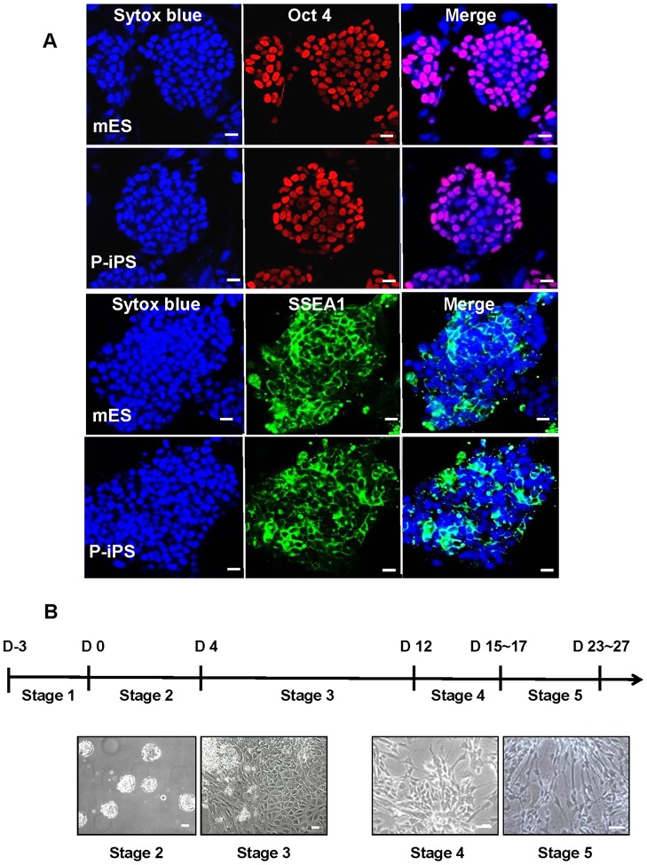
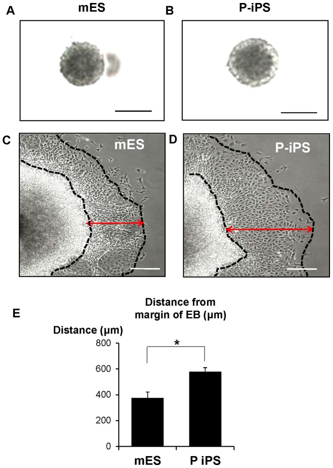
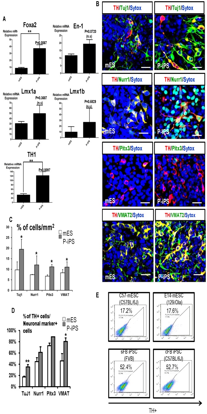
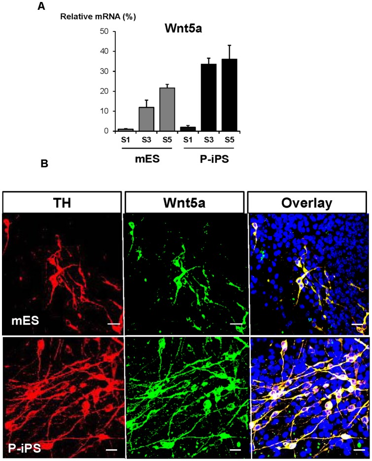
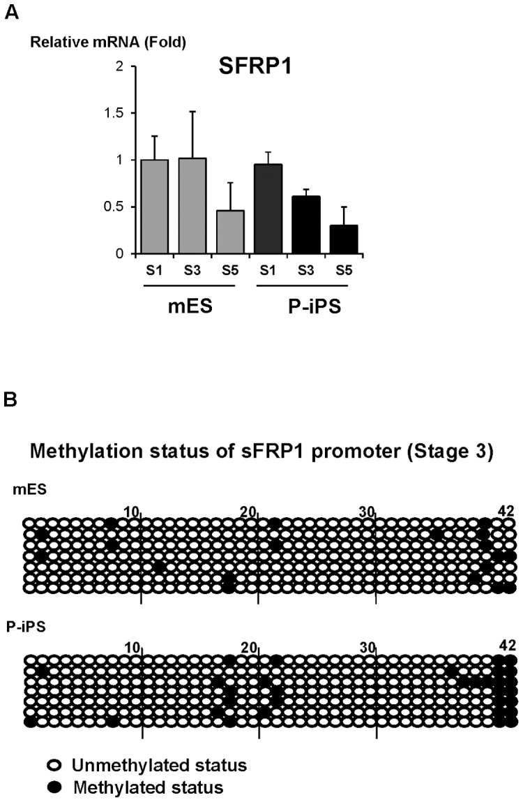
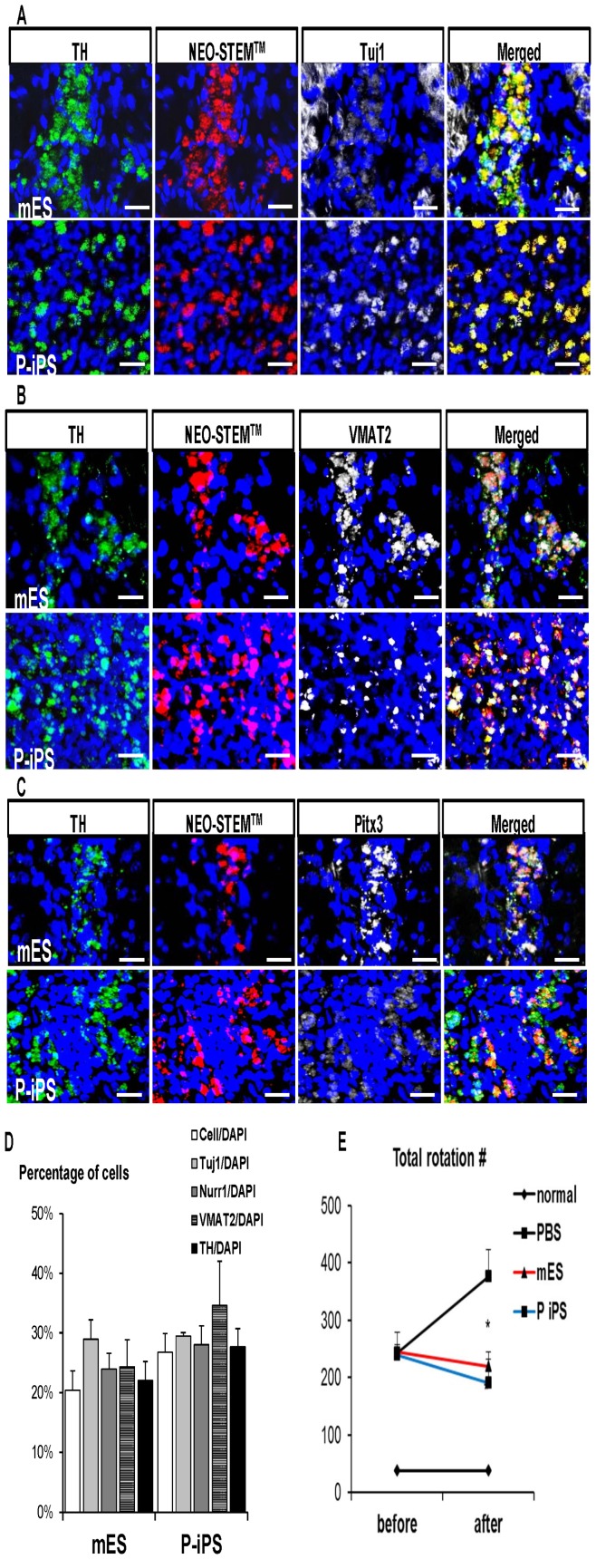
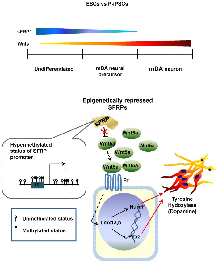
In patients with Parkinson's disease (PD), stem cells can serve as therapeutic agents to restore or regenerate injured nervous system. Here, we differentiated two types of stem cells; mouse embryonic stem cells (mESCs) and protein-based iPS cells (P-iPSCs) generated by non-viral methods, into midbrain dopaminergic (mDA) neurons, and then compared the efficiency of DA neuron differentiation from these two cell types. In the undifferentiated stage, P-iPSCs expressed pluripotency markers as ES cells did, indicating that protein-based reprogramming was stable and authentic. While both stem cell types were differentiated to the terminally-matured mDA neurons, P-iPSCs showed higher DA neuron-specific markers' expression than ES cells. To investigate the mechanism of the superior induction capacity of DA neurons observed in P-iPSCs compared to ES cells, we analyzed histone modifications by genome-wide ChIP sequencing analysis and their corresponding microarray results between two cell types. We found that Wnt signaling was up-regulated, while SFRP1, a counter-acting molecule of Wnt, was more suppressed in P-iPSCs than in mESCs. In PD rat model, transplantation of neural precursor cells derived from both cell types showed improved function. The present study demonstrates that P-iPSCs could be a suitable cell source to provide patient-specific therapy for PD without ethical problems or rejection issues.
Conflict of interest statement
Figures
Similar articles
-
Improved cell therapy protocols for Parkinson's disease based on differentiation efficiency and safety of hESC-, hiPSC-, and non-human primate iPSC-derived dopaminergic neurons.Stem Cells. 2013 Aug;31(8):1548-62. doi: 10.1002/stem.1415. Stem Cells. 2013. PMID: 23666606 Free PMC article.
-
SFRP1 and SFRP2 dose-dependently regulate midbrain dopamine neuron development in vivo and in embryonic stem cells.Stem Cells. 2012 May;30(5):865-75. doi: 10.1002/stem.1049. Stem Cells. 2012. PMID: 22290867 Free PMC article.
-
Docosahexaenoic acid promotes dopaminergic differentiation in induced pluripotent stem cells and inhibits teratoma formation in rats with Parkinson-like pathology.Cell Transplant. 2012;21(1):313-32. doi: 10.3727/096368911X580572. Epub 2011 Jun 7. Cell Transplant. 2012. PMID: 21669041
-
Preparation, characterization, and banking of clinical-grade cells for neural transplantation: Scale up, fingerprinting, and genomic stability of stem cell lines.Prog Brain Res. 2017;230:133-150. doi: 10.1016/bs.pbr.2017.02.007. Epub 2017 Apr 7. Prog Brain Res. 2017. PMID: 28552226 Review.
-
[Cell replacement therapy for Parkinson's disease using iPS cells].Rinsho Shinkeigaku. 2013;53(11):1009-12. doi: 10.5692/clinicalneurol.53.1009. Rinsho Shinkeigaku. 2013. PMID: 24291862 Review. Japanese.
Cited by
-
A Compendium of Preparation and Application of Stem Cells in Parkinson's Disease: Current Status and Future Prospects.Front Aging Neurosci. 2016 May 31;8:117. doi: 10.3389/fnagi.2016.00117. eCollection 2016. Front Aging Neurosci. 2016. PMID: 27303288 Free PMC article. Review.
-
PA6 Stromal Cell Co-Culture Enhances SH-SY5Y and VSC4.1 Neuroblastoma Differentiation to Mature Phenotypes.PLoS One. 2016 Jul 8;11(7):e0159051. doi: 10.1371/journal.pone.0159051. eCollection 2016. PLoS One. 2016. PMID: 27391595 Free PMC article.
-
Applications of Induced Pluripotent Stem Cells in Studying the Neurodegenerative Diseases.Stem Cells Int. 2015;2015:382530. doi: 10.1155/2015/382530. Epub 2015 Jul 9. Stem Cells Int. 2015. PMID: 26240571 Free PMC article. Review.
-
Protein-Induced Pluripotent Stem Cells Ameliorate Cognitive Dysfunction and Reduce Aβ Deposition in a Mouse Model of Alzheimer's Disease.Stem Cells Transl Med. 2017 Jan;6(1):293-305. doi: 10.5966/sctm.2016-0081. Epub 2016 Aug 15. Stem Cells Transl Med. 2017. PMID: 28170178 Free PMC article.
-
In vivo distribution of U87MG cells injected into the lateral ventricle of rats with spinal cord injury.PLoS One. 2018 Aug 16;13(8):e0202307. doi: 10.1371/journal.pone.0202307. eCollection 2018. PLoS One. 2018. PMID: 30114270 Free PMC article.
References
-
- Freed CR, Breeze RE, Rosenberg NL, Schneck SA, Kriek E, et al. (1992) Survival of implanted fetal dopamine cells and neurologic improvement 12 to 46 months after transplantation for Parkinson's disease. The New England journal of medicine 327: 1549–1555. - PubMed
-
- Lindvall O, Brundin P, Widner H, Rehncrona S, Gustavii B, et al. (1990) Grafts of fetal dopamine neurons survive and improve motor function in Parkinson's disease. Science 247: 574–577. - PubMed
-
- Dunnett SB, Bjorklund A, Lindvall O (2001) Cell therapy in Parkinson's disease – stop or go? Nat Rev Neurosci 2: 365–369. - PubMed
-
- Winkler C, Kirik D, Bjorklund A (2005) Cell transplantation in Parkinson's disease: how can we make it work? Trends Neurosci 28: 86–92. - PubMed
-
- Bjorklund A, Dunnett SB, Brundin P, Stoessl AJ, Freed CR, et al. (2003) Neural transplantation for the treatment of Parkinson's disease. Lancet neurology 2: 437–445. - PubMed
Publication types
MeSH terms
Substances
LinkOut - more resources
Full Text Sources
Other Literature Sources
