. 2014 Jan 22;9(1):e85836.
doi: 10.1371/journal.pone.0085836. eCollection 2014.

In vitro and in vivo activity of a novel antifungal small molecule against Candida infections

Affiliations

In vitro and in vivo activity of a novel antifungal small molecule against Candida infections

Sarah Sze Wah Wong et al. PLoS One. .

Abstract

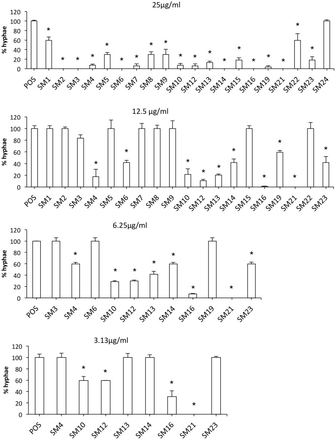
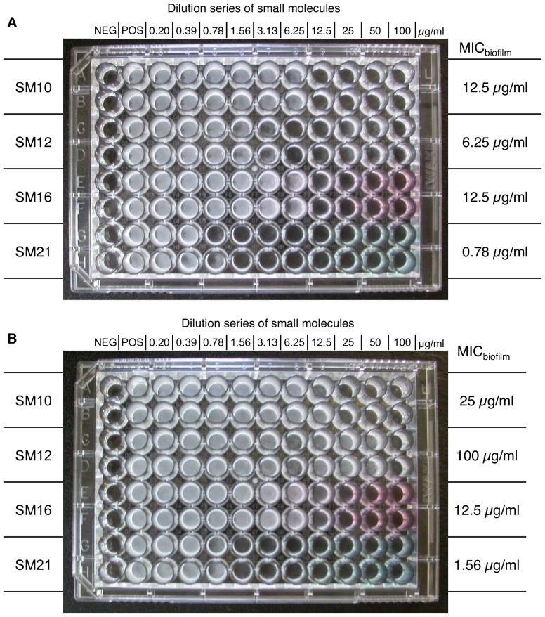
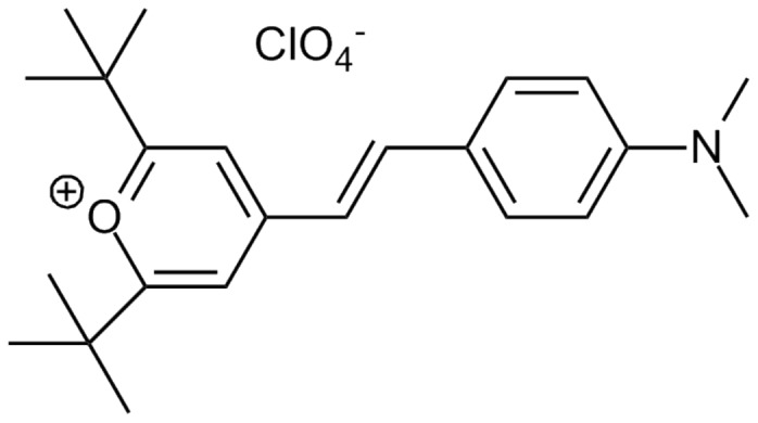
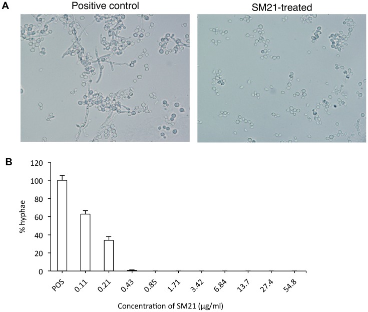
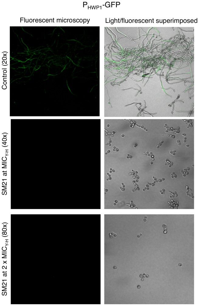
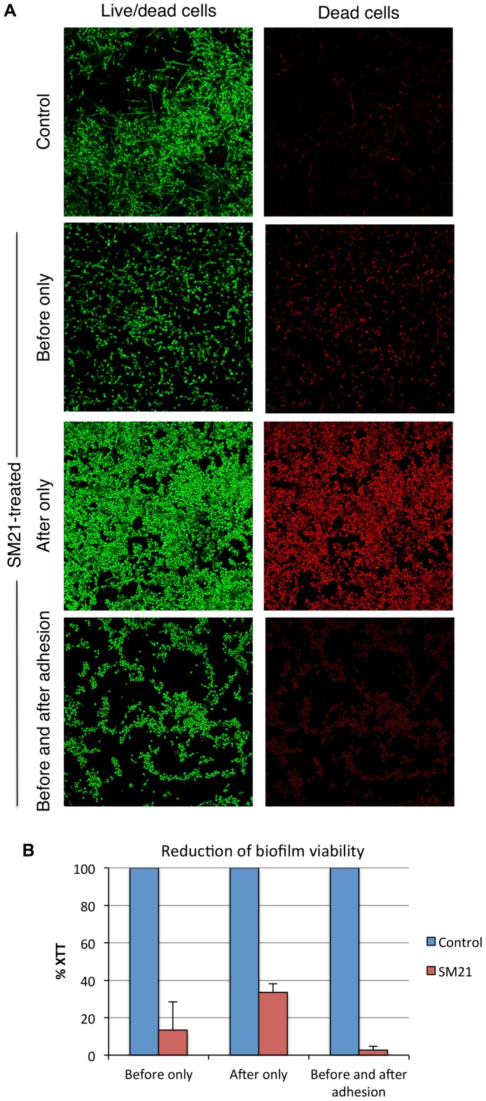
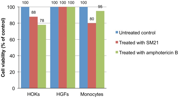
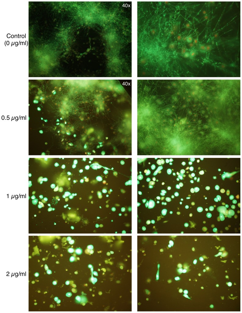
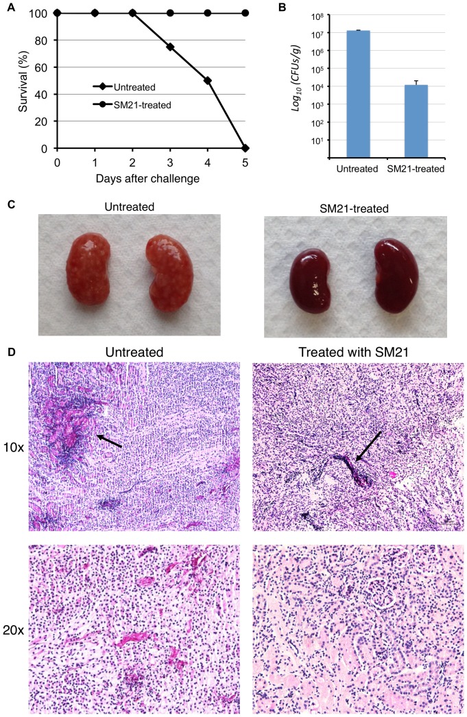
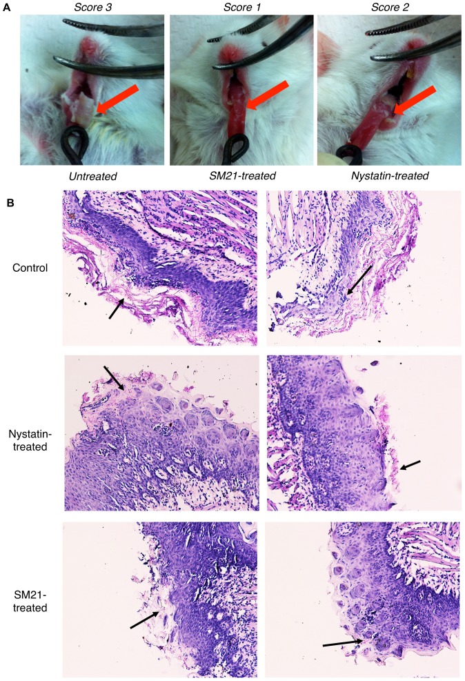
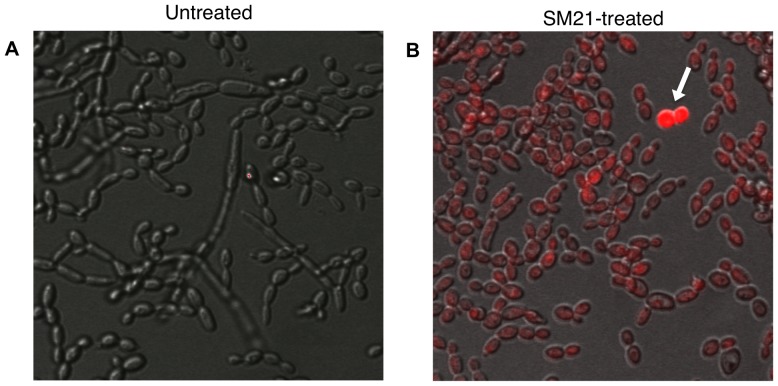
Candida is the most common fungal pathogen of humans worldwide and has become a major clinical problem because of the growing number of immunocompromised patients, who are susceptible to infection. Moreover, the number of available antifungals is limited, and antifungal-resistant Candida strains are emerging. New and effective antifungals are therefore urgently needed. Here, we discovered a small molecule with activity against Candida spp. both in vitro and in vivo. We screened a library of 50,240 small molecules for inhibitors of yeast-to-hypha transition, a major virulence attribute of Candida albicans. This screening identified 20 active compounds. Further examination of the in vitro antifungal and anti-biofilm properties of these compounds, using a range of Candida spp., led to the discovery of SM21, a highly potent antifungal molecule (minimum inhibitory concentration (MIC) 0.2-1.6 µg/ml). In vitro, SM21 was toxic to fungi but not to various human cell lines or bacterial species and was active against Candida isolates that are resistant to existing antifungal agents. Moreover, SM21 was relatively more effective against biofilms of Candida spp. than the current antifungal agents. In vivo, SM21 prevented the death of mice in a systemic candidiasis model and was also more effective than the common antifungal nystatin at reducing the extent of tongue lesions in a mouse model of oral candidiasis. Propidium iodide uptake assay showed that SM21 affected the integrity of the cell membrane. Taken together, our results indicate that SM21 has the potential to be developed as a novel antifungal agent for clinical use.


Conflict of interest statement

Competing Interests: The authors have declared that no competing interests exist.

Figures

Figure 1
Figure 1. Strategy for screening for novel antifungal small molecules.
We screened for Y-H inhibitors in a library containing 50,240 small molecules and found 20 active compounds. These 20 primary hits were further validated by assessing their activity in a dose-dependent manner, which led to the identification of eight potent Y-H inhibitors. The antifungal properties of the eight compounds were analysed in antifungal susceptibility tests, and the four most potent molecules were selected. Finally, the anti-biofilm activity of the four hits was evaluated, and SM21 was chosen for comprehensive in vitro and in vivo assays. HTS, high-throughput screening; AST, antifungal susceptibility test; ABT, anti-biofilm test.
Figure 2
Figure 2. Dose-dependent Y-H inhibition of C. albicans SC5314 by the 20 primary hits under hyphal-inducing conditions.
Cell morphology of C. albicans SC5314 was observed by light microscopy after incubation in the presence of different concentrations of the 20 primary hits under hyphal-inducing conditions (Lee's medium and incubation at 37°C). The degree of Y-H inhibition was quantified by calculating the percentage of hyphal cells in one hundred cells in a single sample. The average percentage of hyphae in the treated samples was normalised to the average percentage of hyphae in the positive control (taken as 100%). Starting with the highest concentration, those small molecules showing no Y-H inhibition were progressively eliminated in subsequent tests, which used lower concentrations. The assay identified eight potent Y-H inhibiting small molecules. The standard deviations are shown for each sample, and asteriks indicate p-value <0.05.
Figure 3
Figure 3. Anti-biofilm properties of SM10, SM12, SM16 and SM21.
The small molecules were added to the C. albicans SC5314 biofilm (A) before and after the adhesion phase; (B) after the adhesion phase; and incubated in 37°C for 24 h. The MICbiofilm was determined as the concentration where the viability of the biofilm was reduced by 50% as compared with the positive control. SM21 had the lowest MICbiofilm among the four hits in both cases, indicating its potent anti-biofilm property. NEG, negative control; POS, positive control.
Figure 4
Figure 4. Structure of SM21.
The molecular weight of SM21 (ChemBridge ID# 6633321) is 438 g/mol.
Figure 5
Figure 5. Y-H inhibition by SM21.
C. albicans cell morphology was observed by light microscopy after incubation in the presence of SM21. (A) SM21-mediated Y-H inhibition against strain CL1, a C. albicans clinical isolate. Under hyphal inducing conditions, hyphae could be observed in the positive control (no SM21), whereas, no hyphae were observed in the SM21-treated samples. (B) Dose-dependent effect of SM21 on Y-H inhibition of C. albicans SC5314 (1×10⁶ CFUs/ml). POS, positive control (no SM21). The degree of Y-H inhibition of each concentration was quantified by calculating the mean percentage of hyphal cells in one hundred cells in quadruplicate. The average percentage of hyphae in the treated samples was normalised to the average percentage of hyphae in the positive control (taken as 100%). MICY-H of SM21 was taken at 0.43 µg/ml, where only minimal amount of hyphae was observed. mean differences between the tests and control were all statistically significant (p-value <0.05).
Figure 6
Figure 6. Inhibition of HWP1 expression by SM21.
The inhibition of HWP1 expression of SM21 was examined by C. albicans strain PHWP1-GFP at MICY-H (0.43 µg/ml) and 2×MICY-H (0.86 µg/ml). Fluorescence, which indicated GFP expression under the control of the HWP1 promoter, was observed by confocal microscopy. In the control, multiple hyphae layers were observed under hypha-inducing conditions, however, only the fluorescence from the top layer could be captured by the confocal microscopy. After treatment with SM21 at MICY-H or 2×MICY-H, no trace of fluorescence was detected.
Figure 7
Figure 7. Effect of SM21 on several bacterial species.
Disk diffusion assays revealed that SM21 was harmless to E. coli, A. actinomycetemcomitans, S. mutans, S. mitis, S. sanguinis, and L. acidophilus. P – PBS, C – chlorhexidine (0.2%), CA – caspofungin (5 µg), A – amphotericin B (10 µg), SM – SM21 (2 µg).
Figure 8
Figure 8. Effect of SM21 on C. albicans biofilm formation on denture acrylic.
(A) Confocal images of C. albicans biofilm after treatment with SM21 and staining with fluorescent labels that distinguish between live and dead cells. All cells were labeled with the green fluorescence, while only the dead cells were labeled with red fluorescence. (B) Biofilm cell viability, quantified by XTT reduction assay, was reduced by 85% and 66%, respectively, when SM21 was added before or after the adhesion phase. The reduced biofilm viability was maximised (97%) when SM21 was added both before and after the adhesion phase. The standard deviations of each sample are shown in the graph, and all the mean differences between the control and test (SM21) were statistically significant (p-value<0.05).
Figure 9
Figure 9. Effect of SM21 on a multidrug-resistant isolate.
SM21 (SM) produced a clear inhibition zone in a disk diffusion assay against T-1549, a C. guilliermondii strain with multidrug resistance to amphotericin B (AMB), caspofungin (CASP) and fluconazole.
Figure 10
Figure 10. Cytotoxicity of SM21 against three primary cell lines.
HOK – human oral keratinocyte, HGF – human gingival fibroblast, AMB – amphotericin B. Both SM21 (0.2 µg/ml) and amphotericin B (0.2 µg/ml) reduced the viability of HOKs and monocytes. SM21 caused a reduction of 12% in cell viability compared with the untreated controls, which is lower than the 22% reduction observed for amphotericin B. Treatment with SM21 or amphotericin B caused, respectively, a 20% or 5% reduction in monocyte viability compared with the controls. Neither SM21 nor amphotericin B affected the viability of HGFs.
Figure 11
Figure 11. Effect of SM21 treatment in Candida–HOK co-culture model.
The viability of co-cultured HOKs and yeast cells in the presence of SM21 was assessed by confocal laser scanning microscopy using fluorescent probes. In the untreated control, many hyphae were observed, and most of the HOKs were dead (the nuclei of the dead cells were labeled with red fluorescence). Samples containing 0.5 µg/ml SM21 still included many hyphae, but they contained more live HOKs than the untreated control. Samples containing 1 µg/ml or 2 µg/ml SM21 included few yeast cells and no hyphae, and most of the HOKs were alive. Magnification = 20× (unless specified otherwise).
Figure 12
Figure 12. Effect of SM21 treatment on a mouse model of systemic candidiasis.
(A) Survival rate of the mice. After the experimental period (day 5), the survival rates for untreated and SM21-treated mice were 0% and 100%, respectively. (B) Fungal burden in the kidneys of the mice (Error bars indicate standard deviation). SM21 significantly reduced the renal fungal burden in the mice by an order of magnitude of 3 (p-value of mean difference <0.05). (C) Surfaces of the kidneys of untreated mice were covered with Candida lesions, while the kidneys of the SM21-treated mice appeared healthy. (D) PAS staining of the kidneys. Candida hyphae (indicated by black arrows) were easily spotted in the kidneys of untreated mice, whereas few were detected in the kidneys of SM21-treated mice.
Figure 13
Figure 13. Effect of SM21 treatment on a mouse model of oral candidiasis.
(A) The degree of tongue lesions in the oral candidiasis mouse model was evaluated by a scoring system of 0 – 3 (0 denotes healthy tongue surface and 3 denotes the most severe stage). A thick oral thrush (score = 3) was observed in untreated mice. Nystatin-treated mice displayed less oral thrush on the tongue surface than the untreated controls, but a considerable amount of oral thrush was observed at the back of the tongue (score = 2). SM21-treated mice displayed the least severe tongue lesions (score = 1). (B) PAS staining of tongue sections. Abundant hyphae (indicated by black arrows) covered most of the tongue surface in the untreated mice, whereas comparatively fewer hyphae were observed in nystatin- and SM21-treated mice.
Figure 14
Figure 14. Confocal microscopic analysis of propidium-iodide uptake assays.
(A) Untreated control samples showed hyphal elements of live C. albicans cells. (B) C. albicans treated with sub-MIC of SM21 (0.1 µg/ml) showed propidium iodide uptake (labeled by red fluorescence), indicating cell membrane damage. Some of the cells (denoted by white arrow) were non-viable.

Similar articles

Cited by

References

    1. Wisplinghoff H, Bischoff T, Tallent SM, Seifert H, Wenzel RP, et al. (2004) Nosocomial bloodstream infections in US hospitals: analysis of 24,179 cases from a prospective nationwide surveillance study. Clinical infectious diseases: an official publication of the Infectious Diseases Society of America 39: 309–317. - PubMed
    1. Odds FC (1988) Candida and candidosis: a review and bibliography. Bailliere Tindall.
    1. Webb BC, Thomas CJ, Willcox MD, Harty DW, Knox KW (1998) Candida-associated denture stomatitis. Aetiology and management: a review. Part 2. Oral diseases caused by Candida species. Australian dental journal 43: 160–166. - PubMed
    1. Akpan A, Morgan R (2002) Oral candidiasis. Postgraduate medical journal 78: 455–459. - PMC - PubMed
    1. Douglas LJ (2003) Candida biofilms and their role in infection. Trends in microbiology 11: 30–36. - PubMed

Publication types

MeSH terms