. 2014 Jan 20;9(1):e85849.
doi: 10.1371/journal.pone.0085849. eCollection 2014.

Pro-inflammatory mediators and apoptosis correlate to rt-PA response in a novel mouse model of thromboembolic stroke

Affiliations

Pro-inflammatory mediators and apoptosis correlate to rt-PA response in a novel mouse model of thromboembolic stroke

Saema Ansar et al. PLoS One. .

Abstract

Background: A recent study suggests that patients with persistent occlusion of the middle cerebral artery (MCA) following treatment with recombinant tissue plasminogen activator (rt-PA) have better outcomes than patients with MCA occlusion not receiving rt-PA. We performed a study to elucidate possible mechanisms of this finding in a new model of thromboembolic stroke closely mimicking human pathophysiology.

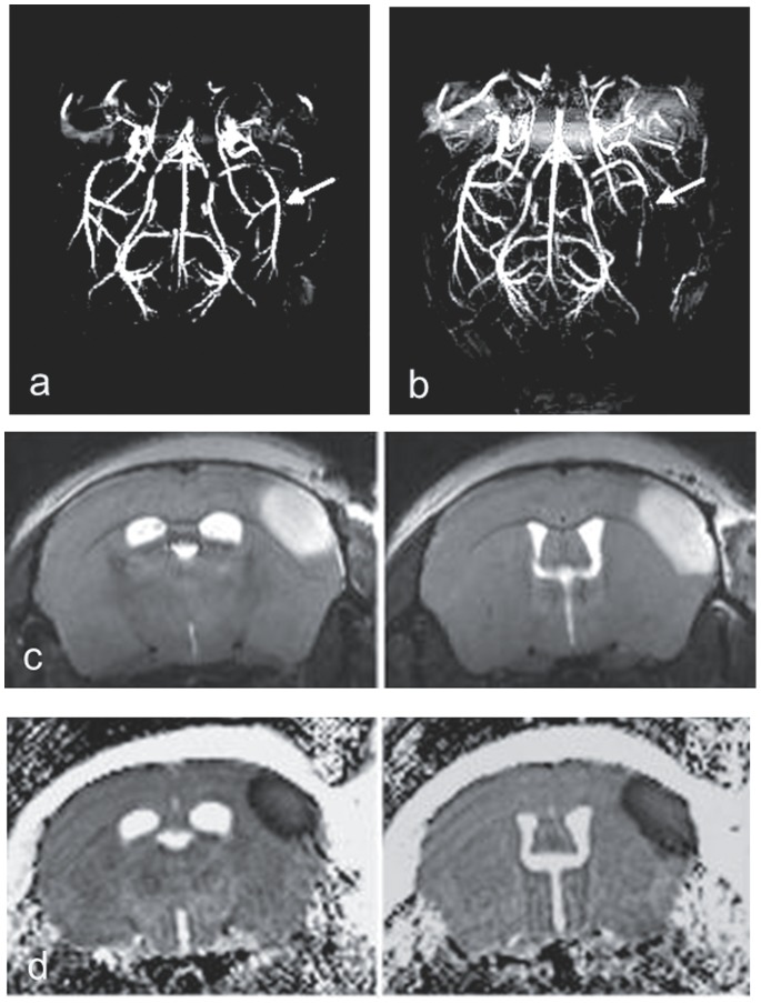
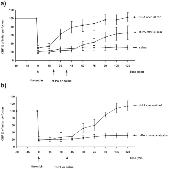
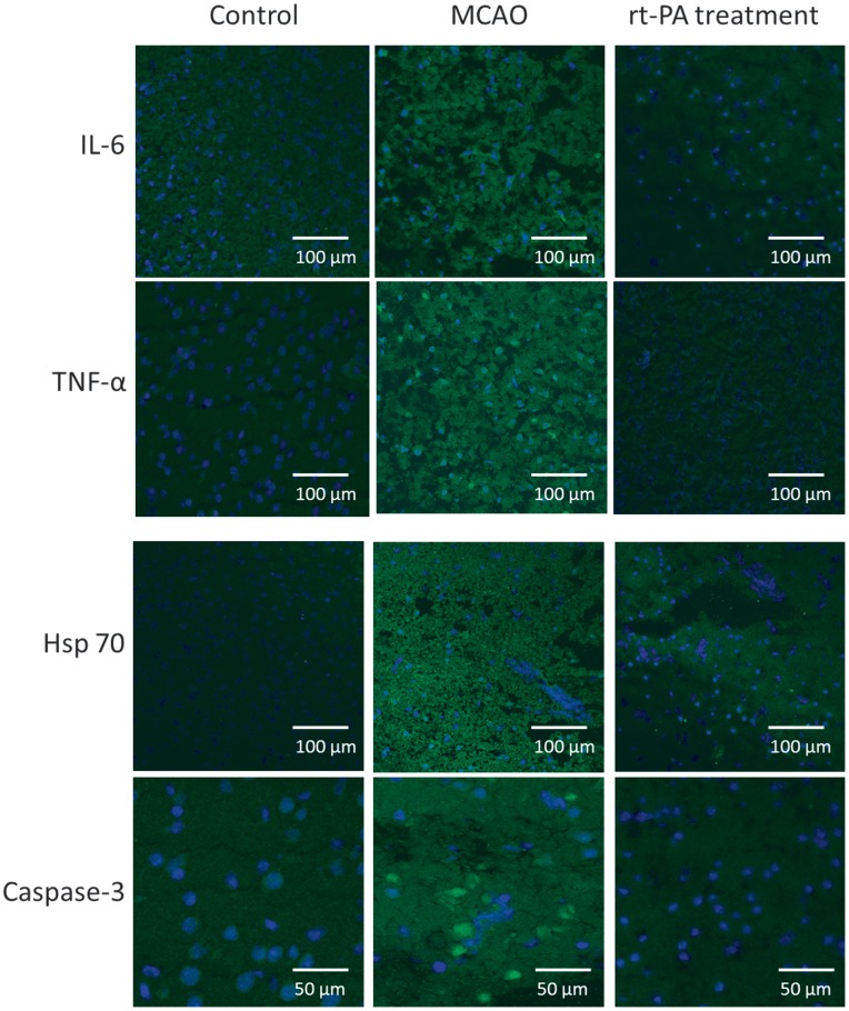
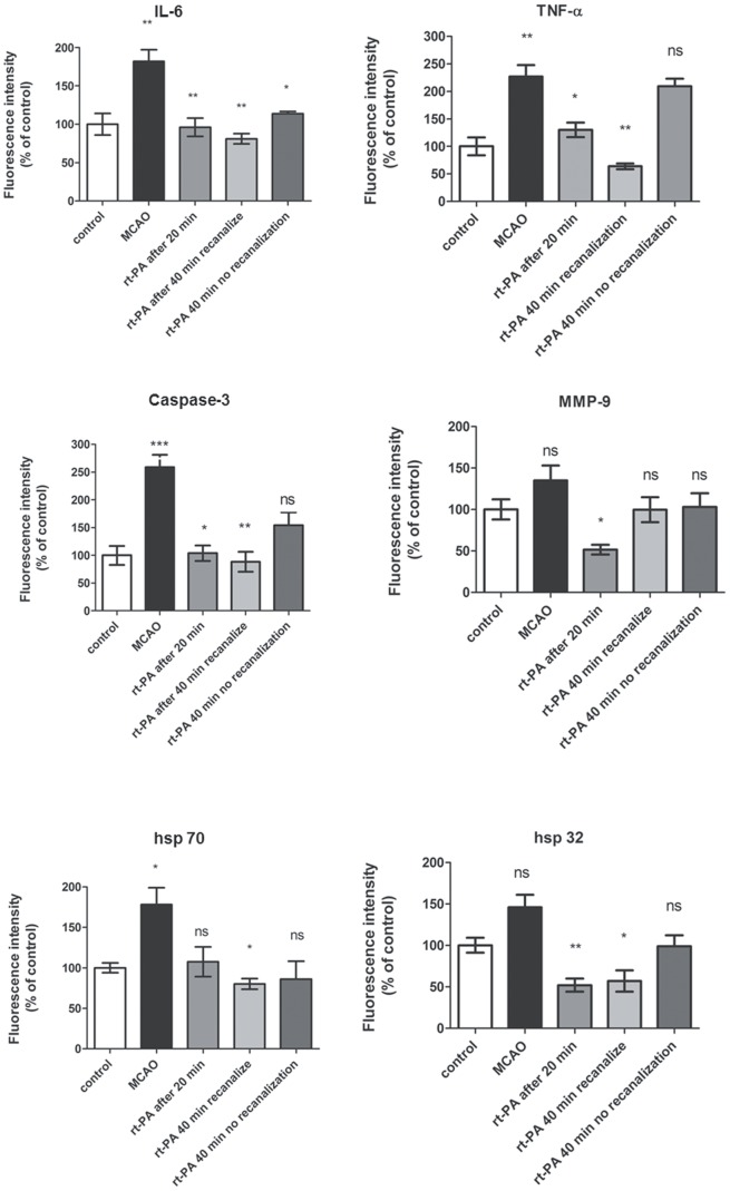
Methods: Thromboembolic stroke was induced by local injection of thrombin directly into the right MCA of C57 black/6J mice. Rt-PA was administered 20 and 40 min after clot formation. The efficiency of rt-PA to induce thrombolysis was measured by laser Doppler. After 24 h, all animals were euthanized and interleukin (IL)-6, tumor necrosis factor-alpha (TNF-α), matrix metalloproteinase (MMP)-9, Caspase-3, hsp 32 and hsp 70 protein levels were investigated by immunofluorescence. Presence of hemorrhage was verified and infarct volume was measured using histology.

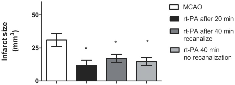
Results: Thrombin injection resulted in clot formation giving rise to cortical brain infarction. Early rt-PA treatment starting at 20 min after the clot formation resulted in 100% recanalization. However, rt-PA-induced thrombolysis dissolved the clot in only 38% of the animals when administered 40 min after clot formation. Protein levels of IL-6, TNF-α, MMP-9, Caspase-3, hsp 32 and hsp 70 were increased after MCAO, whereas treatment with rt-PA attenuated the expressions of inflammatory markers in those animals where the thrombolysis was successful. In addition, the infarct size was significantly reduced with rt-PA treatment compared to non-treated MCAO, regardless of whether MCA thrombolysis was successful.

Conclusions: The present study demonstrates a clear correlation of the protein expression of inflammatory mediators, apoptosis and stress genes with the recanalization data after rt-PA treatment. In this model rt-PA treatment decreases the infarct size regardless of whether vessel recanalization is successful.


Conflict of interest statement

Competing Interests: The authors have declared that no competing interests exist.

Figures

Figure 1
Figure 1. High resolution MR angiography of mouse brain before (a) and after (b) right MCA occlusion (arrows).
T2-weighted images (c) and ADC-maps (d) demonstrating infarction.
Figure 2
Figure 2. Effect of treatment with rt-PA on CBF after MCAO in mice.
(a), MCAO+rt-PA after 20 min and 40 min, (b), MCAO+rt-PA after 40 min. Data were obtained by laser Doppler flowmetry and are expressed as mean ± s.e.m. values, n = 5–9 *P≤0.05, **P≤0.01, ***P<0.001 significant difference between treated groups and MCAO groups. Statistical analyses were performed using Kruskal-Wallis non-parametric test together with Dunn’s post-hoc test.
Figure 3
Figure 3. Size of infarct volume.
Treatment with rt-PA significantly reduced the infarct volume compared to MCAO. Data are expressed as mean± s.e.m.; n = 5–9 *P≤0.05, significant difference between treated groups and MCAO groups. Statistical analyses were performed using Kruskal-Wallis non-parametric test together with Dunn’s post-hoc test.
Figure 4
Figure 4. Effect of administration of rt-PA on protein expression.
Sections from the infarction core showing IL-6, TNF-α, Caspase 3, MMP-9, and hsp 70 protein expressions. The images represent the control group (A, D, G, J, M and P), MCAO (B, E, H, K, N, Q) and MCAO+rt-PA treatment (C, F, I, L, O, R). There are significant increases in IL-6, TNF-α, Caspase 3 and hsp 70 protein levels in the MCAO group compared to the control group. Treatment with rt-PA prevented the increase in expression of these proteins in those animals that recanalized after treatment. There was a slight increase in hsp 32 and MMP-9 protein expression in ischemic brain tissue as compared to control. Data were obtained with confocal microscopy.
Figure 5
Figure 5. Bar graphs showing semi-quantification of fluorescence intensity for IL-6, TNF-α, Caspase 3, MMP-9, hsp 70 and hsp 32 proteins.
Data are presented as the mean percentage relative to control ± s.e.m.; n 5 = 9 *P≤0.05, significant difference between control groups and MCAO, # P≤0.05, significant difference between treated groups and MCAO groups. Statistical analyses were performed using Kruskal-Wallis non-parametric test together with Dunn’s post-hoc test.

Similar articles

Cited by

References

    1. Iadecola C, Anrather J (2011) The immunology of stroke: from mechanisms to translation. Nat Med 17: 796–808. - PMC - PubMed
    1. Lakhan SE, Kirchgessner A, Hofer M (2009) Inflammatory mechanisms in ischemic stroke: therapeutic approaches. J Transl Med 7: 97. - PMC - PubMed
    1. Muir KW, Tyrrell P, Sattar N, Warburton E (2007) Inflammation and ischaemic stroke. Curr Opin Neurol 20: 334–342. - PubMed
    1. Candelario-Jalil E, Yang Y, Rosenberg GA (2009) Diverse roles of matrix metalloproteinases and tissue inhibitors of metalloproteinases in neuroinflammation and cerebral ischemia. Neuroscience 158: 983–994. - PMC - PubMed
    1. Amantea D, Nappi G, Bernardi G, Bagetta G, Corasaniti MT (2009) Post-ischemic brain damage: pathophysiology and role of inflammatory mediators. FEBS J 276: 13–26. - PubMed

Publication types

MeSH terms