The rapid growth of fibroids during early pregnancy
- PMID: 24465797
- PMCID: PMC3896432
- DOI: 10.1371/journal.pone.0085933
The rapid growth of fibroids during early pregnancy
Abstract
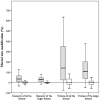
Several studies aimed to disentangle whether pregnancy influences the growth of uterine fibroids but results were inconsistent. In this study, we speculated that fibroid enlargement during pregnancy may not be linear and we hypothesized that this phenomenon may mainly occur during initial pregnancy. To test this hypothesis, we set up a prospective cohort study of women with fibroids undergoing IVF. Cases were women achieving a viable pregnancy. Controls were the subsequent women with fibroids but failing to become pregnant. Twenty-five cases and 25 controls were recruited. The total number of fibroids in the two groups was 46 and 41, respectively. The mean ± SD diameter of the fibroids was 17 ± 10 and 20 ± 11 mm, respectively (p = 0.18). A statistically significant enlargement emerged exclusively in pregnant women. The median (Interquartile Range) modification of the diameter of the lesions in cases and controls was +34% (+6%/+65%) and +2% (-6%/+12%), respectively (p<0.001). The median (Interquartile Range) modification of the volume of the lesions was +140% (+23%/+357%) and 0% (-18%/+37%), respectively (p<0.001). In pregnant women, we failed to document any significant correlation between the magnitude of the growth and ovarian responsiveness to hyper-stimulation, suggesting that steroids hormones are not the unique factors involved. In conclusion, fibroids undergo a rapid and remarkable growth during initial pregnancy. Reasons behind this phenomenon remain to be clarified. The early rise in steroids hormones during early pregnancy may not be sufficient to explain the process. Other pregnancy-related hormones and proteins may play also key roles.
Conflict of interest statement
Figures

Similar articles
-
Ulipristal acetate before in vitro fertilization: efficacy in infertile women with submucous fibroids.Reprod Biol Endocrinol. 2020 May 19;18(1):50. doi: 10.1186/s12958-020-00611-1. Reprod Biol Endocrinol. 2020. PMID: 32430027 Free PMC article. Clinical Trial.
-
Effects of the distance between small intramural uterine fibroids and the endometrium on the pregnancy outcomes of in vitro fertilization-embryo transfer.Gynecol Obstet Invest. 2015;79(1):62-8. doi: 10.1159/000363236. Epub 2014 Nov 22. Gynecol Obstet Invest. 2015. PMID: 25427667
-
The management of uterine fibroids in women with otherwise unexplained infertility.J Obstet Gynaecol Can. 2015 Mar;37(3):277-285. doi: 10.1016/S1701-2163(15)30318-2. J Obstet Gynaecol Can. 2015. PMID: 26001875
-
Management of uterine fibroids in pregnancy: recent trends.Curr Opin Obstet Gynecol. 2015 Dec;27(6):432-7. doi: 10.1097/GCO.0000000000000220. Curr Opin Obstet Gynecol. 2015. PMID: 26485457 Review.
-
The Impact of Noncavity-Distorting Intramural Fibroids on Live Birth Rate in In Vitro Fertilization Cycles: A Systematic Review and Meta-Analysis.J Womens Health (Larchmt). 2020 Feb;29(2):210-219. doi: 10.1089/jwh.2019.7813. Epub 2019 Dec 10. J Womens Health (Larchmt). 2020. PMID: 31821069
Cited by
-
Uterine Fibroids and Pregnancy: A Review of the Challenges from a Romanian Tertiary Level Institution.Healthcare (Basel). 2022 May 6;10(5):855. doi: 10.3390/healthcare10050855. Healthcare (Basel). 2022. PMID: 35627994 Free PMC article.
-
Effect of pregnancy on recurrence of symptomatic uterine myomas in women who underwent myomectomy.Hippokratia. 2018 Jul-Sep;22(3):122-126. Hippokratia. 2018. PMID: 31641332 Free PMC article.
-
Natural History of Uterine Fibroids: A Radiological Perspective.Curr Obstet Gynecol Rep. 2018;7(3):117-121. doi: 10.1007/s13669-018-0243-5. Epub 2018 May 8. Curr Obstet Gynecol Rep. 2018. PMID: 30101039 Free PMC article. Review.
-
Fibroids in Pregnancy: A Case Series.Cureus. 2025 Jul 9;17(7):e87577. doi: 10.7759/cureus.87577. eCollection 2025 Jul. Cureus. 2025. PMID: 40786359 Free PMC article.
-
Overcoming Obstacles During Caesarean Section with a Fibroid in the Uterus, from Diagnosis to Decision: A Case Series.Cureus. 2023 May 29;15(5):e39642. doi: 10.7759/cureus.39642. eCollection 2023 May. Cureus. 2023. PMID: 37388593 Free PMC article.
References
-
- Lev-Toaff AS, Coleman BG, Arger PH, Mintz MC, Arenson RL, et al. (1987) Leiomyomas in pregnancy: sonographic study. Radiology 164: 375–380. - PubMed
-
- Aharoni A, Reiter A, Golan D, Paltiely Y, Sharf M (1988) Patterns of growth of uterine leiomyomas during pregnancy. A prospective longitudinal study. Br J Obstet Gynaecol 95: 510–513. - PubMed
-
- Rosati P, Exacoustòs C, Mancuso S (1992) Longitudinal evaluation of uterine myoma growth during pregnancy. A sonographic study. J Ultrasound Med 11: 511–515. - PubMed
-
- Hammoud AO, Asaad R, Berman J, Treadwell MC, Blackwell S, et al. (2006) Volume change of uterine myomas during pregnancy: do myomas really grow? J Minim Invasive Gynecol 13: 386–390. - PubMed
-
- Neiger R, Sonek JD, Croom CS, Ventolini G (2006) Pregnancy-related changes in the size of uterine leiomyomas. J Reprod Med 51: 671–674. - PubMed
MeSH terms
LinkOut - more resources
Full Text Sources
Other Literature Sources
Medical

