. 2014 Jan 22;9(1):e86071.
doi: 10.1371/journal.pone.0086071. eCollection 2014.

The productive entry pathway of HIV-1 in macrophages is dependent on endocytosis through lipid rafts containing CD4

Affiliations

The productive entry pathway of HIV-1 in macrophages is dependent on endocytosis through lipid rafts containing CD4

Bonnie van Wilgenburg et al. PLoS One. .

Abstract

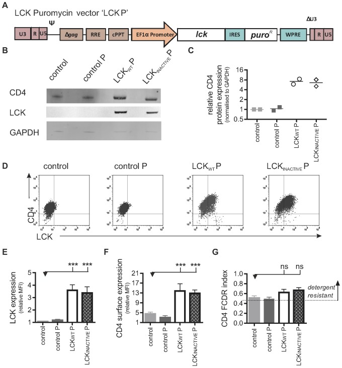
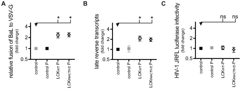
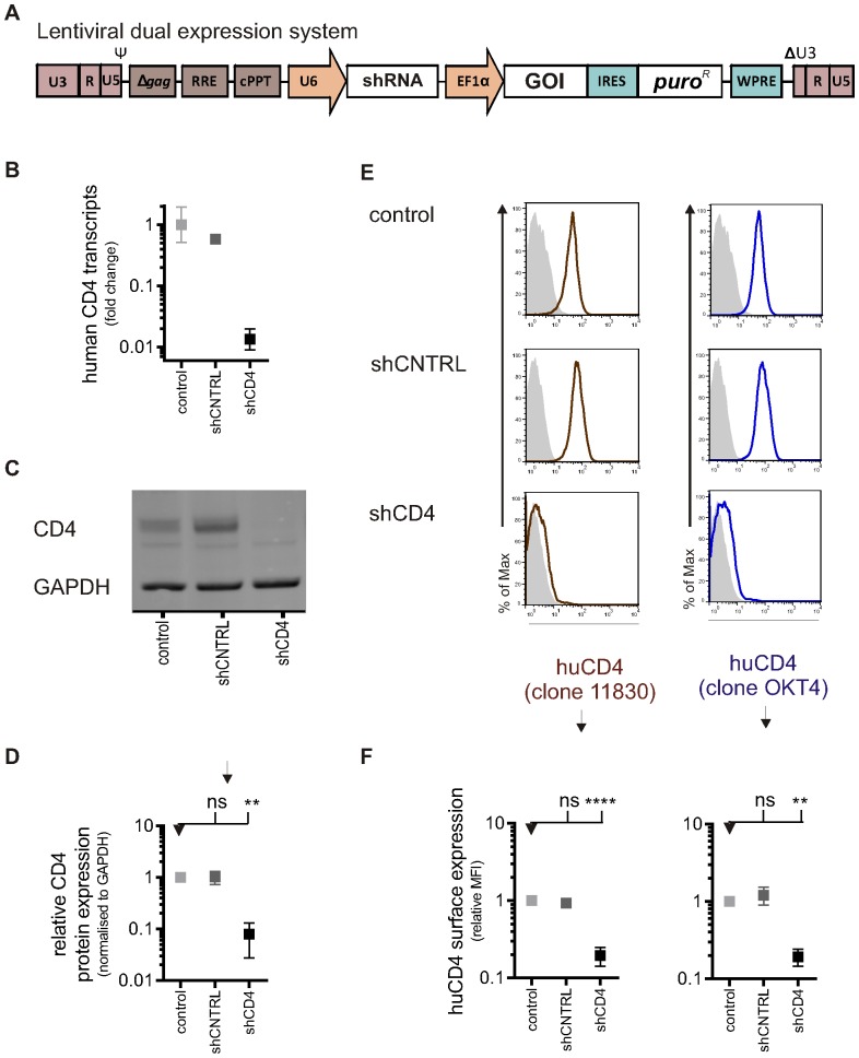
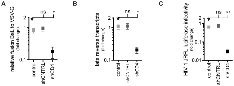
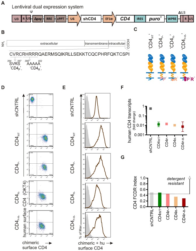
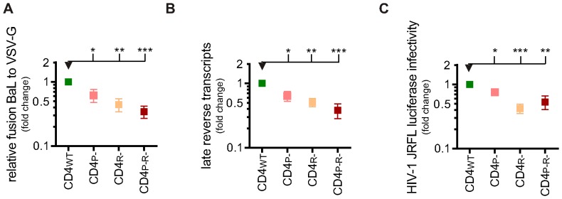
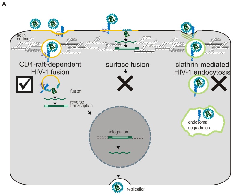
Macrophages constitute an important reservoir of HIV-1 infection, yet HIV-1 entry into these cells is poorly understood due to the difficulty in genetically manipulating primary macrophages. We developed an effective genetic approach to manipulate the sub-cellular distribution of CD4 in macrophages, and investigated how this affects the HIV-1 entry pathway. Pluripotent Stem Cells (PSC) were transduced with lentiviral vectors designed to manipulate CD4 location and were then differentiated into genetically modified macrophages. HIV-1 infection of these cells was assessed by performing assays that measure critical steps of the HIV-1 lifecycle (fusion, reverse transcription, and expression from HIV-1 integrants). Expression of LCK (which tethers CD4 to the surface of T cells, but is not normally expressed in macrophages) in PSC-macrophages effectively tethered CD4 at the cell surface, reducing its normal endocytic recycling route, and increasing surface CD4 expression 3-fold. This led to a significant increase in HIV-1 fusion and reverse transcription, but productive HIV-1 infection efficiency (as determined by reporter expression from DNA integrants) was unaffected. This implies that surface-tethering of CD4 sequesters HIV-1 into a pathway that is unproductive in macrophages. Secondly, to investigate the importance of lipid rafts (as detergent resistant membranes - DRM) in HIV-1 infection, we generated genetically modified PSC-macrophages that express CD4 mutants known to be excluded from DRM. These macrophages were significantly less able to support HIV-1 fusion, reverse-transcription and integration than engineered controls. Overall, these results support a model in which productive infection by HIV-1 in macrophages occurs via a CD4-raft-dependent endocytic uptake pathway.


Conflict of interest statement

Competing Interests: The authors have declared that no competing interests exist.

Figures

Figure 1
Figure 1. Higher levels of CD4 are found in macrophages expressing LCK.
A) Schematic diagram of self-inactivating lentiviral construct used to express wild-type Lck and puroR (LCKWT P). A similar construct was used to express a kinase inactive form of Lck and puroR (LCKINACTIVE P) or to express puroR only (‘control P’). B) Detection of protein expression of LCK and CD4. Control and transgenic PSC-macrophages lysates were analysed by western blotting using anti-LCK and anti-CD4 antibodies. The loading control GAPDH was detected using anti-GAPDH antibody. C) Protein levels were measured with Odyssey software (Li-COR) and CD4 expression was normalised to GAPDH expression. Symbols represent normalised CD4 expression, relative to the PSC-macrophages control group, of two independent experiments. D) Detection of surface CD4 and total LCK expression. Representative two-colour immunofluorescence (dot plot) analysis is shown. Gates were determined by using the two relevant isotype control antibodies. Quantification of total LCK expression (E) and surface CD4 expression (F), expressed as the ratio of the geometric mean fluorescence intensity (MFI) over the isotype control ±SEM of independent experiments (n = 7). G) Detergent resistance of CD4, expressed as mean Flow Cytometric Detergent Resistance (FCDR) index of CD4 (n = 4) in PSC-macrophages, calculated as described in materials and methods.
Figure 2
Figure 2. Increased HIV-1 entry in LCK+ macrophages results in increased reverse transcription, but not successful infection.
A) HIV-1 viral fusion in PSC-macrophages. The percentage of fusion of HIV-1 virions was normalised to the percentage fusion of VSV-G pseudotyped virions. Symbols represent the normalised mean fusion, relative to the PSC-macrophages control group ±SEM of independent experiments. Control-, LCKWT P and LCKINACTIVE P (n = 3) and control P PSC-macrophages (n = 1). B) Reverse transcription of HIV-1 in PSC-macrophages. Late (pol) products were detected by qPCR after 30 h of infection. Symbols represent the relative mean number of copies of HIV-1 DNA±SEM of independent experiments (n = 3), normalised to the number of cells using a β-actin control. C) HIV-1 productive infection in PSC-macrophages. Infection was measured by detecting luciferase activity in PSC-macrophages three days after transduction with NL4.3.Luc.R-E- virus pseudotyped with HIV-1 or VSV-G envelope. HIV-1 NL4.3.Luc.R-E- transduced cells were normalised to VSV-G NL4.3.Luc.R-E- transduced cells. Symbols represent the mean luciferase detection relative to the PSC-macrophages control group ±SEM of independent experiments (n = 3).
Figure 3
Figure 3. CD4 knock-down in genetically modified stem cell-derived macrophages.
A) Schematic diagrams of self-inactivating lentiviral constructs used to express a shRNA (non-specific short-hairpin control (shCNTRL) or a short hairpin targeting CD4 (shCD4)) and puromycin resistance gene (puroR). The Gene Of Interest (GOI) linked to expression of PuromycinR can easily be cloned downstream from the EF1α promoter. B) Detection of CD4 transcripts. RNA was isolated from control and transgenic PSC-macrophages and analysed by RT-qPCR. Symbols represent the relative mean number of copies of CD4 mRNA±SEM of technical replicates (n = 3) using pooled RNA from three independent experiments. C) Detection of protein expression of CD4. Control and transgenic PSC-macrophages lysates were analysed by western blotting using anti-CD4 antibodies. GAPDH, a loading control, was detected using anti-GAPDH antibody. Representative blot is shown.D) Protein levels were measured with Odyssey software (Li-COR) and CD4 expression was normalised to GAPDH expression. Symbols represent mean normalised CD4 expression relative to the PSC-macrophages control group, ±SEM (n = 3 independent experiments). E) Detection of surface protein expression of CD4. Control and transgenic PSC-macrophages were tested for surface CD4 expression by flow cytometry using two different clones of anti-CD4 antibodies. Representative histograms showing CD4 surface staining with mAb clone 11830 (red/brown line, left panel) and with mAb clone OKT4 (blue line, right panel), both compared to isotype control (shaded gray). The expected phenotype (presence or absence of endogenous human CD4) in cells expressing this lentiviral vector is indicated by the symbols. F) Quantification of CD4 expression with mAb clone 11830 (left) and with mAb clone OKT4 (right) relative to the PSC-macrophages control group. The bars reflect the ratio of the geometric mean fluorescence intensity (MFI) over the isotype control ±SEM of independent experiments (n = 4).
Figure 4
Figure 4. HIV-1 infection is reduced in CD4-knock-down macrophages.
A) HIV-1 viral fusion in PSC-macrophages. The percentage of fusion of HIV-1 virions was normalised to the percentage fusion of VSV-G-pseudotyped virions. Symbols represent the normalised mean fusion relative to the PSC-macrophages CD4WT group (shown in Figure 7) of four independent experiments ±SEM (n = 4). B) Reverse transcription of HIV-1 in PSC-macrophages. Late (pol) products were detected by qPCR after 30 h of infection. Symbols represent the relative mean number of copies of HIV-1 DNA±SEM of independent experiments (n = 4), normalised to the number of cells using a β-actin control. C) HIV-1 productive infection in PSC-macrophages Infection was measured by detecting luciferase activity in PSC-macrophages three days after transduction with NL4.3.Luc.R-E- virus pseudotyped with HIV-1 or VSV-G envelope. HIV-1 NL4.3.Luc.R-E- transduced cells were normalised to VSV-G NL4.3.Luc.R-E- transduced cells. Symbols represent the mean luciferase detection relative to the PSC-macrophages CD4WT group (shown in Figure 7) ±SEM of independent experiments (n = 4).
Figure 5
Figure 5. CD4 localization to detergent resistant membranes is reduced in CD4- mutants compared to wild-type CD4 in macrophages.
A) Schematic diagram of self-inactivating dual-expression lentiviral vector used to knock-down endogenous CD4 and express puroR and chimeric CD4. B) Amino acid sequence of cytoplasmic tail of CD4 showing location of regions involved with DRM localization and the mutants inserted to produce CD4P-, CD4R- and CD4P-R-. C) Symbols used to highlight the structure of each mutant chimeric CD4. Chimeric CD4 contains domain 1+2 and the cytoplasmic tail of human CD4 (depicted in blue), but domain 3+4 and transmembrane domain of rat CD4 (depicted in orange). D) Validation of the design of the lentiviral constructs expressing the chimeric CD4, TZM-bl cells were transduced with lentiviral vectors encoding shCD4 and wild-type (WT) or mutant versions of chimeric CD4 (P-, R-, or P-R-) or with a lentiviral vector expressing a control shRNA only (shCNTRL). Two-colour immunofluorescence (dot plot), showing the mean fluorescence intensities of mAb clones OKT4 and OX68, plotted on the Y-axis and X-axis, respectively. Gates were determined by using the two relevant isotype control antibodies. anti-CD4 mAb clone OKT4 recognises endogenous CD4 (domain 3 of human CD4) and anti-CD4 mAb clone OX68 recognises chimeric CD4 (domain 3+4 of rat CD4). E) Detection of surface expression of CD4 in PSC-macrophages. Histogram showing CD4 surface staining of PSC-macrophages with mAB clone 11830 (red/brown line) compared to the matched isotype control (shaded grey). anti-CD4 mAb clone 11830 recognises both endogenous human CD4 and chimeric CD4 (domain 1). F) Detection of CD4 transcripts. RNA was isolated from PSC-macrophages and analysed by RT-qPCR using specific primers. Symbols represent the relative mean number of copies of human endogenous CD4 mRNA relative to the control group (shown in Figure 4A) ±SEM of technical replicates (n = 3) using pooled RNA from three independent experiments. G) Detergent resistance of CD4. Bars reflect the mean Flow Cytometric Detergent Resistance (FCDR) index of CD4 in PSC-macrophages, calculated as described in materials and methods.
Figure 6
Figure 6. HIV-1 infection is reduced when CD4 is excluded from DRM in macrophages.
A) HIV-1 viral fusion in PSC-macrophages. The percentage of fusion of HIV-1 virions was normalised to the percentage fusion of VSV-G pseudotyped virions. Symbols represent the normalised mean fusion relative to the PSC-macrophages CD4WT group of four independent experiments ±SEM (n = 4). B) Reverse transcription of HIV-1 in PSC-macrophages. Late (pol) products were detected using qPCR after 30 h of infection. Symbols represent the relative mean number of copies of HIV-1 DNA of four independent experiments ±SEM (n = 4), normalised to the number of cells using a β-actin control. C) HIV-1 productive infection in PSC-macrophages. Infection was measured by detecting luciferase activity in PSC-macrophages three days after transduction with NL4.3.Luc.R-E- virus pseudotyped with HIV-1 or VSV-G envelope. HIV-1 NL4.3.Luc.R-E- transduced cells were normalised to VSV-G NL4.3.Luc.R-E- transduced cells.Symbols represent the mean luciferase detection relative to the PSC-macrophages CD4WT group of six independent experiments, ±SEM (n = 6).
Figure 7
Figure 7. Proposed model of HIV-1 entry in macrophages.
A) Virions can be taken up non-specifically via endocytic routes (left and right), or via fusion with the cell surface (middle). Plasma membrane fusion is likely a dead-end pathway, as few reverse transcripts reach the nucleus, presumably due to the barrier formed by the cortical actin. Macrophages have low levels of surface CD4, thereby favouring virion uptake through endocytosis over plasma membrane fusion. Endocytic routes naturally overcome the actin barrier and facilitate productive infection, providing that the virion engages with CD4/CCR5 within the endosome and fuse from within this compartment prior to degradation. Virions are more likely to fuse from a raft-dependent uptake pathway (left) and escape endosomal degradation or recycling (not depicted in figure) compared to other endocytic routes, such as clathrin-mediated endocytosis (right), as the co-receptor CCR5 associates with lipid raft microdomains in macrophages.

Similar articles

Cited by

References

    1. Harbison MA, Gillis JM, Pinkston P, Byrn RA, Rose RM, et al. (1990) Effects of recombinant soluble CD4 (rCD4) on HIV-1 infection of monocyte/macrophages. The Journal of infectious diseases 161: 1–6. - PubMed
    1. Collman R, Godfrey B, Cutilli J, Rhodes A, Hassan NF, et al. (1990) Macrophage-tropic strains of human immunodeficiency virus type 1 utilize the CD4 receptor. Journal of Virology 64: 4468–4476. - PMC - PubMed
    1. Jordan CA, Watkins BA, Kufta C, Dubois-Dalcq M (1991) Infection of brain microglial cells by human immunodeficiency virus type 1 is CD4 dependent. Journal of Virology 65: 736–742. - PMC - PubMed
    1. Ho WZ, Lioy J, Song L, Cutilli JR, Polin RA, et al. (1992) Infection of cord blood monocyte-derived macrophages with human immunodeficiency virus type 1. Journal of Virology 66: 573–579. - PMC - PubMed
    1. Collin M, Herbein G, Montaner L, Gordon S (1993) PCR analysis of HIV1 infection of macrophages: virus entry is CD4-dependent. Research in virology 144: 13–19. - PubMed

Publication types

MeSH terms

Substances