. 2014 Jan 21;9(1):e86173.
doi: 10.1371/journal.pone.0086173. eCollection 2014.

Targeting cancer cells with reactive oxygen and nitrogen species generated by atmospheric-pressure air plasma

Affiliations

Targeting cancer cells with reactive oxygen and nitrogen species generated by atmospheric-pressure air plasma

Hak Jun Ahn et al. PLoS One. .

Abstract

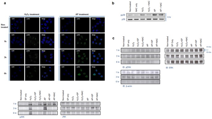
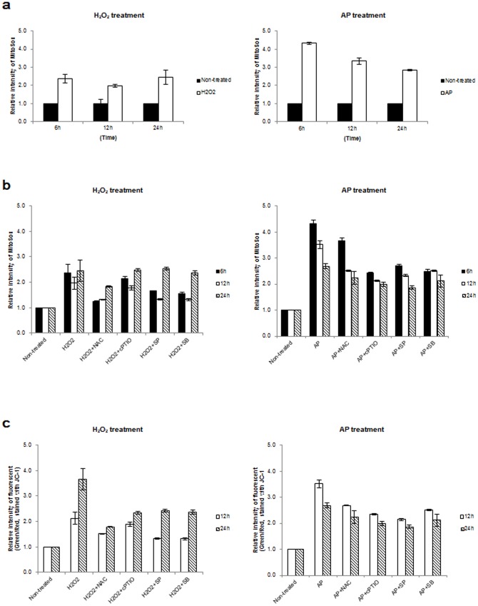
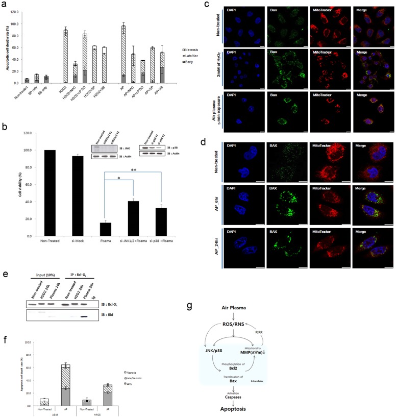
The plasma jet has been proposed as a novel therapeutic method for cancer. Anticancer activity of plasma has been reported to involve mitochondrial dysfunction. However, what constituents generated by plasma is linked to this anticancer process and its mechanism of action remain unclear. Here, we report that the therapeutic effects of air plasma result from generation of reactive oxygen/nitrogen species (ROS/RNS) including H2O2, Ox, OH-, •O2, NOx, leading to depolarization of mitochondrial membrane potential and mitochondrial ROS accumulation. Simultaneously, ROS/RNS activate c-Jun NH2-terminal kinase (JNK) and p38 kinase. As a consequence, treatment with air plasma jets induces apoptotic death in human cervical cancer HeLa cells. Pretreatment of the cells with antioxidants, JNK and p38 inhibitors, or JNK and p38 siRNA abrogates the depolarization of mitochondrial membrane potential and impairs the air plasma-induced apoptotic cell death, suggesting that the ROS/RNS generated by plasma trigger signaling pathways involving JNK and p38 and promote mitochondrial perturbation, leading to apoptosis. Therefore, administration of air plasma may be a feasible strategy to eliminate cancer cells.


Conflict of interest statement

Competing Interests: The authors have declared that no competing interests exist.

Figures

Figure 1
Figure 1. The microplasma jet system and its optical emission spectrum.
(a) Photograph of the fabricated microplasma jet system. The inset is a schematic presentation of the micro plasma jet nozzle. (b) Photograph of air micro plasma jet generated at atmospheric pressure. (c) Optical emission spectrum of air micro plasma jet during discharge.
Figure 2
Figure 2. Generation of ROS and RNS by air plasma.
(a) Levels of extracellular H2O2 were determined in culture or non-culture (Medium only) supernatants, with (AP) or without (Non-treated) air plasma treatment. The culture supernatant was harvested at the indicated times following air plasma treatment (AP). AP 0 (min) indicates supernatant harvested immediately after plasma treatment. The concentration of H2O2 in the culture supernatant was determined by comparison to a H2O2 standard calibration curve, following incubation with Amplex UltraRed reagent (n = 5). (b) Generation and/or penetration across the plasma membrane of ROS including H2O2, OH, and •O2 in the intracellular matrix following air plasma jet (AP) were determined using the ROS-sensitive probe H2DCFDA (n = 5). The fluorescence of untreated cells (Non-treated) was arbitrarily set to 1. (c) Levels of extracellular NO were determined in non-culture (in the absence of HeLa cells, upper) and culture (in the presence of HeLa cells, bottom) supernatants at the indicated times after air plasma jet exposure by the Griess assay (n = 10). (d) Levels of intracellular NO were evaluated using DAF-FM. Data are shown as the mean ± S.E.M. (n = 10).
Figure 3
Figure 3. Treatment of antioxidants reduced level of extra- and intracellular ROS/RNS generated by air plasma.
(a) Levels of extracellular H2O2 were determined in culture or non-culture (Medium only) supernatants with air plasma treatment in the presence (AP + NAC) or absence (AP) of the antioxidant NAC (n = 5). NAC was added 1h prior to plasma treatment. The culture supernatant was harvested at the indicated times following air plasma treatment. AP 0 (min) indicates supernatant harvested immediately after plasma treatment. (b) Generation of ROS including H2O2, OH, and •O2 in the intracellular matrix following air plasma jet (AP) were determined using the ROS-sensitive probe H2DCFDA (n = 5). Cells were pretreated with the antioxidants NAC or cPTIO for 1h and then exposed to air plasma (AP+NAC or AP+cPTIO) or H2O2 (H2O2+NAC or H2O2+cPTIO). At indicated times following air plasma treatment, cells were harvested. The fluorescence of untreated cells (Non-treated) was arbitrarily set to 1. (c) Levels of extracellular NO were determined in non-culture (in the absence of HeLa cells, upper) and culture (in the presence of HeLa cells, bottom) supernatants at the indicated times after air plasma jet exposure by the Griess assay (n = 10). Medium in the presence or absence of HeLa cells was pretreated with NAC or cPTIO for 1h prior to exposure to plasma or H2O2. (d) Levels of intracellular NO were evaluated using DAF-FM. Data are shown as the mean ± S.E.M. (n = 10).
Figure 4
Figure 4. Air plasma-induced phosphorylation of JNK and p38.
(a) Phosphorylation of JNK following treatment with air plasma jet or H2O2 was analyzed via immunofluorescence staining and immunoblotting using anti-phospho-JNK antibody. The equivalent amount of total JNK proteins is shown as a quantitative loading control for JNK phosphorylation. (b) Phosphorylation of p38 induced by air plasma was visualized by immunoblotting using anti-phospho-p38 antibody. The comparable total p38 proteins are shown as a loading control for p38 phosphorylation. (c) Variation of phosphorylation status of ERK was not detected following treatment with air plasma or H2O2.
Figure 5
Figure 5. Collapse of the mitochondrial transmembrane potential (Δψm) following plasma treatment.
(a) Mitochondrial ROS production was evaluated using the mitochondria-specific probe Mitosox following H2O2 or air plasma treatment. The mitochondrial ROS in untreated cells (Non-treated) was arbitrarily set to 1. The cells was harvested at the indicated times following air plasma treatment and analyzed by flow cytometry. (b) Cells were pretreated with the antioxidants (NAC or cPTIO) or kinase inhibitors (SP600125 or SB203580) for 1 h and then exposed to air plasma (AP+NAC, AP+cPTIO, AP+SP or AP+SB) or H2O2 (H2O2+NAC, H2O2+cPTIO, H2O2+SP or H2O2+SB). Cells were harvested at indicated times following plasma treatment, and levels of mitochondrial ROS were measured (n = 5). The fluorescence of untreated cells (Non-treated) was arbitrarily set to 1. (c) The mitochondrial membrane potential was evaluated using the mitochondria-specific probe JC-1 with or without air plasma or H2O2, (n = 5). The fluorescence in untreated HeLa cells was arbitrarily set to 1.
Figure 6
Figure 6. Air plasma induces cell death by activation of JNK and p38 via generating ROS and RNS.
(a) Antioxdizing agents (NAC and cPTIO) or kinase inhibitors (SP600125 and SB203580) partially rescued plasma-induced cell death. Data are shown as the mean ± S.E.M. (n = 10). (b) Plasma-induced cell death was partially abrogated in JNK1/2 (siJNK1/2) or p38 (sip38) knockdown cells. To monitor plasma-induced cell death, ATP-based cell viability assay was performed in the presence of control, JNK1/2, or p38 siRNA. Data are shown as the mean ± S.E.M. in triplicate from three independent experiment (n = 3). Cell viability of untreated HeLa cell population was arbitrarily set to 100%. Immunoblotting with anti-JNK and p38 antibodies was done to confirm knockdown. *p<0.01, **p<0.05. (c)–(d) Air plasma induced Bax translocation to the mitochondria. The plasma- or H2O2-treated cells were further incubated for 6–24 h. After 6–24 h incubation, cells were fixed by 3.7% formaldehyde. Bax (green) was stained anti-bax antibody and MitoTracker was used for staining of mitochondria (Red). Bax (green) and mitochondrial (red, MitoTracker) fluorescence were assessed, 6 h (d) and 24 h ((c) and (d)) after exposure to air plasma for 5 min by fluorescence confocal microscopy. Bax was diffusely distributed in untreated cells (non-treated). However, after treatment with plasma, Bax was localized to mitochondria, based on the overlap of the Bax and MitoTracker fluorescence images (Merge, yellow). DAPI was used for nuclear staining (blue). White bar was mean magnification of image (10 μM). (e) Bid formed a proapoptotic complex with Bcl-xL following plasma treatment. (f) Air plasma induced differential cell death in human lung adenocarcinoma A549 and normal lung fibroblast MRC5 cell lines. A549 and MRC5 cells were treated with air plasma jets and then incubated further for 24 h. After harvesting and staining cells with anti-annexin V-FITC and PI, cell death was evaluated by flow cytometry. The values represent the mean (s.e.m) from three independent experiments. (g) A proposed model for air plasma jet-induced apoptosis in HeLa cells through ROS/RNS production, trespassing on cells, activation of signal transduction pathways, and mitochondrial damage.

References

    1. Kalghatgi S, Kelly CM, Cerchar E, Torabi B, Alekseev O, et al. (2011) Effects of non-thermal plasma on mammalian cells. PLoS one 6: e16270. - PMC - PubMed
    1. Ahn HJ, Kim KI, Kim G, Moon E, Yang SS, et al. (2011) Atmospheric-pressure plasma jet induces apoptosis involving mitochondria via generation of free radicals. PLoS one 6: e28154. - PMC - PubMed
    1. Hong YF, Kang JG, Lee HY, Uhm HS, Moon E, et al. (2009) Sterilization effect of atmospheric plasma on Escherichia coli and Bacillus subtilis endospores. Lett Appl Microbiol 48: 33–7. - PubMed
    1. Rupf S, Lehmann A, Hannig M, Schäfer B, Schubert A, et al. (2010) Killing of adherent oral microbes by a non-thermal atmospheric plasma jet. J Med Microbiol 59: 206–12. - PubMed
    1. Yamamori T, Yasui H, Yamazumi M, Wada Y, Nakamura Y, et al. (2012) Ionizing radiation induces mitochondrial reactive oxygen species production accompanied by upregulation of mitochondrial electron transport chain function and mitochondrial content under control of the cell cycle checkpoint. Free Radic Biol Med 15: 260–70. - PubMed

Publication types

MeSH terms