

Ureter smooth muscle cell orientation in rat is predominantly longitudinal
- PMID: 24465961
- PMCID: PMC3897663
- DOI: 10.1371/journal.pone.0086207
Ureter smooth muscle cell orientation in rat is predominantly longitudinal
Abstract
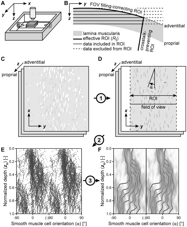
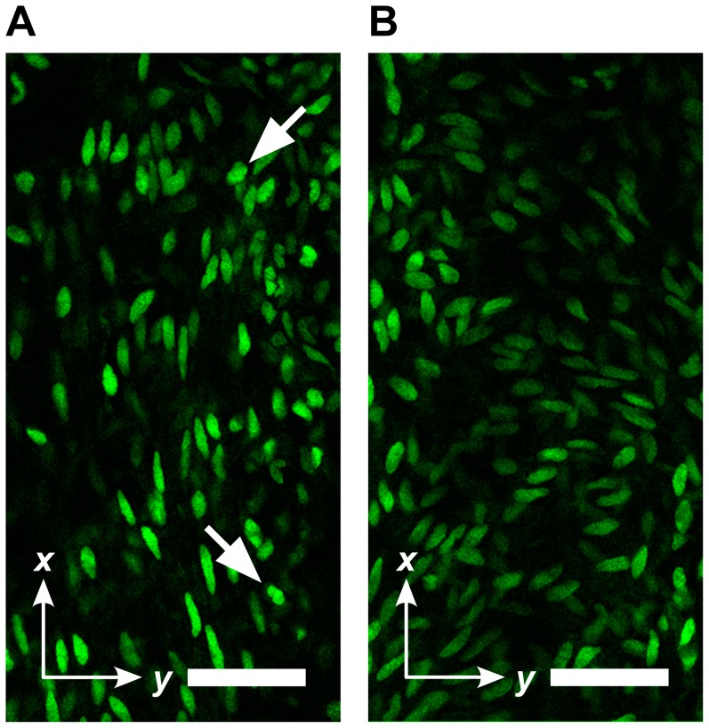
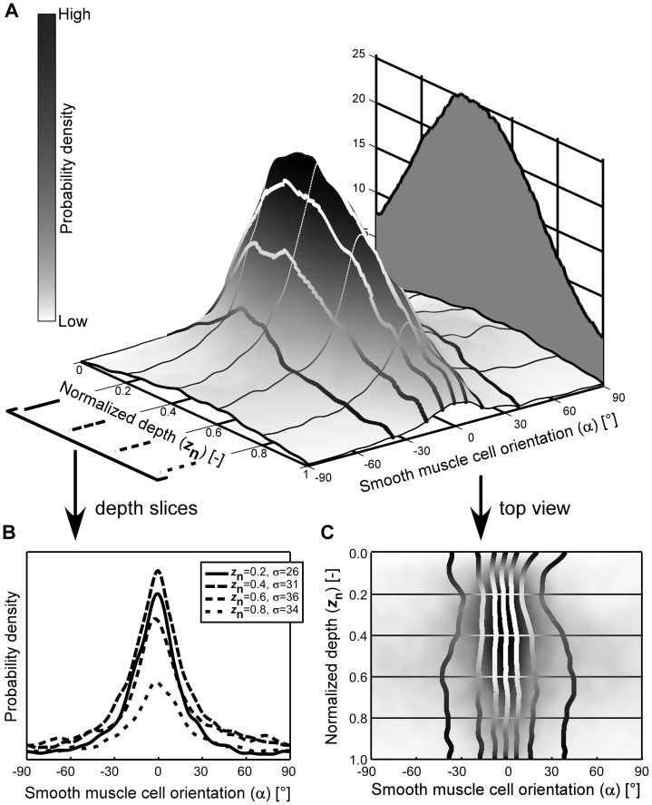
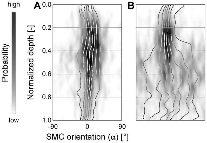
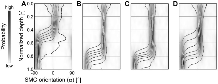
In ureter peristalsis, the orientation of the contracting smooth muscle cells is essential, yet current descriptions of orientation and composition of the smooth muscle layer in human as well as in rat ureter are inconsistent. The present study aims to improve quantification of smooth muscle orientation in rat ureters as a basis for mechanistic understanding of peristalsis. A crucial step in our approach is to use two-photon laser scanning microscopy and image analysis providing objective, quantitative data on smooth muscle cell orientation in intact ureters, avoiding the usual sectioning artifacts. In 36 rat ureter segments, originating from a proximal, middle or distal site and from a left or right ureter, we found close to the adventitia a well-defined longitudinal smooth muscle orientation. Towards the lamina propria, the orientation gradually became slightly more disperse, yet the main orientation remained longitudinal. We conclude that smooth muscle cell orientation in rat ureter is predominantly longitudinal, though the orientation gradually becomes more disperse towards the proprial side. These findings do not support identification of separate layers. The observed longitudinal orientation suggests that smooth muscle contraction would rather cause local shortening of the ureter, than cause luminal constriction. However, the net-like connective tissue of the ureter wall may translate local longitudinal shortening into co-local luminal constriction, facilitating peristalsis. Our quantitative, minimally invasive approach is a crucial step towards more mechanistic insight into ureter peristalsis, and may also be used to study smooth muscle cell orientation in other tube-like structures like gut and blood vessels.
Conflict of interest statement
Figures
Similar articles
-
[Origin of motion in the human ureter: mechanics, energetics and kinetics of the myosin molecular motors].Urologia. 2012 Apr-Jun;79(2):123-9. doi: 10.5301/RU.2012.9110. Urologia. 2012. PMID: 22427244 Italian.
-
Circular smooth muscle contributes to esophageal shortening during peristalsis.World J Gastroenterol. 2012 Aug 28;18(32):4317-22. doi: 10.3748/wjg.v18.i32.4317. World J Gastroenterol. 2012. PMID: 22969194 Free PMC article.
-
The IgCAM CLMP regulates expression of Connexin43 and Connexin45 in intestinal and ureteral smooth muscle contraction in mice.Dis Model Mech. 2018 Feb 22;11(2):dmm032128. doi: 10.1242/dmm.032128. Dis Model Mech. 2018. PMID: 29361518 Free PMC article.
-
Excitation-Contraction Coupling in Ureteric Smooth Muscle: Mechanisms Driving Ureteric Peristalsis.Adv Exp Med Biol. 2019;1124:103-119. doi: 10.1007/978-981-13-5895-1_4. Adv Exp Med Biol. 2019. PMID: 31183824 Review.
-
Pyeloureteric peristalsis: role of atypical smooth muscle cells and interstitial cells of Cajal-like cells as pacemakers.J Physiol. 2006 Nov 1;576(Pt 3):695-705. doi: 10.1113/jphysiol.2006.116855. Epub 2006 Aug 31. J Physiol. 2006. PMID: 16945969 Free PMC article. Review.
Cited by
-
A method for three-dimensional quantification of vascular smooth muscle orientation: application in viable murine carotid arteries.Biomech Model Mechanobiol. 2016 Apr;15(2):419-32. doi: 10.1007/s10237-015-0699-4. Epub 2015 Jul 15. Biomech Model Mechanobiol. 2016. PMID: 26174758 Free PMC article.
-
Ureter development and associated congenital anomalies.Nat Rev Nephrol. 2025 Jun;21(6):366-382. doi: 10.1038/s41581-025-00951-4. Epub 2025 Mar 31. Nat Rev Nephrol. 2025. PMID: 40164775 Review.
References
-
- Henle J (1866) Handbuch der Systematischen Anatomie des Menschen; Zweiter band: Eingeweidelehre. Braunschweig: Friedrich Vieweg und Sohn. 836 p.
-
- Ebner VW (1902) A. Koelliker's Handbuch der Gewebelehre des Menschen. Leipzig: Wilhelm Engelmann. 1020 p.
-
- Bouvin MJ (1869) Over den bouw en de beweging der ureteres [PhD Thesis]: Hoogeschool te Utrecht.
-
- Toldt C (1877) Lehrbuch der Gewebelehre mit Vorzugsweiser Berücksichtigung der Menschlichen Körpers. Stuttgart: Ferdinand Enke. 655 p.
Publication types
MeSH terms
LinkOut - more resources
Full Text Sources
Other Literature Sources
