. 2014 Jan 23;9(1):e86240.
doi: 10.1371/journal.pone.0086240. eCollection 2014.

Alteration of daily and circadian rhythms following dopamine depletion in MPTP treated non-human primates

Affiliations

Alteration of daily and circadian rhythms following dopamine depletion in MPTP treated non-human primates

Karim Fifel et al. PLoS One. .

Abstract

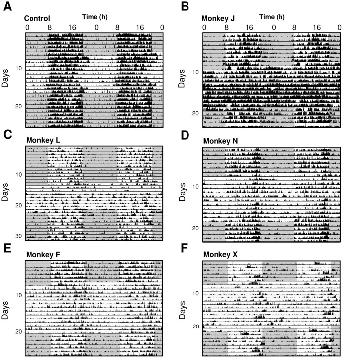
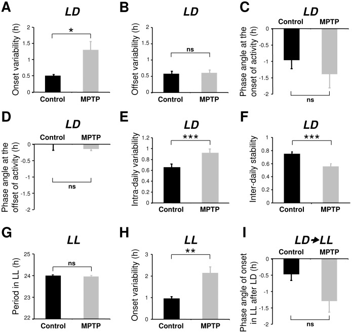
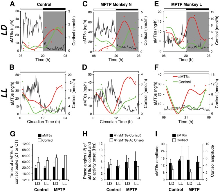
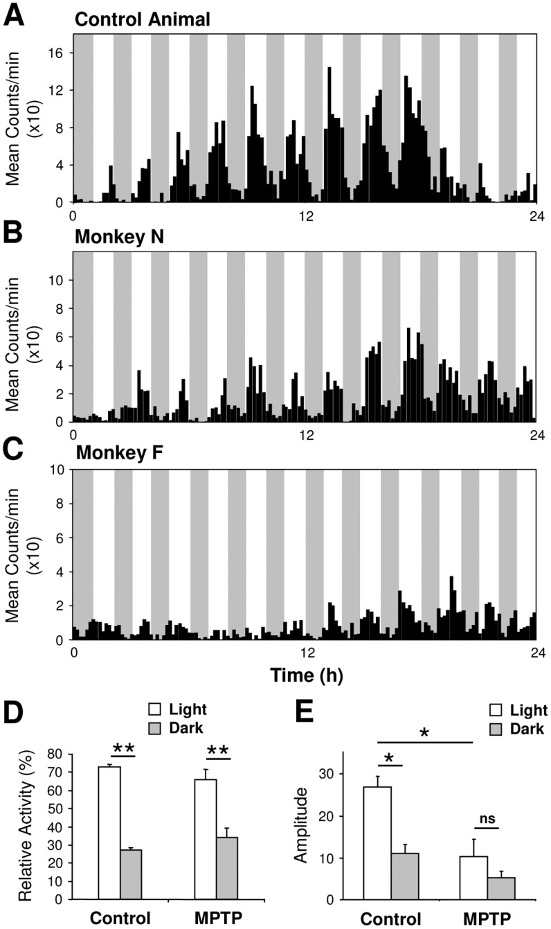
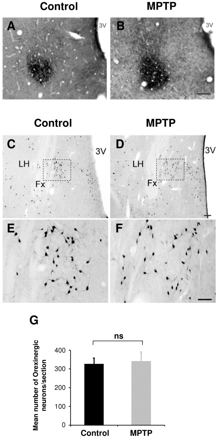
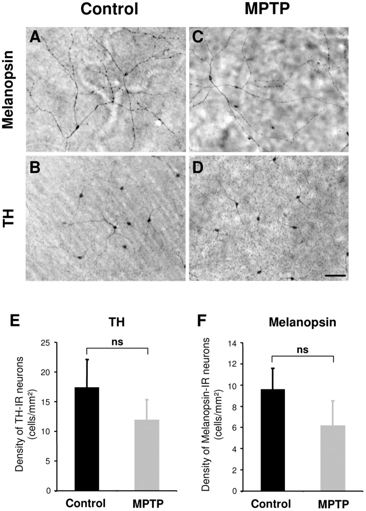
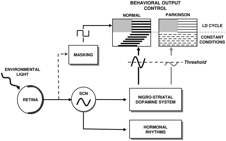
Disturbances of the daily sleep/wake cycle are common non-motor symptoms of Parkinson's disease (PD). However, the impact of dopamine (DA) depletion on circadian rhythms in PD patients or non-human primate (NHP) models of the disorder have not been investigated. We evaluated alterations of circadian rhythms in NHP following MPTP lesion of the dopaminergic nigro-striatal system. DA degeneration was assessed by in vivo PET ([(11)C]-PE2I) and post-mortem TH and DAT quantification. In a light∶dark cycle, control and MPTP-treated NHP both exhibit rest-wake locomotor rhythms, although DA-depleted NHP show reduced amplitude, decreased stability and increased fragmentation. In all animals, 6-sulphatoxymelatonin peaks at night and cortisol in early morning. When the circadian system is challenged by exposure to constant light, controls retain locomotor rest-wake and hormonal rhythms that free-run with stable phase relationships whereas in the DA-depleted NHP, locomotor rhythms are severely disturbed or completely abolished. The amplitude and phase relations of hormonal rhythms nevertheless remain unaltered. Use of a light-dark masking paradigm shows that expression of daily rest-wake activity in MPTP monkeys requires the stimulatory and inhibitory effects of light and darkness. These results suggest that following DA lesion, the central clock in the SCN remains intact but, in the absence of environmental timing cues, is unable to drive downstream rhythmic processes of striatal clock gene and dopaminergic functions that control locomotor output. These findings suggest that the circadian component of the sleep-wake disturbances in PD is more profoundly affected than previously assumed.


Conflict of interest statement

Competing Interests: The authors have declared that no competing interests exist.

Figures

Figure 1
Figure 1. Following MPTP treatment, the circadian locomotor rest-activity rhythm is abolished under constant light.
(A) Pre-MPTP treatment, daily locomotor activity is synchronized to the LD cycle (dark phase shown by shading) and in constant light (LL) the circadian rhythm free runs with a period of 23.97 hrs (Monkey Y). (B) Same animal, 4.8 months post-MPTP treatment, rest-wake activity is synchronized to the LD cycle but becomes arrhythmic in LL. Compared to pre-treatment, periodogram analyses (panels to right of actograms) confirm a quantitative decrease in amplitude during LD and the absence of rhythmicity during LL. The averaged 24 h locomotor activity profiles (C–D) illustrate the consolidated activity during the light phase in LD cycle both pre- and post-MPTP and during LL pre-MPTP but a complete loss of rhythmicity post-MPTP.
Figure 2
Figure 2. Double-plotted actograms illustrating alteration of circadian expression of locomotor activity in constant light (LL) in an untreated control and in 5 MPTP treated animals in addition to that shown in Figure 1.
Note that while daily locomotor rest activity rhythms are observed for all animals during the LD cycle preceding and subsequent to LL. In MPTP-treated animals the circadian rhythm of locomotor activity was strongly altered (D, E, F) or abolished (B, C) in LL. C and F were highly symptomatic cases, D and E were mildly symptomatic and B was asymptomatic. The dark phase is shown by shaded areas and light phase by light areas.
Figure 3
Figure 3. Variability of onsets, offsets, period, intra-daily variability, inter-daily stability and phase angle in control (n = 10) and MPTP-treated animals (n = 7).
After MPTP treatment there was a significant increase of variability at the onset but not in the offset of locomotor activity in LD (A, B). Control and MPTP-treated animals show an approximately 1 hour phase delay in activity following lights on (C) and no phase angle difference in activity offset at lights off (D). The intra-daily variability increased (E) while the inter-daily stability decreased (F) after MPTP treatment. In LL the still rhythmic MPTP-treated animals showed an increased activity onsets variability (H), but with no significant difference in period (G). Upon release from LD to LL, MPTP animals had no significant difference in the phase angle of activity onset compared to controls (I) showing that the entrainment of activity rhythm under LD regime was not affected by MPTP. *P<0.05, **P<0.01, ***P<0.001.
Figure 4
Figure 4. Averaged 24-activity profiles for all control and MPTP-treated monkeys with individual traces for all recordings (n = 39 assays) in LD (upper panel) and in LL (lower panel) conditions (A).
Data for LD always correspond to the recording period immediately preceding LL. Control (or pre-treatment) animals show a robust daily and circadian rhythmicity with a consolidated activity during the light phase in LD and the subjective day in LL. In MPTP-treated animals, the daily rhythmicity of locomotor activity in the LD cycle is clearly evident but becomes progressively less robust in non-symptomatic, mildly symptomatic and highly symptomatic animals. When transferred to constant LL conditions, circadian rhythmicity deteriorates and highly symptomatic animals are arrhythmic with a continuous distribution of activity throughout the 24 h period. Note that the loss of rhythmicity is also recorded in one asymptomatic animal under LL (also shown in Fig. 2B ). Relationship between the amplitude of the rhythm (χ2 periodogram) and clinical score in LD and LL (B). A complete loss of rhythmicity is mainly, but not exclusively observed for the highest clinical scores whereas some subjects with severe motor symptoms still maintained rhythmicity. Values are shown as mean ± SEM. MPTP treatment induced loss of dopamine neurons in the mesencephalon and dopaminergic depletion of the striatum (C). Examples of [11C]-PE2I scans of the basal ganglia before (C1) and after MPTP treatment showing 69% decrease in [11C]-PE2I binding potential in the striatum (C2) and 86% in a more extreme case (C3). The reduction of TH immunostaining in the mesencephalon of MPTP-treated monkeys (C5, 65%, C6, 72%, C7, >80%) compared to a control (C4). a: Substantia nigra (SN); b: Ventral tegmental area (VTA), Scale bar: 1000 µm.
Figure 5
Figure 5. MPTP-treated monkeys maintain 24 h hormonal rhythms of melatonin and cortisol in both entrained (LD) and constant conditions (LL).
Representative individual 24(dual harmonic regression fit) of urinary concentration of 6 sulphatoxymelatonin (red circles, line) and cortisol (green circles, line) compared to the mean relative activity profile (black line). Gray shading indicates the dark phase. In LD, controls and MPTP-treated animals show clear rhythms of locomotor activity, aMT6s and cortisol (A, C, E). In LL, control (B), rhythmic (D) and arrhythmic (F) MPTP animals also show a clear circadian rhythmicity of aMT6s and cortisol. The times of the aMT6s and cortisol peaks (G), the phase angles of aMT6s to both cortisol and activity rhythms (H) and the amplitudes of aMT6s and cortisol rhythms (I) were similar in LD and LL in both controls and MPTP-treated subjects (controls n = 4; MPTP n = 3).
Figure 6
Figure 6. MPTP-treated monkeys show normal masking responses to darkness in a 1 h light–1 h dark regime.
The control monkey (A) shows a clear 24 h rhythmicity coupled with a strong masking effect of darkness on locomotor activity. MPTP-treated monkeys show attenuated amplitude of the rhythm (B, mildly symptomatic, C, highly symptomatic) but nevertheless a strong masking effect of darkness proportionately similar to that of controls. (D) Mean relative total activity during light and dark phases. (E) Amplitude of the rhythm analysed separately for light and dark phases (controls n = 4; MPTP n = 5). *P<0.05, **P<0.001.
Figure 7
Figure 7. The orexinergic system in the lateral hypothalamic area and vasoactive intestinal peptide (VIP) containing neurons in the suprachiasmatic nucleus (SCN) are not affected after MPTP treatment.
Example section in the hypothalamic SCN illustrating staining of VIP immunopositive neurons in a control (A) and an MPTP-treated animal (B). Scale bar = 100 µm. Example sections of orexin-A immunopositive neurons in the lateral hypothalamus of a control (C, E) and MPTP-treated animal (D, F). The regions indicated dashed square in C and D are shown at higher magnification in E and F. (G) There was no significant difference in the number of orexin expressing neurons between control (n = 1) and MPTP-treated animals (n = 3). All orexin-A expressing neurons in the hypothalamus were counted on 5–7 sections per animal (distance between sections 150–200 µm). Scale bar: C, D = 200 µm, E, F = 100 µm. LH = Lateral hypothalamus; Fx = Fornix; 3V = Third ventricle.
Figure 8
Figure 8. In the retina, the morphology of melanopsin and TH immunoreactive neurons appear normal after MPTP treatment.
The photomicrographs are from representative flat-mounted retina immunostained for melanopsin (A, C) or TH (B, D) in control (A, B) and MPTP-treated animals (C, D). Scale bar: 100 µm. E and F illustrate histograms of the densities of TH and melanopsin immunoreactive neurons. (Controls n = 2; MPTP n = 3).
Figure 9
Figure 9. Model of the consequences of dopamine depletion in the nigrostriatal system.
In healthy subjects the LD cycle synchronizes daily output rhythms (activity, hormones) and fine tunes these rhythms through acute masking effects. In constant conditions, rhythmic outputs free-run under the control of the circadian clock. In the case of dopamine depletion in the nigrostriatal system, the excitatory and inhibitory effects of environmental light and dark are sufficient to sustain expression of a daily locomotor rhythm. In constant conditions, the endogenous clock is intact and drives rhythmic hormonal outputs. In contrast, rhythmic locomotor behavior is abolished if the rhythmic dopamine function driven by the SCN falls below a hypothetical threshold.

Similar articles

Cited by

References

    1. Chaudhuri KR, Healy DG, Schapira AH (2006) Non-motor symptoms of Parkinson's disease: diagnosis and management. Lancet Neurol 5: 235–245. - PubMed
    1. Chaudhuri KR, Schapira AH (2009) Non-motor symptoms of Parkinson's disease: dopaminergic pathophysiology and treatment. Lancet Neurol 8: 464–474. - PubMed
    1. Carlsson A (1972) Biochemical and pharmacological aspects of Parkinsonism. Acta Neurol Scand Suppl 51: 11–42. - PubMed
    1. Arnulf I, Leu-Semenescu S (2009) Sleepiness in Parkinson's disease. Parkinsonism Relat Disord 15: S101–S104. - PubMed
    1. Wisor JP, Nishino S, Sora I, Uhl GH, Mignot E, et al. (2001) Dopaminergic role in stimulant-induced wakefulness. J Neurosci 21: 1787–1794. - PMC - PubMed

Publication types

MeSH terms

LinkOut - more resources