. 2014 Jan 22;9(1):e86339.
doi: 10.1371/journal.pone.0086339. eCollection 2014.

Low structural variation in the host-defense peptide repertoire of the dwarf clawed frog Hymenochirus boettgeri (Pipidae)

Affiliations

Low structural variation in the host-defense peptide repertoire of the dwarf clawed frog Hymenochirus boettgeri (Pipidae)

Severine Matthijs et al. PLoS One. .

Abstract

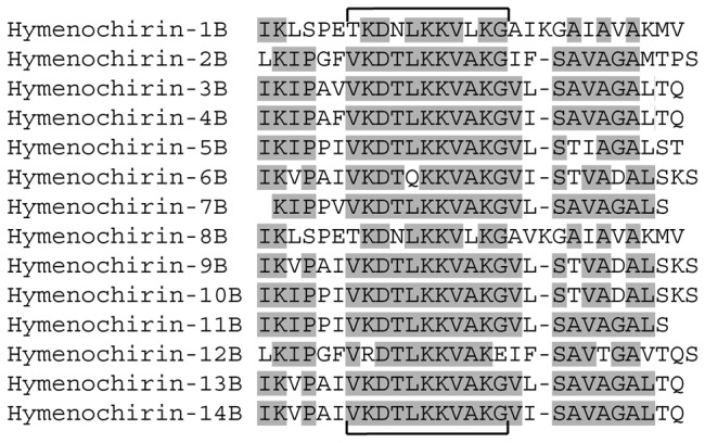
THE skin secretion of many amphibians contains peptides that are able to kill a broad range of microorganisms (antimicrobial peptides: AMPs) and potentially play a role in innate immune defense. Similar to the toxin arsenals of various animals, amphibian AMP repertoires typically show major structural variation, and previous studies have suggested that this may be the result of diversifying selection in adaptation to a diverse spectrum of pathogens. Here we report on transcriptome analyses that indicate a very different pattern in the dwarf clawed frog H. boettgeri. Our analyses reveal a diverse set of transcripts containing two to six tandem repeats, together encoding 14 distinct peptides. Five of these have recently been identified as AMPs, while three more are shown here to potently inhibit the growth of gram-negative bacteria, including multi-drug resistant strains of the medically important Pseudomonas aeruginosa. Although the number of predicted peptides is similar to the numbers of related AMPs in Xenopus and Silurana frog species, they show significantly lower structural variation. Selection analyses confirm that, in contrast to the AMPs of other amphibians, the H. boettgeri peptides did not evolve under diversifying selection. Instead, the low sequence variation among tandem repeats resulted from purifying selection, recent duplication and/or concerted gene evolution. Our study demonstrates that defense peptide repertoires of closely related taxa, after diverging from each other, may evolve under differential selective regimes, leading to contrasting patterns of structural diversity.


Conflict of interest statement

Competing Interests: The authors have declared that no competing interests exist.

Figures

Figure 1
Figure 1. Structure of hymenochirin transcript 1 and comparison to other precursors and AMPs.
A Structure of hymenochirin transcript 1 (obtained from a breeding skin gland cDNA library of Hymenochrius boettgeri males) and of two AMP precursor proteins from Xenopus laevis and Silurana tropicalis. preproPGLa-Xl1: X. laevis PGLa precursor; preproCPF-St7: S. tropicalis CPF precursor. Region coloration distinguishes UTR (white), and sequences encoding signal peptide (dark green), spacer (light green) and antimicrobial peptides (blue). Dashed lines indicate known exon boundaries in Xenopus and Silurana AMP precursors. B Comparative alignment of the deduced amino acid sequence of the coding part of hymenochirin transcript 1 with two X. laevis and S. tropicalis AMP precursors and the previously published hymenochirins . Predicted signal peptides are printed in lower case, antimicrobial peptides are printed in bold. Amino acids shared between the hymenochirin precursor and at least one of the other precursors are indicated in light grey. Residues identical between all hymenochirin peptides but not present in preproPGLa-Xl1 or preproCPF-St7 are labeled in dark grey. Small arrowheads indicate putative cleavage sites.
Figure 2
Figure 2. Overview of hymenochirin transcripts: amino acid sequences, structure and encoded peptides.
A Deduced amino acid sequences of the hymenochirin transcripts. Previously published hymenochirins are marked in grey; predicted novel encoded hymenochirins are marked in black. Small arrowheads indicate putative cleavage sites for the hymenochirins. Names of encoded hymenochirins are indicated on the right. B Comparative schematic representation of repeat sequences in the transcripts. The number of cDNA sequences represented by each transcript is indicated between brackets. Each repeat is represented by one larger and one smaller block (repeat sections), corresponding to exons 2 and 3 in S. tropicalis and X. laevis AMP genes. The numbers in the blocks correspond to unique repeat sections as used in the phylogenetic analyses. Hymenochirins encoded by the corresponding transcripts are indicated on the right; previously published hymenochirins are labeled grey, the novel hymenochirins are labeled black.
Figure 3
Figure 3. Alignment of all known and predicted hymenochirin peptides.
Amino acids shared by more than 50% of the peptides are marked in grey. Brackets delineate a conserved central sequence motif.
Figure 4
Figure 4. Comparison of pairwise sequence similarities between AMPs of H. boettgeri, S. tropicalis and X. laevis.
Box plots comparing the distribution of pairwise sequence similarities (in %) between the 14 hymenochirins of H. boettgeri and all known AMP peptides of S. tropicalis and X. laevis respectively. Boxes indicate median, and 25- and 75- percentiles, and whiskers indicate minimum and maximum values.
Figure 5
Figure 5. Helical wheel projections of the four peptides that were synthesized and tested for antimicrobial activity.
A hymenochirin-6B; B hymenochirin-7B; C hymenochirin-10B; D hymenochirin-12B. Only the region of the peptide predicted to have an alpha-helical structure with a confidence level of 5 or more (PSIPRED v3.0 [30]) is shown in the projection. Amino acids are shaded according to the Combined Consensus Scale with hydrophobic residues in white, nearly neutral residues in light grey, polar residues in dark grey, charged residues in black with+or - sign indicating charge. Helical wheels are adapted from http://cti.itc.virginia.edu/~cmg/Demo/wheel/wheelApp.html. Circle sizes indicate the relative distance from the N-terminal end of the peptide; smaller circles are further away.
Figure 6
Figure 6. Figure 6. Phylogenetic relationships among pipid AMPs (exon 2).
Phylogenetic relationships as inferred by Bayesian analysis of a data set consisting of exon 2 of amphibian cck genes and AMP genes in X. laevis and S. tropicalis, aligned with the corresponding repeat sections of the hymenochirin precursor proteins. Repeat numbers correspond to those in Figure 2. The depicted tree represents the Bayesian consensus phylogram rooted with cck genes. Branches are shown in bold if Bayesian posterior probability is above 0.95 and RAxML bootstrap is more than 75%.
Figure 7
Figure 7. Figure 7. Origin and loss of AMPs and CCK-like peptide in the family Pipidae.
A The three recently postulated phylogenetic hypotheses for the family Pipidae and their implications for the origin (vertical bars) and loss (crosses) of an AMP gene repertoire. B Summarized gene tree illustrating the evolution of the pipid AMP gene family.

References

    1. Bevins CL, Zassloff M (1990) Peptides from frog skin. Annu Rev Biochem 59: 395–414. - PubMed
    1. Basir YJ, Knoop FC, Dulka J, Conlon JM (2000) Multiple antimicrobial peptides and peptides related to bradykinin and neuromedin N isolated from skin secretions of the pickerel frog, Rana palustris . Biochim Biophys Acta 1543: 95–105. - PubMed
    1. Li L, Bjourson AJ, He J, Cai G, Rao P, et al. (2003) Bradykinins and their cDNA from piebald odorous frog, Odorrana schmackeri, skin. Peptides 24: 863–872. - PubMed
    1. Zasloff M (2002) Antimicrobial peptides of multicellular organisms. Nature 415: 389–395. - PubMed
    1. Brogden KA (2005) Antimicrobial peptides: pore formers or metabolic inhibitors in bacteria? Nat Rev Microbiol 3: 238–250. - PubMed

Publication types

MeSH terms

Substances

LinkOut - more resources