. 2014 Jan 21;9(1):e86348.
doi: 10.1371/journal.pone.0086348. eCollection 2014.

CD4+ T-cell help is required for effective CD8+ T cell-mediated resolution of acute viral hepatitis in mice

Affiliations

CD4+ T-cell help is required for effective CD8+ T cell-mediated resolution of acute viral hepatitis in mice

Tanja Trautmann et al. PLoS One. .

Abstract

Cytotoxic CD8+ T cells are essential for the control of viral liver infections, such as those caused by HBV or HCV. It is not entirely clear whether CD4+ T-cell help is necessary for establishing anti-viral CD8+ T cell responses that successfully control liver infection. To address the role of CD4+ T cells in acute viral hepatitis, we infected mice with Lymphocytic Choriomeningitis Virus (LCMV) of the strain WE; LCMV-WE causes acute hepatitis in mice and is cleared from the liver by CD8+ T cells within about two weeks. The role of CD4+ T-cell help was studied in CD4+ T cell-lymphopenic mice, which were either induced by genetic deficiency of the major histocompatibility (MHC) class II transactivator (CIITA) in CIITA-/- mice, or by antibody-mediated CD4+ cell depletion. We found that CD4+ T cell-lymphopenic mice developed protracted viral liver infection, which seemed to be a consequence of reduced virus-specific CD8+ T-cell numbers in the liver. Moreover, the anti-viral effector functions of the liver-infiltrating CD8+ T cells in response to stimulation with LCMV peptide, notably the IFN-γ production and degranulation capacity were impaired in CIITA-/- mice. The impaired CD8+ T-cell function in CIITA-/- mice was not associated with increased expression of the exhaustion marker PD-1. Our findings indicate that CD4+ T-cell help is required to establish an effective antiviral CD8+ T-cell response in the liver during acute viral infection. Insufficient virus control and protracted viral hepatitis may be consequences of impaired initial CD4+ T-cell help.


Conflict of interest statement

Competing Interests: The authors have declared that no competing interests exist.

Figures

Figure 1
Figure 1. Increased and persistent LCMV infection in CIITA−/− mice.
C57BL/6 and CIITA−/− mice were infected with 106 Focus Forming Units (FFU) LCMV-WE. (A) At the indicated time-points after infection, spleens were homogenized and the virus titer was determined by the Focus Forming Assay. Black dots represent the mean titer of C57BL/6 mice, white squares represent the mean titer of CIITA−/− mice. The broken line represents the limit of detection. (B) At the indicated time-points after infection, the infection rate of the liver was determined as in (A). (C) At the indicated time-points after infection, the infection rate of the liver was determined by quantitative RT-PCR analysis for the LCMV Z RNA. (D) Frozen liver sections taken at the indicated time-points after infection were stained for the LCMV nucleoprotein with VL4 antibody (green); nuclei were stained with Hoechst 33258 (blue). (E) At the indicated time-points after infection, serum ALT levels were determined. The graphed lines represent the mean and s.e.m.
Figure 2
Figure 2. Protracted LCMV infection in livers of wild-type C57BL/6 mice depleted of CD4+ T cells.
C57BL/6 mice were treated twice per week with depleting CD4 antibody (GK1.5) or isotype-matched control antibody to obtain CD4-depleted or CD4-replete mice that were subsequently infected with 106 FFU LCMV-WE. All samples were taken at day 18 after infection. (A) Spleens were homogenized and the virus titer was determined by FFA. Each dot (black: C57BL/6 mice; white: CIITA−/− mice) represents the average FFU of one sample tested in duplicate; the broken line represents the limit of detection. (B) Livers were homogenized and the infection rate was determined as in (A). (C) The infection rate of the livers was determined by quantitative RT-PCR analysis for the LCMV Z RNA. (D) Serum ALT levels were determined. (E) Frozen liver sections were stained for the LCMV nucleoprotein with VL4 antibody (green); nuclei were stained with Hoechst 33258 (blue).
Figure 3
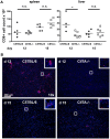
Figure 3. Reduced numbers of CD8+ T cells in the livers of LCMV-infected CIITA−/− mice.
C57BL/6 and CIITA−/− mice were infected with 106 FFU LCMV-WE. (A) At day 12 or 15 after infection, the numbers of CD8+ T cells in spleen and liver of C57BL/6 and CIITA−/− mice were determined. Each dot represents the absolute number of CD8+ T cells per spleen or liver of one individual mouse. (B) Frozen liver sections taken at day 12 or 15 after infection were stained for CD8 T cells (red); nuclei were stained with Hoechst 33258 (blue).
Figure 4
Figure 4. Preserved chemokine production and non-specific leukocyte recruitment to livers of infected CIITA−/− mice.
C57BL/6 and CIITA−/− mice were infected with 106 FFU LCMV-WE. (A) At the indicated time-points after infection, expression of the CXCR3 chemokine ligands CXCL9, CXCL10 and CXCL11 in infected livers were determined by qPCR. Black dots represent C57BL/6 mice, white squares represent CIITA−/− mice. (B) At the indicated time-points after infection, the numbers of leukocytes in infected livers were determined. Each dot represents the absolute number of liver-infiltrating mononuclear cells of one individual mouse.
Figure 5
Figure 5. Reduced numbers of LCMV-specific CD8+ T cells in the livers of LCMV-infected CIITA−/− mice.
C57BL/6 and CIITA−/− mice were infected with 106 FFU LCMV-WE. At day 12 or 15 after infection, the numbers of LCMV-gp33 specific CD8+ T cells in spleen and liver of C57BL/6 and CIITA−/− mice were determined by staining with LCMV-gp33 loaded H-2Db dextramer. Each dot represents the percentage of dextramer+ CD8+ T cells among all CD8+ T cells in spleen or liver of individual mice.
Figure 6
Figure 6. Reduced IFN-γ production by CD8+ T cells of CIITA−/− mice.
C57BL/6 and CIITA−/− mice were infected with 106 FFU LCMV-WE. At day 12 or 15 after infection, the effector function of LCMV-specific CD8+ T cells in spleen and liver of C57BL/6 and CIITA−/− mice was determined by intracellular staining of CD8+ T cells for IFN-γ after stimulation with LCMV-gp33 peptide. Each dot represents the percentage of IFN-γ+ CD8+ T cells among all CD8+ T cells of spleen or liver of individual mice.
Figure 7
Figure 7. Reduced degranulation capacity of CD8+ T cells of CIITA−/− mice.
C57BL/6 and CIITA−/− mice were infected with 106 FFU LCMV-WE. At day 12 or 15 after infection, the degranulation capacity of CD8+ T cells (A) or LCMV-specific dextramer+ CD8+ T cells (B) in spleen and liver in response to stimulation with LCMV-gp33 peptide was determined by staining for CD107a. Each dot represents the percentage of CD107a+ CD8+ T cells among all CD8+ T cells (A) or CD107a+ dextramer+ CD8+ T cells among all CD8+ T cells (B) of spleen or liver of individual mice.
Figure 8
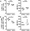
Figure 8. PD-1 expression of LCMV-specific CD8+ T cells in wild-type and CIITA−/− mice.
C57BL/6 and CIITA−/− mice were infected with 106 FFU LCMV WE. At day 15 after infection, the expression of the exhaustion marker PD-1 on LCMV-specific CD8+ T cells in spleen and liver was determined by staining of dextramer+ CD8+ T cells for PD-1. (A) Each dot represents the percentage of PD1+ dextramer+ CD8+ T cells among all dextramer+ CD8+ T cells of spleen or liver of individual mice. (B) Each dot represents the mean fluorescence intensity (MFI) of PD-1 staining of all dextramer+ CD8+ T cells in spleen or liver of individual mice.

References

    1. Blachier M, Leleu H, Peck-Radosavljevic M, Valla DC, Roudot-Thoraval F (2013) The burden of liver disease in Europe: a review of available epidemiological data. J Hepatol 58(3): 593–608. - PubMed
    1. Ganem D, Prince AM (2004) Hepatitis B virus infection–natural history and clinical consequences. N Engl J Med 350: 1118–1129. - PubMed
    1. Lauer GM, Walker BD (2001) Hepatitis C virus infection. N Engl J Med 345: 41–52. - PubMed
    1. Hughes SA, Wedemeyer H, Harrison PM (2011) Hepatitis delta virus. Lancet 378: 73–85. - PubMed
    1. Kamar N, Bendall R, Legrand-Abravanel F, Xia NS, Ijaz S, et al. (2012) Hepatitis E. Lancet. 379: 2477–2488. - PubMed

Publication types

Substances