. 2014 Jan 17;9(1):e86365.
doi: 10.1371/journal.pone.0086365. eCollection 2014.

Human lactate dehydrogenase a inhibitors: a molecular dynamics investigation

Affiliations

Human lactate dehydrogenase a inhibitors: a molecular dynamics investigation

Yun Shi et al. PLoS One. .

Abstract

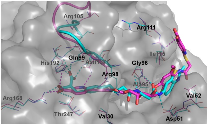
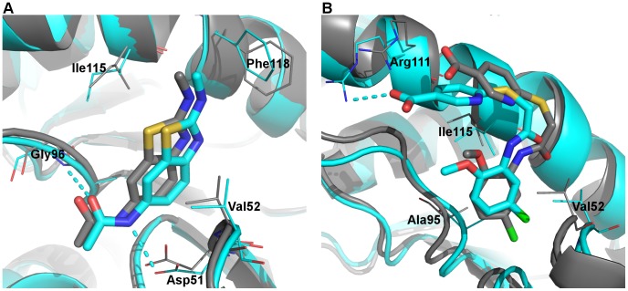
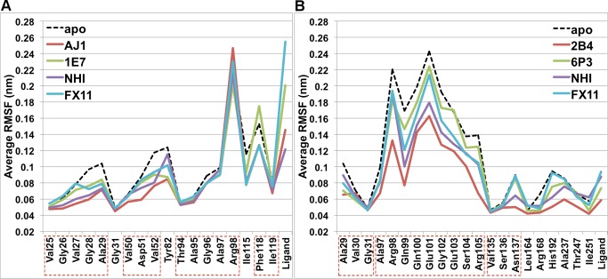
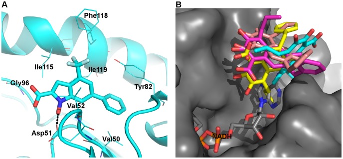
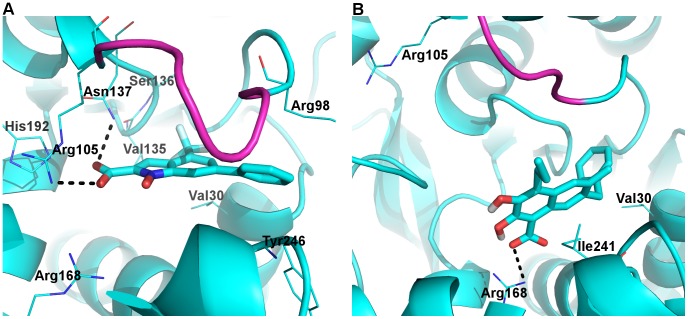
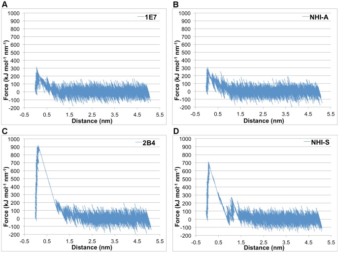
Lactate dehydrogenase A (LDHA) is an important enzyme in fermentative glycolysis, generating most energy for cancer cells that rely on anaerobic respiration even under normal oxygen concentrations. This renders LDHA a promising molecular target for the treatment of various cancers. Several efforts have been made recently to develop LDHA inhibitors with nanomolar inhibition and cellular activity, some of which have been studied in complex with the enzyme by X-ray crystallography. In this work, we present a molecular dynamics (MD) study of the binding interactions of selected ligands with human LDHA. Conventional MD simulations demonstrate different binding dynamics of inhibitors with similar binding affinities, whereas steered MD simulations yield discrimination of selected LDHA inhibitors with qualitative correlation between the in silico unbinding difficulty and the experimental binding strength. Further, our results have been used to clarify ambiguities in the binding modes of two well-known LDHA inhibitors.


Conflict of interest statement

Competing Interests: The grant from NSERC requires that we specify a company who might be interested in the results of the research, Alectos Therapeutics Inc. This commercial company was originally formed from research conducted at Simon Fraser University, however the authors are not affiliated with, nor receive funding from, this entity. There are no patents, products in development or marketed products to declare. This does not alter the authors' adherence to all the PLOS ONE policies on sharing data and materials.

Figures

Figure 1
Figure 1. Structure of human LDHA (PDB 1I10).
Amino acid residues are shown in cartoons and NADH/oxamate are shown in sticks. A) Tetrameric structure of human LDHA. Chains A, B, C, and D are colored green, yellow, magenta, and cyan, respectively. B) Close-up view of the binding site from chain A. The active site mobile loop is colored red.
Figure 2
Figure 2. Structures of LDHA binders of interest.
A-site and S-site binding moieties are indicated by boxes with blue dashed lines and red dashed lines, respectively.
Figure 3
Figure 3. Comparison of monomeric and tetrameric MD models of LDHA:PYR-NADH.
Representative structures of the monomeric (carbon atoms in magenta) and the tetrameric (carbon atoms in cyan) forms are overlaid with the crystal structure (PDB 1I10, carbon atoms in grey). Selected binding site residues are shown in thin lines, while pyruvate and NADH are shown in thick sticks. Other atoms are colored: oxygen, red; nitrogen, blue; phosphate, orange.
Figure 4
Figure 4. Root-mean-squared fluctuations (RMSF) of dual-site binding systems.
“1E4-on” represents data from trajectories where 1E4 had strong ionic interactions with Arg111, while “1E4-off” indicates data from trajectories without such interactions. Contiguous residues are labeled by boxes with red dashed lines.
Figure 5
Figure 5. Comparison of the binding of dual-site inhibitors.
Representative MD snapshots of LDHA:0SN (carbon atoms in cyan) and LDHA:1E4 (carbon atoms in magenta) are superimposed. Selected binding site residues are labeled and shown in thin lines, while 0SN and 1E4 are shown in thick sticks. The mobile loop is represented by a ribbon, and the solvent-accessible surface of LDHA is indicated by a grey transparent surface. Dashed lines represent polar interactions. Other atoms are colored: oxygen, red; nitrogen, blue; sulfur, yellow; chlorine, green; fluorine, pale cyan.
Figure 6
Figure 6. Binding of AJ1 and 1E7.
Representative MD structures (cartoon and carbon atoms in cyan) and corresponding crystal structures (cartoon and carbon atoms in grey) are overlaid for A) LDHA:AJ1 and B) LDHA:1E7. Selected binding site residues are labeled and shown in thin lines, while ligands are shown in thick sticks. Dashed lines represent polar interactions. Other atoms are colored: oxygen, red; nitrogen, blue; sulfur, yellow; chlorine, green.
Figure 7
Figure 7. Root-mean-squared fluctuations (RMSF) of A-site binding and S-site binding systems.
Contiguous residues are labeled by boxes with red dashed lines.
Figure 8
Figure 8. Binding of NHI and FX11 at the A-site.
A) A representative MD snapshot of LDHA:NHIA, with coloring scheme identical to Figure 6. Black dashed lines represent polar interactions. B) Overlay of representative MD structures from four monomers of LDHA:FX11A (carbon atoms in cyan, magenta, yellow, and pink, respectively) and PDB 1I10 (carbon atoms in grey). Grey surface indicates the solvent-accessible surface of LDHA, showing that two of the FX11 structures are completely outside the binding groove.
Figure 9
Figure 9. Binding of NHI and FX11 at the S-site.
Representative MD snapshots of A) LDHA:NHIS and B) LDHA:FX11S are shown. The color scheme is identical to Figure 6 with hydrogen atoms in white, while the mobile loop is in magenta.
Figure 10
Figure 10. Examples of force-distance curves for the pulling simulation.
One of the 12 replicate steered MD runs is shown for A) LDHA:1E7, B) LDHA:NHIA, C) LDHA:2B4, and D) LDHA:NHIS.

References

    1. Hanahan D, Weinberg RA (2011) Hallmarks of cancer: the next generation. Cell 144: 646–674. - PubMed
    1. Ward PS, Thompson CB (2012) Metabolic reprogramming: a cancer hallmark even warburg did not anticipate. Cancer Cell 21: 297–308. - PMC - PubMed
    1. Warburg O (1956) On respiratory impairment in cancer cells. Science 124: 269–270. - PubMed
    1. Gatenby RA, Gillies RJ (2004) Why do cancers have high aerobic glycolysis? Nat Rev Cancer 4: 891–899. - PubMed
    1. Banga I, Szent-Gyorgyi A, Vargha L (1932) The coenzyme of lactic acid oxidation. Hoppe Seylers Z Physiol Chem 210: 228–235.

Publication types