. 2014 Jan 20;9(1):e86373.
doi: 10.1371/journal.pone.0086373. eCollection 2014.

Endocytosis of Fgf8 is a double-stage process and regulates spreading and signaling

Affiliations

Endocytosis of Fgf8 is a double-stage process and regulates spreading and signaling

Charanya Rengarajan et al. PLoS One. .

Abstract

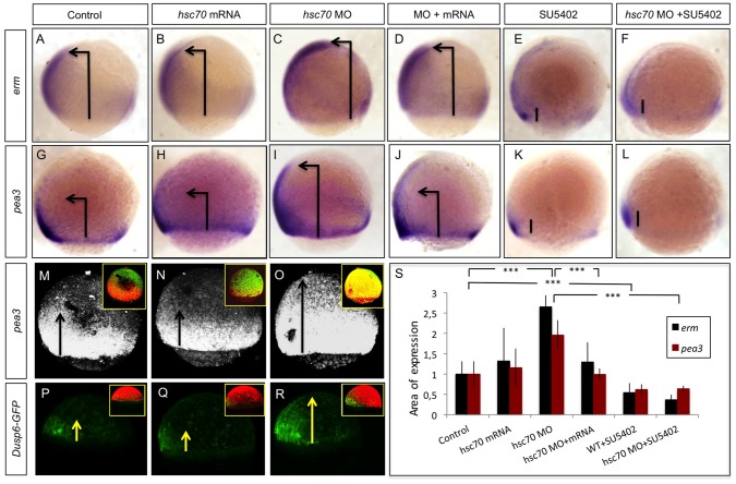
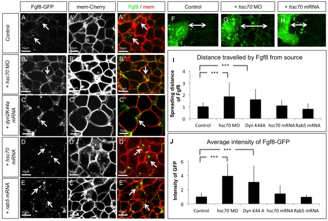
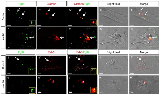
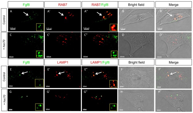
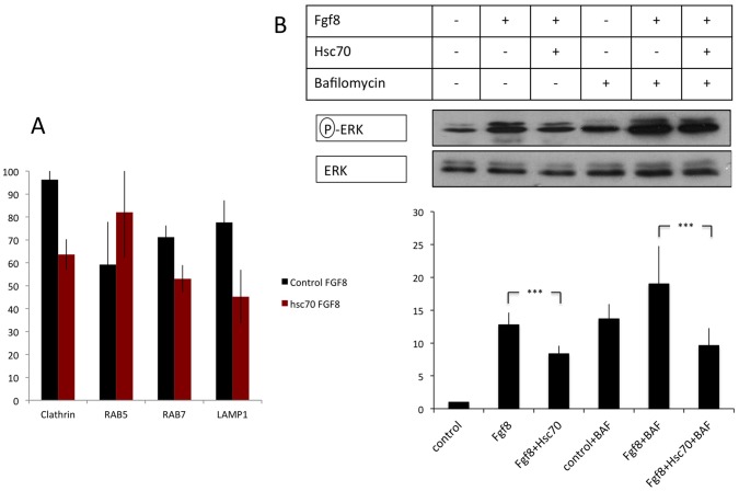
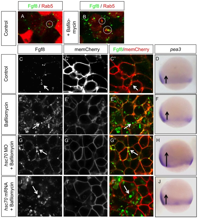
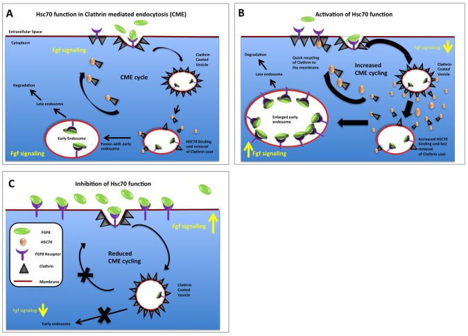
Tightly controlled concentration gradients of morphogens provide positional information and thus regulate tissue differentiation and morphogenesis in multicellular organisms. However, how such morphogenetic fields are formed and maintained remains debated. Here we show that fibroblast growth factor 8 (Fgf8) morphogen gradients in zebrafish embryos are established and maintained by two essential mechanisms. Firstly, Fgf8 is taken up into the cell by clathrin-mediated endocytosis. The speed of the uptake rate defines the range of the morphogenetic gradient of Fgf8. Secondly, our data demonstrate that after endocytosis the routing of Fgf8 from the early endosome to the late endosome shuts down signaling. Therefore, intracellular endocytic transport regulates the intensity and duration of Fgf8 signaling. We show that internalization of Fgf8 into the early endosome and subsequent transport towards the late endosome are two independent processes. Therefore, we hypothesize that Fgf8 receiving cells control both, the propagation width and the signal strength of the morphogen.


Conflict of interest statement

Competing Interests: The authors have declared that no competing interests exist.

Figures

Figure 1
Figure 1. Hsc70 regulates the Fgf8 signaling range.
(A–L) In situ hybridization for Fgf target genes erm and pea3 at 75% epiboly stage. Embryos were mounted laterally with animal pole to the top and dorsal region towards the left. The area of the expression domains of Fgf target genes erm and pea3 were investigated in embryos injected with indicated constructs and the border of the expression domains were marked by arrows. (E, F, K, L) show embryos co-treated with 16 µM of the Fgf signaling inhibitor SU5402. Fluorescent ISH for target gene expression pea3 is shown in (M–O). As an in-vivo reporter for Fgf signaling, the expression of Dusp6-EGFP in live zebrafish embryos at 50% epiboly stage was investigated (P–R). The area of expression of target genes erm and pea3 (A–L) was quantified in 10 embryos for each experiment (S).
Figure 2
Figure 2. Fgf8 uptake is regulated by Hsc70.
Confocal analysis of live embryos at 50% epiboly stage at the animal pole. Fgf8-GFP DNA was injected along with the red membrane marker mCherry at the one cell stage to determine subcellular localization of Fgf8 in embryos co-injected with indicated constructs (A–E”). The range of Fgf8 propagation in the receiving tissue was analyzed using confocal microscopy (F–H) and the distance spread by Fgf8 was quantified (I). Quantification of the average intensity of Fgf8-GFP is demonstrated in embryos (J). For quantification 7 different embryos were used for each experiment.
Figure 3
Figure 3. Analysis of Fgf8 internalization Clathrin coated vesicles and early endosomes in fish fibroblasts.
Confocal analysis of Fgf8-GFP co-localization with indicated early endocytic markers in zebrafish PAC2 cells. Columns 1–3 show confocal images and columns 4–5 shows bright field images merged with the confocal image. Co-localization of Fgf8 with Clathrin (A–D’) and with Rab5 (E–H’) was investigated. Furthermore, insets show the increasing size of Rab5 positive early endosomes upon Hsc70 overexpression (E and G).
Figure 4
Figure 4. Analysis of Fgf8 internalization in late endosomes and lysosomes in fish fibroblasts.
Confocal analysis of Fgf8-GFP localization in zebrafish PAC2 cells. Columns 1–3 show confocal images and columns 4–5 shows bright field images merged with the confocal image. Co-localization with late endosomal markers was investigated: with Rab7 (A–D’) and the lysosomal Lamp1 (E–H’).
Figure 5
Figure 5. Quantification of localization and signaling during endocytosis.
Co-localization of Fgf8 is found with Clathrin, Rab7 and Lamp1, whereas co-localization of Rab5 is reduced (A). However, after stimulation of Hsc70, co-localization of Fgf8 and Rab5 is strongly increased. Three independent experiments were quantified. Cultivation of HEK293 cells after transfection with Fgf8, and Hsc70 with and without treatment of Bafilomycin A1 (B). Three independent experiments were analyzed and the ratio between activated Erk1/2 and Erk total was calculated. Cells transfected with Fgf8 showed a significant 12-fold increase in Erk1/2 phosphorylation compared to the un-transfected control. Co-transfection of Hsc70 and Fgf8 led to 8-fold increase of phosphorylation of Erk. Treatment with Bafilomycin A1 leads to a 14-fold increase of double phosphorylated Erk1/2 level. Activation by co-transfection of Fgf8 leads to a significant 19-fold increase, and co-transfection of Hsc70 and Fgf8 decreased this activation to a 10-fold activation of double phosphorylated Erk.
Figure 6
Figure 6. Inhibition of Fgf8 transport from early to late endosome does not alter signaling range.
Analysis of localization of Fgf8-GFP during inhibition of endocytic transport from early to late endosomes by treatment with Bafilomycin A1. Embryos were injected with Fgf8-GFP along with the early endosomal marker Rab5 or membrane marker mCherry at one cell stage, treated with 100 nM Bafilomycin A1 at 30% epiboly stage for a period of 1 hr and subjected to live imaging using confocal microscopy at 50% epiboly stage. At 75% epiboly stage embryos were fixed and stained for erm expression by ISH. Circle highlight co-localization of Rab5 and Fgf8, white arrows point to typical Fgf8 localizations, and balck arrows visualize the extend of the pea3 expression domain.
Figure 7
Figure 7. Graphical summary of Hsc70 function regarding Fgf8 endocytosis in zebrafish.
Detailed explanation in text.

References

    1. Wiedlocha A, Sorensen V (2004) Signaling, internalization, and intracellular activity of fibroblast growth factor. Current topics in microbiology and immunology 286: 45–79. - PubMed
    1. Dorey K, Amaya E (2010) FGF signalling: diverse roles during early vertebrate embryogenesis. Development 137: 3731–3742. - PMC - PubMed
    1. Itoh N, Ornitz DM (2011) Fibroblast growth factors: from molecular evolution to roles in development, metabolism and disease. Journal of biochemistry 149: 121–130. - PMC - PubMed
    1. Bokel C, Brand M (2013) Generation and interpretation of FGF morphogen gradients in vertebrates. Current opinion in genetics & development 23: 415–422. - PubMed
    1. Schlessinger J, Plotnikov AN, Ibrahimi OA, Eliseenkova AV, Yeh BK, et al. (2000) Crystal structure of a ternary FGF-FGFR-heparin complex reveals a dual role for heparin in FGFR binding and dimerization. Molecular cell 6: 743–750. - PubMed

Publication types

LinkOut - more resources