. 2014 Jan 22;9(1):e86378.
doi: 10.1371/journal.pone.0086378. eCollection 2014.

Evidence from a mouse model that epithelial cell migration and mesenchymal-epithelial transition contribute to rapid restoration of uterine tissue integrity during menstruation

Affiliations

Evidence from a mouse model that epithelial cell migration and mesenchymal-epithelial transition contribute to rapid restoration of uterine tissue integrity during menstruation

Fiona L Cousins et al. PLoS One. .

Abstract

Background: In women dynamic changes in uterine tissue architecture occur during each menstrual cycle. Menses, characterised by the shedding of the upper functional layer of the endometrium, is the culmination of a cascade of irreversible changes in tissue function including stromal decidualisation, inflammation and production of degradative enzymes. The molecular mechanisms that contribute to the rapid restoration of tissue homeostasis at time of menses are poorly understood.

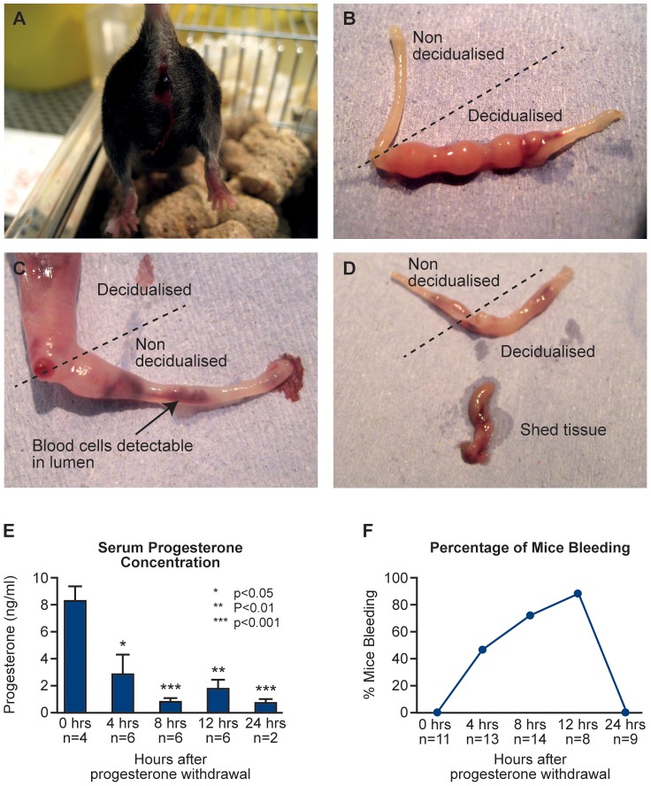
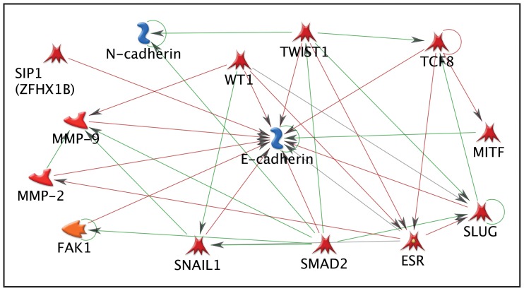
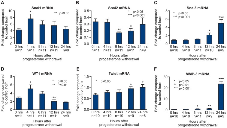
Methodology: A modified mouse model of menses was developed to focus on the events occurring within the uterine lining during endometrial shedding/repair. Decidualisation, vaginal bleeding, tissue architecture and cell proliferation were evaluated at 4, 8, 12, and 24 hours after progesterone (P4) withdrawal; mice received a single injection of bromodeoxyuridine (BrdU) 90 mins before culling. Expression of genes implicated in the regulation of mesenchymal to epithelial transition (MET) was determined using a RT2 PCR profiler array, qRTPCR and bioinformatic analysis.

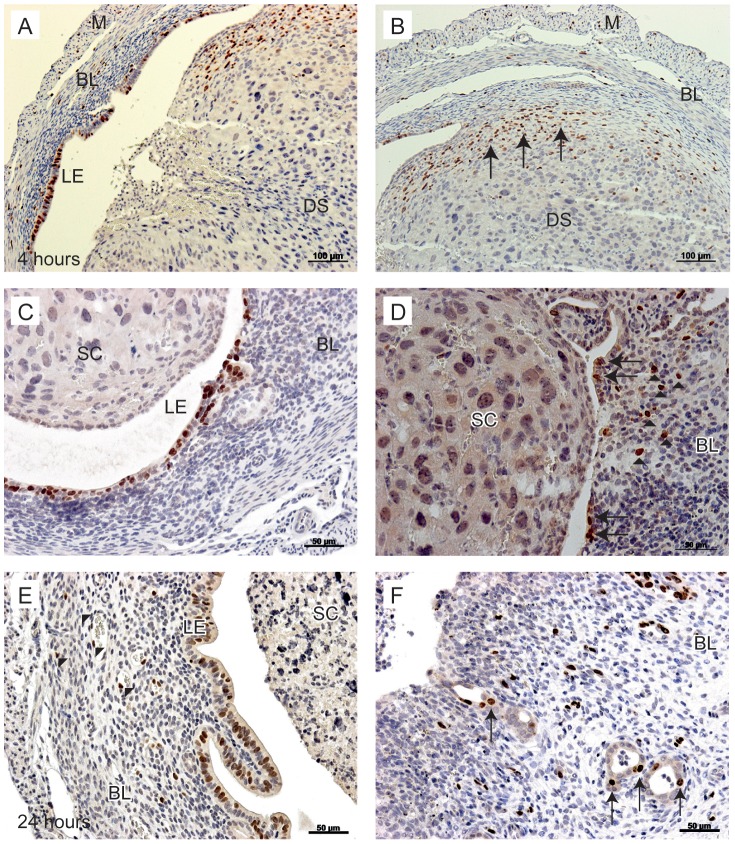
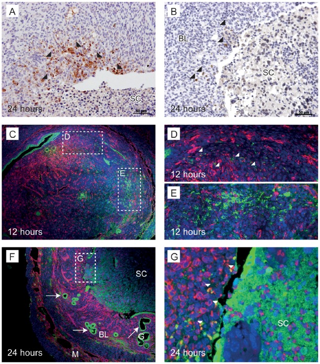
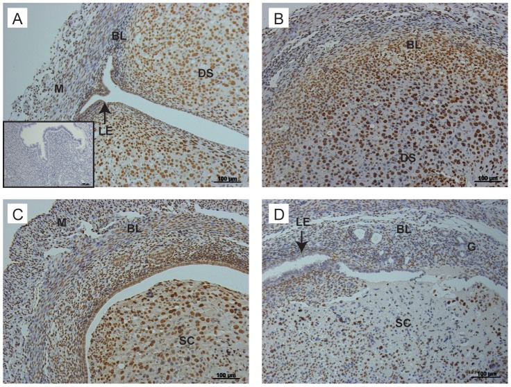
Principal findings: Mice exhibited vaginal bleeding between 4 and 12 hours after P4 withdrawal, concomitant with detachment of the decidualised cell mass from the basal portion of the endometrial lining. Immunostaining for BrdU and pan cytokeratin revealed evidence of epithelial cell proliferation and migration. Cells that appeared to be in transition from a mesenchymal to an epithelial cell identity were identified within the stromal compartment. Analysis of mRNAs encoding genes expressed exclusively in the epithelial or stromal compartments, or implicated in MET, revealed dynamic changes in expression, consistent with a role for reprogramming of mesenchymal cells so that they could contribute to re-epithelialisation.

Conclusions/significance: These studies have provided novel insights into the cellular processes that contribute to re-epithelialisation post-menses implicating both epithelial cell migration and mesenchymal cell differentiation in restoration of an intact epithelial cell layer. These insights may inform development of new therapies to induce rapid healing in the endometrium and other tissues and offer hope to women who suffer from heavy menstrual bleeding.


Conflict of interest statement

Competing Interests: The authors have declared that no competing interests exist.

Figures

Figure 1
Figure 1. Summary of time line for mouse model of menstruation and regeneration.
Colour coding pink = ‘proliferative phase’, blue = ‘secretory phase’, red = ‘menstrual phase’. Ovex; ovariectomy, E; β-oestradiol, P; progesterone. β-oestradiol concentrations in brackets (ng/100 µl), P4 pellet (1 mg/ml). One uterine horn was stimulated on day 15 via oil injection into the luminal cavity; “menses” was induced by P4 pellet removal on day 19. BrdU was injected 90 minutes prior to tissue recovery at 4, 8, 12 and 24 hours after the removal of the P4 secreting pellet.
Figure 2
Figure 2. Gross morphology, bleeding and progesterone concentrations.
A, Mouse at 12 hours after P4 withdrawal showing blood in vagina; B, The non decidualised control (left) and the decidualised horn (right) upon dissection (12 hours after withdrawal); C, Blood cells are detectable in lumen of the non decidualised horn following vaginal lavage (24 hours after withdrawal); D, Shed tissue expelled from cervix (24 hours after withdrawal), decidualised horn shows regression; E, Serum concentrations of progesterone (ng/ml). Statistical analysis was carried out by Student t test, comparing each time-point to the 0 hour time-point *p<0.05 **p<0.01 and ***p<0.001; F. Percentage of mice bleeding between 4 and 24 hours. This was calculated as a percentage of mice that were identified to be bleeding at each time-point of the total number of mice examined at each time-point.
Figure 3
Figure 3. Proliferation of uterine cells between 4 and 24 hours after P4 withdrawal.
To identify proliferating cells, animals were injected with BrdU 90 minutes prior to tissue recovery. A; Proliferating luminal epithelial cells detected in tissues 4 hours after progesterone withdrawal. B; In the same tissue, stromal cells in the basal layer are positive for BrdU (arrows). C; At 12 hours, luminal epithelial cells were positive for BrdU, no BrdU positive cells were identified in the shed cell mass. D; In the same tissue, stromal cells close to the luminal edge were positive for BrdU (arrowheads), new epithelial cells were identified lining the lumen in an area of tissue where the decidualised tissue had shed (arrows). E; At 24 hours after withdrawal, endothelial cells were positive for BrdU (arrowheads), the intact luminal epithelium was also positive for BrdU. F; In another sample at 24 hours, the stromal compartment was exposed to the lumen (arrowheads); note stromal cells positive for BrdU are interspersed throughout the basal layer and evidence of glandular epithelial cell proliferation was also detected (arrows). BL; Basal layer, LE; luminal epithelium, DS; decidualised stromal cells, M; myometrium, SC; shed cells. Scale bars are equal to 100 µm or 50 µm where indicated.
Figure 4
Figure 4. Immunostaining for pan-cytokeratin illustrates re-epithelialisation of the endometrium consistent with cell migration.
Pan-cytokeratin, used as a marker for epithelial cells, was observed in the luminal epithelium and the glandular epithelium. A; The shed decidualised cell mass was observed to be detaching from the underlying stromal cell compartment at 24 hours after progesterone withdrawal. B; The denuded, underlying stroma, as indicated by the arrowheads, next to a region of luminal epithelial cells. C; The luminal epithelium next to an area of denuded basal stroma (arrowheads). D; Round epithelial cells appear to be “rolling” along an area of the denuded basal stroma. LE; luminal epithelium, G: glandular epithelium, SC; shed cells. Scale bars are equal to 200 µm, 100 µm or 20 µm where indicated.
Figure 5
Figure 5. Dynamic changes in concentration of mRNAs specific to stromal and epithelial cell compartments.
Comparison between concentrations of mRNAs encoded by genes typically expressed in stromal (Cdh2, Wnt4, vimentin) and epithelial (Cdh1, Wnt7a, Krt8) cells at 0 hours (full decidualisation) and following P4 withdrawal 4, 8, 12 and 24 hours prior to tissue recovery. Statistical analysis was performed by Student t test, comparing each time-point to the 0 hour time-point: *p<0.05, **p<0.01, ***p<0.001.
Figure 6
Figure 6. Cytokeratin positive cells were identified lining the luminal epithelium and glands as well as in the stroma with the latter appearing to be in transition between stromal and epithelial cell identity.
A; A subset of stromal cells adjacent to the luminal surface were immunopositive for pan-cytokeratin at 24 hours after progesterone withdrawal (arrowheads). B; In another sample, positive pan-cytokeratin stromal cells (arrowheads) were detected adjacent to the luminal surface of a denuded area of endometrium. C; Immunofluorescence for epithelial cells stained for cytokeratin (green) and mesenchymal cells stained for vimentin (red) in the mouse endometrium. 12 hours after progesterone withdrawal, vimentin positive decidualised cells were observed, budding into the lumen. D; Vimentin and cytokeratin positive cells were observed in the stroma, close to the myometrium (arrowheads). E; stromal cells were positive for cytokeratin. F and G; 24 hours after progesterone withdrawal an area of shed endometrium is observed. Co-localisation of vimentin and cytokeratin (white arrowheads) was detected close to the surface of the underlying stroma. BL; basal layer, G; glandular epithelium, SC; shed cells, M; myometrium. Scale bars are equal to 50 µm where indicated.
Figure 7
Figure 7. Metacore analysis of genes detected in mouse uterus that were associated with regulation of E cadherin and N cadherin.
To filter data, the full gene array list was input into Metacore™ software and any gene that was found to have no known interaction with E and N cadherin was excluded. Arrows indicate direct effects on other genes in the pathway. Green arrows indicate activation, whereas red arrows show inhibitory action.
Figure 8
Figure 8. Endometrial remodelling was associated with dynamic changes in concentrations of mRNAs expressed in stromal and epithelial cell types as well as those encoded by genes implicated in MET.
mRNA concentrations for candidate genes involved in mesenchymal to epithelial transition and tissue remodelling; Snai1 (Snail), Snai2 (Slug), Snai3 (Smuc), Wt1, Twist and Mmp3 following progesterone withdrawal. mRNA expression for the decidualised horn (black bars) was normalised against the control 0 hour horn. Statistical analysis was performed by Student t test, comparing each time-point to the decidualised 0 hour time-point, *p<0.05, **p<0.01, ***p<0.001.
Figure 9
Figure 9. Immunostaining for WT1 illustrates dynamic changes in cellular localisation during breakdown and repair.
A; Immunopoistive staining for WT1 was detected in decidualised stromal cells, stromal cells of the basal layer and the luminal epithelium at the time of progesterone withdrawal. B; At 4 hours, strong immunostaining was maintained in the basal and decidual layers of the tissue. C; By 8 hours, strong immunopositive staining was localised to areas close to the luminal epithelium. D; By 12 hours, fewer immunopositive cells were observed, these were limited to the stroma, no immunostaining for WT1 was detected in the luminal epithelium. LE; luminal epithelium, G: glandular epithelium, SC; shed cells, DS; decidualised stroma, M; myometrium. Inset; negative control. Scale bars are equal to 100 µm where indicated.

References

    1. Jabbour HN, Kelly RW, Fraser HM, Critchley HO (2006) Endocrine regulation of menstruation. Endocr Rev 27: 17–46. - PubMed
    1. Emera D, Romero R, Wagner G (2011) The evolution of menstruation: a new model for genetic assimilation: explaining molecular origins of maternal responses to fetal invasiveness. Bioessays 34: 26–35. - PMC - PubMed
    1. Garry R, Hart R, Karthigasu KA, Burke C (2009) A re-appraisal of the morphological changes within the endometrium during menstruation: a hysteroscopic, histological and scanning electron microscopic study. Hum Reprod 24: 1393–1401. - PubMed
    1. Garry R, Hart R, Karthigasu KA, Burke C (2011) Structural changes in endometrial basal glands during menstruation. BJOG 117: 1175–1185. - PubMed
    1. Ferenczy A (1976) Studies on the cytodynamics of human endometrial regeneration. II. Transmission electron microscopy and histochemistry. Am J Obstet Gynecol 124: 582–595. - PubMed

Publication types