. 2014 Jan 21;9(1):e86691.
doi: 10.1371/journal.pone.0086691. eCollection 2014.

Beta-catenin is vital for the integrity of mouse embryonic stem cells

Affiliations

Beta-catenin is vital for the integrity of mouse embryonic stem cells

Angelo Raggioli et al. PLoS One. .

Abstract

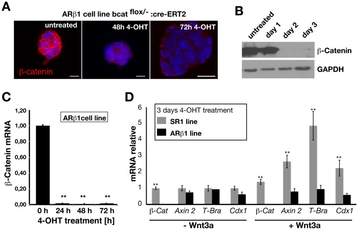
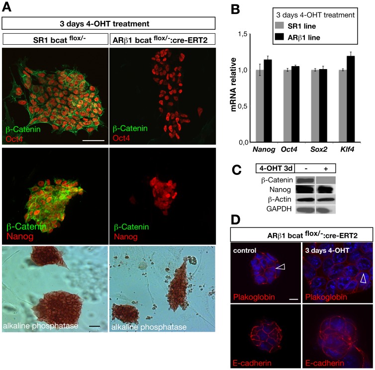
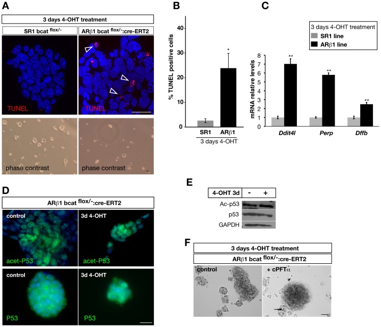
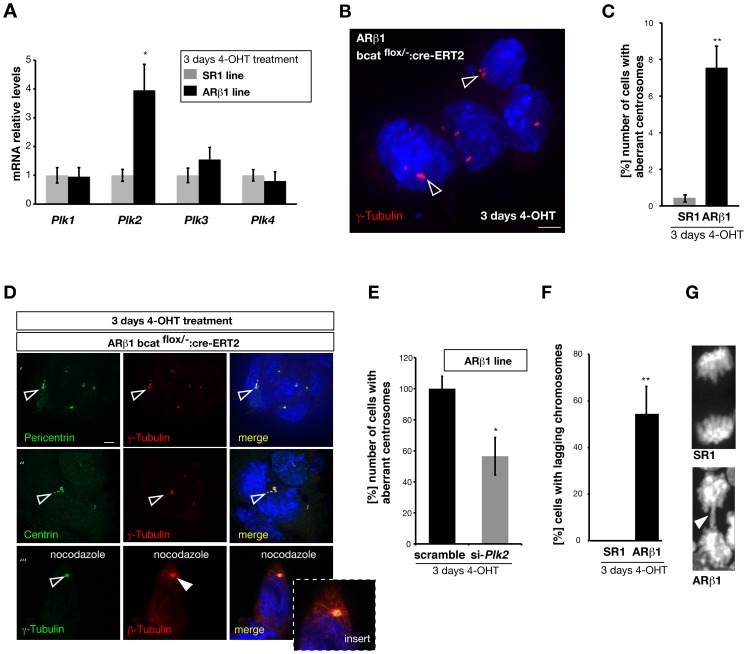
β-Catenin mediated Wnt-signaling is assumed to play a major function in embryonic stem cells in maintaining their stem cell character and the exit from this unique trait. The complexity of β-catenin action and conflicting results on the role of β-catenin in maintaining the pluripotent state have made it difficult to understand its precise cellular and molecular functions. To attempt this issue we have generated new genetically modified mouse embryonic stem cell lines allowing for the deletion of β-catenin in a controlled manner by taking advantage of the Cre-ER-T2 system and analyzed the effects in a narrow time window shortly after ablation. By using this approach, rather then taking long term cultured β-catenin null cell lines we demonstrate that β-catenin is dispensable for the maintenance of pluripotency associated genes. In addition we observed that the removal of β-catenin leads to a strong increase of cell death, the appearance of multiple clustered functional centrosomes most likely due to a mis-regulation of the polo-like-kinase 2 and furthermore, alterations in chromosome segregation. Our study demonstrates the importance of β-catenin in maintaining correct cellular functions and helps to understand its role in embryonic stem cells.


Conflict of interest statement

Competing Interests: The authors have declared that no competing interests exist.

Figures

Figure 1
Figure 1. Treatment of ARβ1 (βcat del/flox :Cre-ER-T2) ES cells with 4-OHT induces Cre-mediated recombination of the β-catenin genomic locus leading to ablation of β-catenin.
(A) Immunocytochemical staining for β-catenin protein on cultured ARβ1 cells reveals loss of β-catenin protein already at day two and appears to be absent at day three. (B) Western-immunoblotting against β-catenin demonstrates the almost complete loss of protein within 3 days of culturing. (C) β-catenin mRNA levels drop already 24 h after 4-OHT administration as revealed by quantitative RT-PCR (n = 3). ARβ1 ES cells do not respond to Wnt3a mediated canonical Wnt signaling after treatment with 4-OHT. (D) SR1 (βcat del/flox)) and ARβ1 ES cells were treated for three days with 4-OHT and Wnt3a protein to induce canonical Wnt-signaling. Quantitative RT-PCR analysis of Wnt-target genes revealed that in contrast to SR1 cells ARβ1 cells are do not up-regulate significantly Axin 2, T-Brachyury and Cdx 1 (n = 3). *P<0,05, **P<0,01; student’s t-test. Error bars = s.e.m.; scale bar 10 µm.
Figure 2
Figure 2. ARβ1 ES cells maintain expression of pluripotency markers at early time points after ablation of β-catenin.
(A) Treatment with 4-OHT leads to loss of β-catenin protein in ARβ1 cells (right panel, green) but not in SR1 cells (left panel, green). The pluripotency markers Oct-4 and Nanog are present after ablation of β-catenin (right panel, red) demonstrated by immunocytochemical staining. Similar, ARβ1 cells still express alkaline phosphatase three days after ablation of β-catenin (visualized by enzymatic reaction using NBT/BCIP as substrate). (B) Quantitative RT-PCR analysis of the pluripotency genes Nanog, Oct 4, Sox 2 and Klf 4 reveal no statistically significant difference between SR1 and ARβ1 cells three days after treatment with 4-OHT (n = 3). (C) Western-immunoblotting demonstrating the loss of β-catenin in ARβ1 cells and the presence of Nanog protein after 4-OHT-treatment. (D) Ablation of β-catenin does not lead to a loss of cell-adhesion contacts. Staining for plakoglobin and E-Cadherin show localization to the cell membrane and cell-cell contacts (arrowheads). *P<0,05, **P<0,01; student’s t-test. Error bars = s.e.m.; scale bars: 50 µm (A), 10 µm (D).
Figure 3
Figure 3. Removal of β-catenin in ARβ1 ES cells leads to an increase of apoptosis.
(A) TUNEL staining on SR1 and ARβ1 ES cells after treatment with 4-OHT for three days shows that more cells undergo apoptosis in ARβ1 cells when β-catenin is absent (arrowhead). (B) Quantification of the number of TUNEL-positive cells after removal of β-catenin by 4-OHT treatment for three days. The number of cells was quantified by TUNEL staining followed by FACS sorting and revealed an average increase of 21% of apoptotic cells (n = 4). (C) The mRNA levels of the pro-apoptotic genes Ddit4l, Perp and Dffb were analyzed by quantitative RT-PCR in SR1 and ARβ1 ES cells after treatment with 4-OHT. In cells lacking β-catenin the mRNA levels were strongly and statistically significant up-regulated in comparison to the SR1 line (n = 3). (D) Immunocytchemical analysis of p53 and acetylated p53 protein in ARβ1 ES cells treated with 4-OHT for three days (right panel) or non-treated (control, left panel). In both conditions anti-p53 as well as anti-acetyl-p53 (Lys-382) staining was observed with similar cellular localization and intensity. (E) Western-Immunoblotting using anti-p53 and anti-acetyl-p53 (Lys-382) on lysates of ARβ1 ES cells treated with 4-OHT for three days to ablate β-catenin and non-treated cells. In both cell lysates similar amounts of p53 and acetylated p53 were observed. GAPDH protein levels were used to control for the loading amount. (F) The p53 inhibitor c-Pifithrin-α (cPFTα) was used to inhibit p53. ARβ1 ES cells were treated for three days with 4-OHT only (control) or together with cPFTα. Under both conditions a similar fraction of cells started to detach and to undergo cell death as shown under brightfield conditions. *P<0,05, **P<0,01; student’s t-test. Error bars = s.e.m.; scale bars: 25 µm (fluorescence pictures), 50 µm (transmitted light pictures).
Figure 4
Figure 4. Depletion of β-catenin affects Plk2 mRNA levels and centrosome number.
(A) Quantitative RT-PCR analysis of the mRNA levels of the polo-like kinases 1–4 in SR1 and ARβ1 ES cells treated with 4-OHT. In β-catenin deficient cells Plk 2 is up-regulated 4 fold whereas Plk 1, 3 and 4 are not statistically significant altered. (B) Immunocytochemical staining against γ-tubulin (red) in β-catenin deficient ES cells to label centrosomes. After ARβ1 cells were treated for three days with 4-OHT many ES exhibit more than two γ-tubulin-positive centrosomal structures arranged in clusters (open arrowheads). (C) Quantification of the appearance of cells with multiple (more than 2) γ-tubulin-positive structures SR1 and ARβ1 ES cells treated with 4-OHT. We found that about 8% of the β-catenin deficient ARβ1 cells exhibit multiple centrosomal structures compared to 0.2% in SR1 cells (n = 3). (D) Centrosome analysis in β-catenin deficient ARβ1 ES cells (following 4-OHT treatment for 3 days). Immunocytochemical staining show co-localization of both, Pericentrin (lane’) and Centrin (lane’’) with γ-tubulin (open arrowheads) on clustered centrosomal structures. To further validate for functional centrosomes β-catenin deficient ARβ1 cells were treated with Nocodazole (lane’’’). Staining for β-tubulin after cell recovery shows the reassembling of centrosome organized microtubules (arrowhead, red, open arrowhead indicates the γ-tubulin-positive centrosome, green). (E) ARβ1 ES cells treated with 4-OHT were transfected with si-RNA against Plk 2 mRNA or control si-RNA (scrambled). β-catenin deficient ES treated with si-Plk 2 show a reduction in the appearance of aberrant chromosomes namely multiple centrosomes (more than 2 and clustered) by 40% (n = 3). (F) β-catenin deficient ES-cells exhibit chromosomal lagging aberrations at early time-points after β-catenin ablation. Counting of the number of cells with lagging chromosomes after treatment for three days with 4-OHT revealed that about 50% of the mitotic ARβ1 ES cells exhibit this phenotype. Lagging chromosomes were absent in SR1 control cells (n = 3). (G) High magnification of SR1 control and ARβ1 nuclei after treatment with 4-OHT for three days. ARβ1 ES cells exhibit to a high degree lagging chromosomes (arrowhead) visualized with DAPI. *P<0,05, **P<0,01; student’s t-test. Error bars = s.e.m.; scale bars: 5 µm.

References

    1. Evans M (2011) Discovering pluripotency: 30 years of mouse embryonic stem cells. Nat Rev Mol Cell Biol 12: 680–686. - PubMed
    1. Evans MJ, Kaufman MH (1981) Establishment in culture of pluripotential cells from mouse embryos. Nature 292: 154–156. - PubMed
    1. Boiani M, Scholer HR (2005) Regulatory networks in embryo-derived pluripotent stem cells. Nat Rev Mol Cell Biol 6: 872–884. - PubMed
    1. Ng HH, Surani MA (2011) The transcriptional and signalling networks of pluripotency. Nat Cell Biol 13: 490–496. - PubMed
    1. Moon RT, Bowerman B, Boutros M, Perrimon N (2002) The promise and perils of Wnt signaling through beta-catenin. Science 296: 1644–1646. - PubMed

Publication types

LinkOut - more resources