. 2014 Jan 23;9(1):e87159.
doi: 10.1371/journal.pone.0087159. eCollection 2014.

Unilateral auditory cortex lesions impair or improve discrimination learning of amplitude modulated sounds, depending on lesion side

Affiliations

Unilateral auditory cortex lesions impair or improve discrimination learning of amplitude modulated sounds, depending on lesion side

Holger Schulze et al. PLoS One. .

Abstract

A fundamental principle of brain organization is bilateral symmetry of structures and functions. For spatial sensory and motor information processing, this organization is generally plausible subserving orientation and coordination of a bilaterally symmetric body. However, breaking of the symmetry principle is often seen for functions that depend on convergent information processing and lateralized output control, e.g. left hemispheric dominance for the linguistic speech system. Conversely, a subtle splitting of functions into hemispheres may occur if peripheral information from symmetric sense organs is partly redundant, e.g. auditory pattern recognition, and therefore allows central conceptualizations of complex stimuli from different feature viewpoints, as demonstrated e.g. for hemispheric analysis of frequency modulations in auditory cortex (AC) of mammals including humans. Here we demonstrate that discrimination learning of rapidly but not of slowly amplitude modulated tones is non-uniformly distributed across both hemispheres: While unilateral ablation of left AC in gerbils leads to impairment of normal discrimination learning of rapid amplitude modulations, right side ablations lead to improvement over normal learning. These results point to a rivalry interaction between both ACs in the intact brain where the right side competes with and weakens learning capability maximally attainable by the dominant left side alone.


Conflict of interest statement

Competing Interests: The authors have declared that no competing interests exist.

Figures

Figure 1
Figure 1. Histological verification of lesion size and location.
Shown are two examples (A and C) of Nissl-stained horizontal brain sections. A For animals in which no damage to the hippocampus could be detected. B The contour of the lesion was determined manually for later computation of lesion volume (cf. Methods). C Animals that showed damage to the hippocampus (the arrows point to damage of parts of the alveus, stratum oriens and stratum pyramidale of the hippocampus) were excluded from further analysis.
Figure 2
Figure 2. Discrimination learning curves of left and right AC lesion and control groups.
Large panels show medians and quartiles of conditioned response counts as a function of daily training sessions. Each session consisted of 60 trials with 30 pseudo-randomized presentations of each stimulus (30 CR+ equal 100% correct responses, 30 CR- equal 100% false alarms). Panels in top row (blue data points) give learning curves of animals that were trained to discriminate periodicities of 20 Hz ( =  CS+) vs. 40 Hz (CS–), panels in bottom row (red data points) those of animals that were trained to discriminate periodicities of 160 Hz ( =  CS+) vs. 320 Hz (CS–). Yellow panels on the right side show spectrum and waveform of the training stimuli.
Figure 3
Figure 3. Comparison of two measures of learning performance, learning speed (LS) and final discrimination performance (final DP).
LS is identified as the first day with significant performance, i.e. with a significant difference of CS+ and CS– induced hurdle jumps (fourfold table χ2-test, note that a smaller value reflects faster learning), final DP is a measure of how well the animals finally learn to discriminate the stimuli and is defined as the median of the differences between CR+ and CR- across the last 8 training sessions of each individual animal (cf. Methods). Given are median values and quartiles for animals that were trained to discriminate periodicities of 20 Hz vs. 40 Hz (blue data points) or 160 Hz vs. 320 Hz (red data points). Between-group comparisons for LS and final DP were carried out using Bonferroni-corrected Mann-Whitney U-statistics, p-level:.*p<0.05, ***p<0.001.
Figure 4
Figure 4. Effect of lesion size on discrimination performance (DP), final DP and learning speed (LS). A
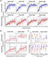
DP of all 83 animals (lines in shades of grey) over all training sessions in the 8 different groups with animals trained to discriminate periodicities of 20 Hz vs. 40 Hz (medians of data and quartiles in blue) and 160 Hz vs. 320 Hz (medians and quartiles in red). B Comparison of the DP of lesioned animals in 160 Hz vs. 320 Hz paradigm divided by lesion size (small lesion: open symbols; large lesions: solid symbols). Given are the DP median values and quartiles (whiskers), the boxes indicate the values used for the calculation of the final DP used in C. Between-group comparisons were carried out using Bonferroni-corrected Mann-Whitney U tests, p-level: *p<0.05, **p<0.01, ***p<0.001. C Comparison of LS and final DP dependent on lesion side and size. Given are median values (arrows), quartiles (boxes) and the full range of values (whiskers) of LS (upper panels) and final DP (lower panels) for animals that were trained to discriminate periodicities of 20 Hz vs. 40 Hz (blue data) or 160 Hz vs. 320 Hz (red data) and received either a sham lesion or a left or right sided lesion of different extension. Between-group comparisons of lesion size effects were carried out using Kruskal-Wallis ANOVA, p-level: *p<0.05, **p<0.01, ***p<0.001.
Figure 5
Figure 5. Effects of lesion size on CR-.
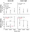
Given are the median values of wrong jumps over the hurdle (false responses) from day 8 to 15 (stable DP phase) for sham control animals and animals with small and large lesions of the left and right AC during slow (upper panels) and fast AM discrimination learning (lower panels). Whiskers indicate the interquartile range. Between-group comparisons of lesion size effects were performed using Kruskal-Wallis ANOVA and post-hoc multiple comparisons of mean ranks were used to investigate differences between two groups; p-levels: *p<0.05, ***p<0.001.

Similar articles

Cited by

References

    1. Budinger E, Heil P, Hess A, Scheich H (2006) Multisensory processing via early cortical stages: Connections of the primary auditory cortical field with other sensory systems. Neuroscience 143: 1065–1083. - PubMed
    1. Budinger E, Scheich H (2009) Anatomical connections suitable for the direct processing of neuronal information of different modalities via the rodent primary auditory cortex. Hear Res 258: 16–27. - PubMed
    1. Brechmann A, Gaschler-Markefski B, Sohr M, Yoneda K, Kaulisch T, et al. (2007) Working memory specific activity in auditory cortex: potential correlates of sequential processing and maintenance. Cereb Cortex 17: 2544–2552. - PubMed
    1. Ohl FW, Scheich H (2005) Learning-induced plasticity in animal and human auditory cortex. Curr Opin Neurobiol 15: 470–477. - PubMed
    1. Scheich H, Brechmann A, Brosch M, Budinger E, Ohl FW (2007) The cognitive auditory cortex: task-specificity of stimulus representations. Hear Res 229: 213–224. - PubMed

Publication types

LinkOut - more resources