. 2013 Dec 10;5(1):136-48.
doi: 10.1364/BOE.5.000136.

Imaging deep skeletal muscle structure using a high-sensitivity ultrathin side-viewing optical coherence tomography needle probe

Affiliations

Imaging deep skeletal muscle structure using a high-sensitivity ultrathin side-viewing optical coherence tomography needle probe

Xiaojie Yang et al. Biomed Opt Express. .

Abstract

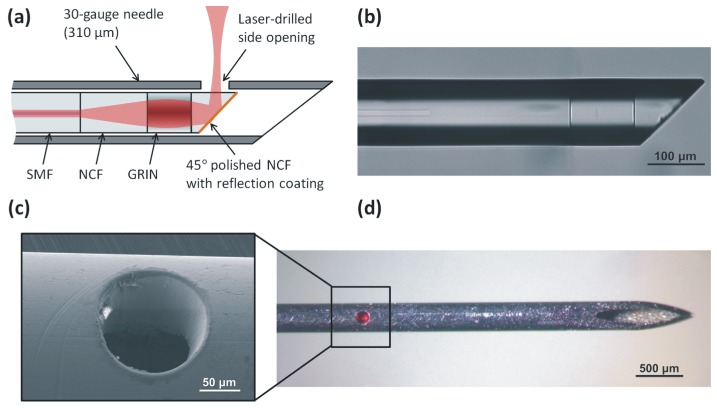
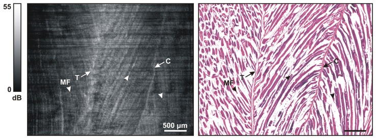
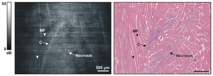
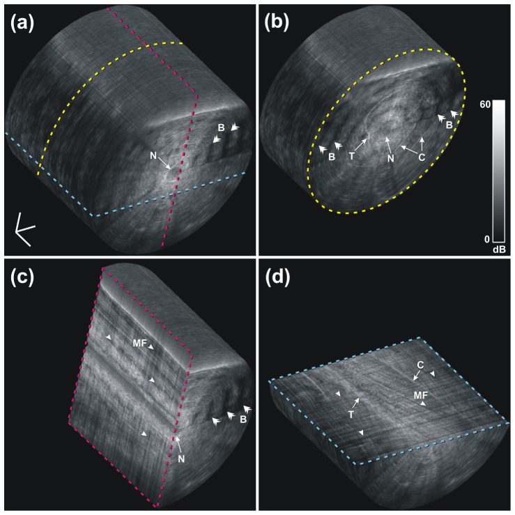
We have developed an extremely miniaturized optical coherence tomography (OCT) needle probe (outer diameter 310 µm) with high sensitivity (108 dB) to enable minimally invasive imaging of cellular structure deep within skeletal muscle. Three-dimensional volumetric images were acquired from ex vivo mouse tissue, examining both healthy and pathological dystrophic muscle. Individual myofibers were visualized as striations in the images. Degradation of cellular structure in necrotic regions was seen as a loss of these striations. Tendon and connective tissue were also visualized. The observed structures were validated against co-registered hematoxylin and eosin (H&E) histology sections. These images of internal cellular structure of skeletal muscle acquired with an OCT needle probe demonstrate the potential of this technique to visualize structure at the microscopic level deep in biological tissue in situ.

Keywords: (060.2370) Fiber optics sensors; (170.4500) Optical coherence tomography; (170.6935) Tissue characterization; (230.3990) Micro-optical devices.


Figures

Fig. 1
Fig. 1
(a) Schematic of the ultrathin OCT needle probe. (b) Microscope image of the angle-polished fiber probe before metallization. (c) SEM image of the laser-drilled side opening. (d) Fully assembled needle probe showing the laser-drilled side opening. In the photo, red light from the aiming laser is visible.
Fig. 2
Fig. 2
Beam profile and sensitivity characterization of the probe. (a) Illustration of the astigmatism introduced by the fiber, resulting in different working distances WDx and WDy in the x- and y-directions. (b) Measured, simulated and fitted FWHM output beam diameters in water, and calculated normalized on-axis intensity (see text for details). (c) Transverse intensity profile of the beam in water at the focus of the x-direction, at a distance of 330 µm from the fiber. (d) Averaged OCT A-scan of a silica/water interface (Peak 4) located at the distance of maximum SNR. The SNR of Peak 4 over a range of distances from the probe is also shown (circles). See text for discussion of the remaining peaks.
Fig. 3
Fig. 3
Schematic of the 1300-nm SSOCT needle imaging system. MZI, Mach-Zehnder interferometer; WDM, wavelength-division multiplexer; VOA, variable optical attenuator; SYNC, synchronization signal; PC, polarization controller.
Fig. 4
Fig. 4
Representative images of normal mouse skeletal muscle. (Left) OCT oblique slice taken from the 3D OCT volumetric data set. The striated appearance indicates the highly organized arrangement of the myofibers (MF and/or arrowhead). Several structures with higher signal intensity indicate tendon (T) and connective tissue (C). (Right) Corresponding H&E histology.
Fig. 5
Fig. 5
Representative images of dystrophic mouse skeletal muscle. (Left) OCT oblique slice taken from the 3D OCT volumetric data set. The striated appearance indicates the highly organized arrangement of myofibers (MF and/or arrowhead). The structure with higher intensity indicates connective tissue (C). Muscle necrosis is visible as a region without striated appearance (Necrosis). (Right) Corresponding H&E histology.
Fig. 6
Fig. 6
A 3D-rendered volumetric OCT data set of normal mouse muscle at an approximate depth of 10 mm in a) and three orthogonal cross sections in b), c) and d). The cross section in d) shows the same image plane as that of Fig. 4, but the brightness and contrast in the visualization software were adjusted differently in this 3D view to enhance the appearance of the full data set, yielding a slightly different dynamic range on the color bar compared to Fig. 4. B, birefringence artifacts; C, connective tissue; MF, myofibers; N, needle tract; T, tendon. The 3D scale bar in a) represents 500 µm in each direction (Media 1).

References

    1. E. N. Marieb, “Human anatomy & physiology,” in Human anatomy & Physiology, K. Ueno, ed. (Daryl Fox, San Francisco, USA, 2001), p. 324.
    1. Bushby K., Finkel R., Birnkrant D. J., Case L. E., Clemens P. R., Cripe L., Kaul A., Kinnett K., McDonald C., Pandya S., Poysky J., Shapiro F., Tomezsko J., Constantin C., DMD Care Considerations Working Group , “Diagnosis and management of Duchenne muscular dystrophy, part 1: diagnosis, and pharmacological and psychosocial management,” Lancet Neurol. 9(1), 77–93 (2010).10.1016/S1474-4422(09)70271-6 - DOI - PubMed
    1. Hoffman E. P., Fischbeck K. H., Brown R. H., Johnson M., Medori R., Loire J. D., Harris J. B., Waterston R., Brooke M., Specht L., Kupsky W., Chamberlain J., Caskey C. T., Shapiro F., Kunkel L. M., “Characterization of dystrophin in muscle-biopsy specimens from patients with Duchenne’s or Becker’s muscular dystrophy,” N. Engl. J. Med. 318(21), 1363–1368 (1988).10.1056/NEJM198805263182104 - DOI - PubMed
    1. Grounds M. D., Radley H. G., Lynch G. S., Nagaraju K., De Luca A., “Towards developing standard operating procedures for pre-clinical testing in the mdx mouse model of Duchenne muscular dystrophy,” Neurobiol. Dis. 31(1), 1–19 (2008).10.1016/j.nbd.2008.03.008 - DOI - PMC - PubMed
    1. Foster F. S., Pavlin C. J., Harasiewicz K. A., Christopher D. A., Turnbull D. H., “Advances in ultrasound biomicroscopy,” Ultrasound Med. Biol. 26(1), 1–27 (2000).10.1016/S0301-5629(99)00096-4 - DOI - PubMed

LinkOut - more resources