. 2014 May;171(10):2685-703.
doi: 10.1111/bph.12608.

The dual PPARα/γ agonist aleglitazar increases the number and function of endothelial progenitor cells: implications for vascular function and atherogenesis

Affiliations

The dual PPARα/γ agonist aleglitazar increases the number and function of endothelial progenitor cells: implications for vascular function and atherogenesis

C M Werner et al. Br J Pharmacol. 2014 May.

Abstract

Background and purpose: Aleglitazar is a dual PPARα/γ agonist but little is known about its effects on vascular function and atherogenesis. Hence, we characterized its effects on circulating angiogenic cells (CAC), neoangiogenesis, endothelial function, arteriogenesis and atherosclerosis in mice.

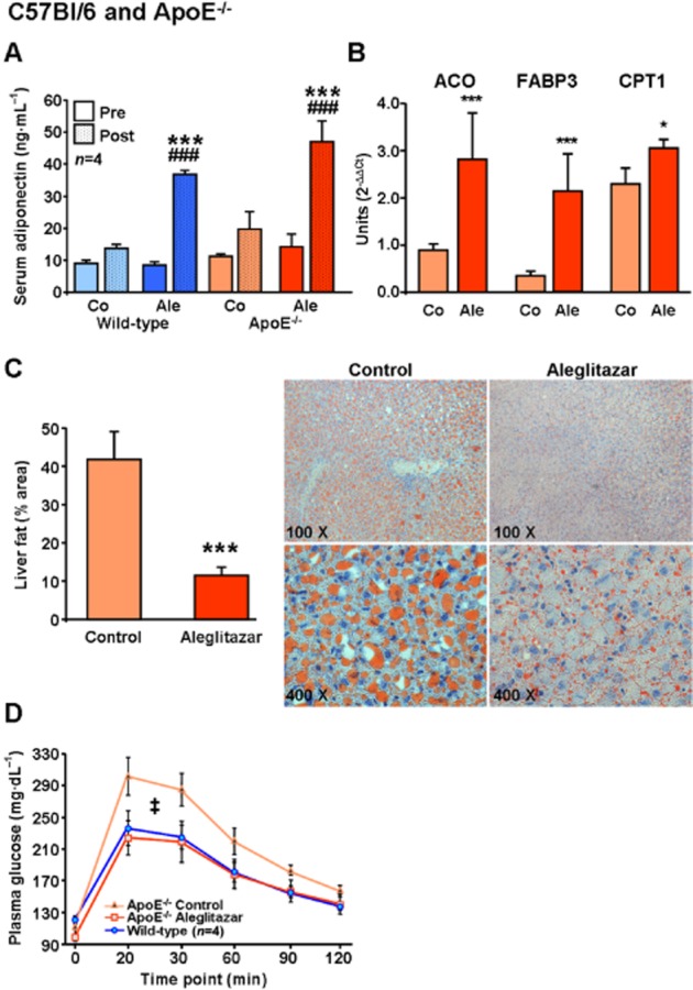
Experimental approach: C57Bl/6 wild-type (WT, normal chow), endothelial NOS (eNOS)(-/-) (normal chow) and ApoE(-/-) (Western-type diet) mice were treated with aleglitazar (10 mg·kg(-1) ·day(-1) , i.p.) or vehicle.

Key results: Aleglitazar enhanced expression of PPARα and PPARγ target genes, normalized glucose tolerance and potently reduced hepatic fat in ApoE(-/-) mice. In WT mice, but not in eNOS(-/-) , aleglitazar up-regulated Sca-1/VEGFR2-positive CAC in the blood and bone marrow and up-regulated diLDL/lectin-positive CAC. Aleglitazar augmented CAC migration and enhanced neoangiogenesis. In ApoE(-/-) mice, aleglitazar up-regulated CAC number and function, reduced markers of vascular inflammation and potently improved perfusion restoration after hindlimb ischaemia and aortic endothelium-dependent vasodilatation. This was associated with markedly reduced formation of atherosclerotic plaques. In human cultured CAC from healthy donors and patients with coronary artery disease with or without diabetes mellitus, aleglitazar increased migration and colony-forming units in a concentration-dependent manner. Furthermore, oxidative stress-induced CAC apoptosis and expression of p53 were reduced, while telomerase activity and expression of phospho-eNOS and phospho-Akt were elevated. Comparative agonist and inhibitor experiments revealed that aleglitazar's effects on CAC migration and colony-forming units were mediated by both PPARα and PPARγ signalling and required Akt.

Conclusions and implications: Aleglitazar augments the number, function and survival of CAC, which correlates with improved vascular function, enhanced arteriogenesis and prevention of atherosclerosis in mice.

Keywords: PPARα/γ agonist; aleglitazar; arteriogenesis; atherosclerosis; circulating angiogenic cells; endothelial NO synthase; endothelial function; endothelial progenitor cells.


Figures

Figure 1
Figure 1
The dual PPARα/γ agonist aleglitazar upregulates adiponectin and PPARα downstream targets, reduces liver fat and improves glucose tolerance in ApoE−/− mice. (A) Serum adiponectin concentrations were measured by elisa before and after treatment of C57Bl/6 WT mice with aleglitazar 10 mg·kg−1 i.p. or vehicle daily for 3 weeks, and in ApoE−/− mice on WTD treated for 6 weeks. (B) Real-time quantitative PCR was used to assess mRNA expression of PPARα target genes ACO, FABP3 and CPT1 in liver tissue of ApoE−/− mice on WTD treated for 6 weeks with aleglitazar or vehicle. The 18 s mRNA was used as loading control and data were analysed using the comparative Ct method. (C) Hepatic steatosis was measured by oil red O staining of liver sections and estimated by calculating the area of the lipid droplets compared with the total area. (D) The time course for serum glucose concentrations after i.p. glucose injection (1.5 mg·kg−1) in untreated C57Bl/6 WT and ApoE−/− mice on WTD treated with aleglitazar 10 mg·kg−1 i.p. or vehicle daily for 6 weeks. Significant differences were calculated for the AUC. n = 6 unless otherwise indicated, *P < 0.05 and ***P < 0.001 versus vehicle-injected control mice, ###P < 0.001 versus aleglitazar-treated mice. ‡P < 0.001 versus C57Bl/6 WT mice and aleglitazar-treated ApoE−/− mice.
Figure 2
Figure 2
Enhancement of CAC number, functional capacity and neoangiogenesis by aleglitazar. (A) Quantification and (B) representative fluorescence microscopy (100 × magnification) of spleen-derived diLDL/lectin positive CAC after treatment of WT mice with aleglitazar 10 mg·kg−1 i.p. daily or vehicle (controls) for 3 weeks. (C) Effects of aleglitazar treatment in WT mice compared with controls on the number of Sca-1/VEGFR2 positive EPC in the blood and (D) the bone marrow as measured by FACS analyses. (E) Effect of aleglitazar 10 mg·kg−1 i.p. daily for 3 weeks in C57Bl/6 WT mice on CAC migration in modified Boyden chambers using SDF-1 (100 ng·mL−1) as a chemoattractant. (F) Quantification of disc neoangiogenesis 2 weeks after s.c. implantation of polyvinyl sponges and after perfusion with fluorescent microspheres (0.2 μm) in WT mice treated with aleglitazar or vehicle. Lower panels show representative microscopic images (20 × magnification) of the vascularized border zone of perfused discs for (G) a control and (H) an aleglitazar-treated animal. n = 6, *P < 0.05 versus control mice.
Figure 3
Figure 3
Aleglitazar improves CAC, restores aortic endothelial function and improves perfusion recovery after hindlimb ischaemia in ApoE−/− mice. Effects of aleglitazar 10 mg·kg−1 i.p. daily for 6 weeks in ApoE−/− mice on WTD on (A) the number of diLDL/lectin positive CAC and (B) CAC migration in modified Boyden chambers; *P < 0.05 and ***P < 0.001 versus control mice. (C) Endothelium-dependent vasorelaxation of aortic rings in response to increasing concentrations of carbachol and (D) endothelium-independent vasorelaxation of aortic rings in response to increasing concentrations of nitroglycerin (shown as percentage of maximal phenylephrine-induced constriction) in untreated C57Bl/6 WT mice (n = 3), ApoE−/− control mice after 6 weeks WTD (n = 6), and ApoE−/− mice on WTD and concomitant aleglitazar treatment (10 mg·kg−1 i.p. daily) for 6 weeks (n = 6). †P < 0.001 WT versus ApoE−/− mice, #P < 0.05 for difference between ApoE−/− groups. (E) Laser Doppler measurement of endothelial-dependent hindlimb perfusion before, directly after, 3 days and 7 days after right FAL in ApoE−/− mice on WTD treated with vehicle (n = 10) or aleglitazar 10 mg·kg−1 i.p. (n = 12) daily for 5 weeks prior to ligation until the end of the study. Bars represent the mean Doppler perfusion ratio of the right ligated leg versus the left unligated leg. Lower panels show representative Doppler images. (F) Microsphere perfusion measurement of endothelial-independent hindlimb blood flow recovery 7 days after right FAL in C57Bl/6 mice on standard diet (treated with vehicle or aleglitazar 10 mg·kg−1 i.p. daily for 2 weeks before ligation until the end of the study) and ApoE−/− mice on WTD (treated with vehicle or aleglitazar 10 mg·kg−1 i.p. daily for 5 weeks before ligation until the end of the study). Bars represent the mean perfusion ratio of the right ligated leg versus the left unligated leg. ***P < 0.001 versus WT control mice, ###P < 0.001 versus ApoE−/− control mice; n = 6.
Figure 4
Figure 4
Aleglitazar retards development of atherosclerosis. (A) Real-time PCR assays of aortic MCP-1 and TNF-α mRNA expression in ApoE−/− mice on a WTD and treated with aleglitazar 10 mg·kg−1 i.p. or vehicle (n = 6 per group) for 8 weeks. Effects of aleglitazar for 6 or 8 weeks on atherosclerotic lesion formation in ApoE−/− mice on a WTD. (B) Histomorphometric quantification of atherosclerotic plaques in the aortic sinus after 6 weeks (n = 6) or 8 weeks (n = 7) aleglitazar 10 mg·kg−1 i.p. daily, shown as percentage of plaque area compared with total lumen area. *P < 0.05, **P < 0.01 and ***P < 0.001 versus vehicle-treated controls. Representative aortic root sections with oil red O staining of atherosclerotic plaques (40 × magnification) after (C) 6 weeks or (D) 8 weeks of aleglitazar treatment are shown.
Figure 5
Figure 5
eNOS is required for aleglitazar's effects on CAC. Effects of vehicle or aleglitazar 10 mg·kg−1 i.p. daily for 3 weeks in eNOS−/− mice on standard diet on (A) the number of diLDL/lectin positive CAC and (B) migration in modified Boyden chambers. Comparison of the effects of 3 weeks aleglitazar treatment in C57Bl/6 and eNOS−/− mice on Sca-1/VEGFR2 positive EPC in (C) the blood and (D) the bone marrow as measured by FACS analyses. *P < 0.05 and **P < 0.01 versus WT control. Repetitive measurements of (E) systolic and (F) diastolic BP at baseline and weekly during treatment with aleglitazar 10 mg kg−1 i.p. daily in eNOS−/− mice, compared with untreated C57Bl/6 mice.
Figure 6
Figure 6
Effects of aleglitazar on survival and apoptosis of human CAC. CAC were isolated from healthy donors by differentiation in cell culture for 4 days and treated as indicated. Effects of aleglitazar treatment for 24 h on (A) CAC migration in modified Boyden chambers and (B) CAC CFU. (C) CAC apoptosis as determined by FACS analysis shown as percentage of annexin V+/propidium iodide cells. Basal and hydrogen peroxide-induced (H2O2, 500 μmol·L−1, 24 h) apoptosis were compared in control and aleglitazar-treated (10 nmol·L−1, 24 h) CAC. (D) Effects of pioglitazone (10 μmol·L−1, 24 h) and aleglitazar (10 nmol·L−1 and 100 nmol·L, 24 h) on CAC telomerase activity as measured by TRAP assays (n = 3). Representative Western blots and quantification of aleglitazar effects compared with pioglitazone (10 μmol·L−1) on CAC protein expression of (E) the ratio of phospho-eNOS/total eNOS and (F) phospho-Akt/total Akt, (G) the apoptosis regulator p53, and (H) the anti-apoptotic protein Bcl-2 (all Western blots normalized to GAPDH). n = 4 unless otherwise indicated, *P < 0.05, **P < 0.01, ***P < 0.001 versus controls (CAC treated for 24 h with 1% DMSO).
Figure 7
Figure 7
Aleglitazar effects on CAC function in cell culture involve both PPARα and PPARγ and require Akt (PKB). (A) Effects of CAC treatment with 10 nmol·L−1 aleglitazar on the expression of the proliferation marker Ki67 in CAC colonies derived from healthy donors (n = 4). Right panels show an example of Ki67 + CAC-CFU (the asterisk marks a mitotic fission). Effects of treatment for 24 h with aleglitazar (10 nmol·L−1), pioglitazone (10 μmol·L−1), fenofibric acid (150 μmol·L−1) and the PPAR-γ antagonist GW9662 (1 μmol/L) on (B) CAC migration in modified Boyden chambers (n = 6) and (C) CAC proliferative capacity as shown by the number of CFU (n = 4). CAC from healthy donors (n = 4) were treated with 10 nmol·L−1 aleglitazar for 24 h and/or with the NOS inhibitor L-NAME (1 mmol·L−1), the phosphoinositide 3-kinase inhibitor LY294002 (10 μmol·L−1) or the direct Akt inhibitor 1L-6-hydroxymethyl-chiro-inositol2-[(R)-2-O-methyl-3-ooctadecylcarbonate] (10 μmol·L−1). Effects of these treatments on (D) CAC migration in modified Boyden chambers and (E) CFU were determined; *P < 0.05, **P < 0.001 and ***P < 0.001 versus controls.
Figure 8
Figure 8
Aleglitazar improves CAC function in patients with CAD. Cultured CAC from healthy donors and from patients with stable CAD with or without DM were treated with vehicle or 10 nmol·L−1 aleglitazar for 24 h and as read-outs of CAC function (A) migration in modified Boyden chambers and (B) the number of CFU were measured; n = 3 per group; *P < 0.05, **P < 0.001 and ***P < 0.001 versus control CAC of healthy donors; ##P < 0.01 and ###P < 0.001 versus CAD without DM; †††P < 0.001 versus CAD with DM.

Similar articles

Cited by

References

    1. Adams V, Lenk K, Linke A, Lenz D, Erbs S, Sandri M, et al. Increase of circulating endothelial progenitor cells in patients with coronary artery disease after exercise-induced ischemia. Arterioscler Thromb Vasc Biol. 2004;24:684–690. - PubMed
    1. Alexander SPH, Benson HE, Faccenda E, Pawson AJ, Sharman JL, Spedding M, Peters JA, Harmar AJ CGTP Collaborators. The Concise Guide to PHARMACOLOGY 2013/14: Nuclear Hormone Receptors. Br J Pharmacol. 2013;170:1652–1675. - PMC - PubMed
    1. Asahara T, Murohara T, Sullivan A, Silver M, van der Zee R, Li T, et al. Isolation of putative progenitor endothelial cells for angiogenesis. Science. 1997;275:964–967. - PubMed
    1. Benameur T, Tual-Chalot S, Andriantsitohaina R, Martinez MC. PPARalpha is essential for microparticle-induced differentiation of mouse bone marrow-derived endothelial progenitor cells and angiogenesis. PLoS ONE. 2010;5:e12392. - PMC - PubMed
    1. Benardeau A, Benz J, Binggeli A, Blum D, Boehringer M, Grether U, et al. Aleglitazar, a new, potent, and balanced dual PPAR alpha/gamma agonist for the treatment of type II diabetes. Bioorg Med Chem Lett. 2009;19:2468–2473. - PubMed

Publication types

MeSH terms