. 2014 Jan 28;16(1):R28.
doi: 10.1186/ar4456.

Fucosyltransferase 1 mediates angiogenesis, cell adhesion and rheumatoid arthritis synovial tissue fibroblast proliferation

Fucosyltransferase 1 mediates angiogenesis, cell adhesion and rheumatoid arthritis synovial tissue fibroblast proliferation

Takeo Isozaki et al. Arthritis Res Ther. .

Abstract

Introduction: We previously reported that sialyl Lewis(y), synthesized by fucosyltransferases, is involved in angiogenesis. Fucosyltransferase 1 (fut1) is an α(1,2)-fucosyltransferase responsible for synthesis of the H blood group and Lewis(y) antigens. However, the angiogenic involvement of fut 1 in the pathogenesis of rheumatoid arthritis synovial tissue (RA ST) has not been clearly defined.

Methods: Assay of α(1,2)-linked fucosylated proteins in RA was performed by enzyme-linked lectin assay. Fut1 expression was determined in RA ST samples by immunohistological staining. We performed angiogenic Matrigel assays using a co-culture system of human dermal microvascular endothelial cells (HMVECs) and fut1 small interfering RNA (siRNA) transfected RA synovial fibroblasts. To determine if fut1 played a role in leukocyte retention and cell proliferation in the RA synovium, myeloid THP-1 cell adhesion assays and fut1 siRNA transfected RA synovial fibroblast proliferation assays were performed.

Results: Total α(1,2)-linked fucosylated proteins in RA ST were significantly higher compared to normal (NL) ST. Fut1 expression on RA ST lining cells positively correlated with ST inflammation. HMVECs from a co-culture system with fut1 siRNA transfected RA synovial fibroblasts exhibited decreased endothelial cell tube formation compared to control siRNA transfected RA synovial fibroblasts. Fut1 siRNA also inhibited myeloid THP-1 adhesion to RA synovial fibroblasts and RA synovial fibroblast proliferation.

Conclusions: These data show that α(1,2)-linked fucosylated proteins are upregulated in RA ST compared to NL ST. We also show that fut1 in RA synovial fibroblasts is important in angiogenesis, leukocyte-synovial fibroblast adhesion, and synovial fibroblast proliferation, all key processes in the pathogenesis of RA.


Figures
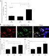
Figure 1
Figure 1
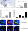
α(1,2)-linked fucosylated proteins are expressed in rheumatoid arthritis (RA). (A) RA synovial tissue (ST) homogenates contained more α(1,2)-linked fucosylated proteins than did osteoarthritis (OA) or normal (NL) STs (normalized to total protein concentration). Results are expressed as a ratio of the amount of fucosylated BSA/total proteins in the ST homogenates using BSA as a standard. (B) Photographs of RA ST. The far left panel shows staining with mouse anti-collagen 1. The middle panel shows staining with Ulex Europeaus Agglutinin 1 lectin (UEA-1) and goat anti-UEA-1. The right panel shows merging of the left panel and middle panel. Yellow indicates α(1,2)-linked fucosylated proteins associated with RA ST fibroblasts and the blue indicates DAPI staining of the tissue (original magnification 100×). (C) α(1,2)-linked fucosylated proteins in RA synovial fibroblast-conditioned medium were significantly higher than in OA and NL synovial fibroblast-conditioned medium. (D) α(1,2)-linked fucosylated proteins in RA synovial fibroblast cell lysates were significantly higher than in NL synovial fibroblast cultures: 2′fucosyllactose-bovine serum albumin (2′FL-BSA) was used as a standard.
Figure 2
Figure 2
Immunohistologic analysis of fucosyltransferase 1 (fut1) expression. (A and B) Photomicrographs of ST samples from patients with rheumatoid arthritis (RA). Cryosections were stained with anti-fut1 (A) or control IgG (B). Original magnification is 400×. Arrows indicate fut1 expression. RA synovial tissues (STs) contain a greater percentage of fut1 lining cells (C) compared to OA and NL ST. A significantly elevated percentage of macrophage staining on RA compared to OA or NL STs was also found (D). Expression of fut1 mRNA in TNF-α stimulated or nonstimulated RA synovial fibroblasts was significantly elevated compared to TNF-α stimulated or nonstimulated NL synovial fibroblasts (E). Means are presented with standard error. *P <0.05 was significant. NS = nonstimulated. (n = number of RA patients or patient ST fibroblasts). (F) Left panel, fut1 straining in RA ST (green); middle panel, cadherin-11 staining in RA ST (red); right panel, merge of the previous two figures. The arrow indicates fut1 and cadherin-11-positive cells respectively (yellow), indicating that fut1 is expressed on fibroblasts in RA ST. (G) Left panel, fut1 straining in RA ST (green); middle panel, CD68 staining in RA ST (red); right panel, merge of the previous two panels. The arrows indicate fut1 on CD68-positive cells, validating that fut1 is expressed on macrophages in RA ST (yellow). The blue background is 4′,6-diamidino-2-phenylindole (DAPI) staining. IgG control staining was performed and showed no fluorescence staining (data not shown; all figures are 400× magnification).
Figure 3
Figure 3
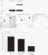
Fucosyltransferase 1 (fut1) expression was decreased using siRNA against fut1 in rheumatoid arthritis (RA) synovial fibroblasts. Cells were stimulated with TNF-α (25 ng/ml) for 24 hours. (A) Fut1 expression in fut1 siRNA-treated RA synovial fibroblasts and control siRNA-treated RA synovial fibroblasts. (B) RA synovial fibroblasts were first transfected with control or fut1 siRNA, and were grown on the top inserts. Human dermal microvascular endothelial cells (HMVECs) were plated in the bottom of the wells of the transwell system for 24 hours. HMVECs were plated on Matrigel with nontreated, control siRNA-transfected or fut1 siRNA-transfected RA synovial fibroblast-conditioned medium for 6 hours. A representative photograph of HMVECs from co-culture with nontreated, control or fut1 siRNA-transfected RA synovial fibroblasts is shown. Arrows indicate tubes. (C) HMVECs incubated with fut1 siRNA-transfected RA synovial fibroblast conditioned media formed significantly fewer tubes on Matrigel compared with control siRNA-treated RA synovial fibroblasts or nontreated RA synovial fibroblasts. Means are presented with standard error of the mean. *P <0.05 was significant (n = number of RA patient synovial fibroblasts).
Figure 4
Figure 4
Fucosyltransferase 1 (Fut1) siRNA inhibited pro-angiogenic mediator production in TNF-α-stimulated rheumatoid arthritis (RA) synovial fibroblasts. (A) Monocyte chemoattract protein 1 (MCP-1/CCL2) mRNA expression in fut1 siRNA-treated RA synovial fibroblasts was 60 ± 9% (mean ± standard error (SE)) of control siRNA-transfected RA synovial fibroblasts, showing a 40% reduction in MCP-1/CCL2 mRNA expression in fut1 siRNA-transfected cells. (B) Epithelial-derived neutrophil-activating peptide 78 (ENA-78/CXCL5) mRNA expression in fut1 siRNA-treated RA synovial fibroblasts was 69 ± 11% (mean ± SE) of control siRNA transfected RA synovial fibroblasts, showing a 31% reduction in ENA-78/CXCL5 mRNA expression fut1 siRNA-transfected cells. (C) Vascular endothelial growth factor (VEGF) mRNA expression in fut1 siRNA-treated RA synovial fibroblasts was 79 ± 7% (mean ± SE) of control siRNA-transfected RA synovial fibroblasts, showing a 21% reduction in VEGF mRNA expression fut1 siRNA-transfected cells. (D) Fut1 siRNA inhibited production of MCP-1/CCL2. (E) Fut1 siRNA inhibited production of ENA-78/CXCL5. (F) Fut1 siRNA inhibited production of VEGF. Means are presented with SE. *P <0.05 was significant (n = number of RA patient synovial fibroblasts).
Figure 5
Figure 5
Fucosyltransferase 1 (fut1) mediates adhesion of THP-1 cells to rheumatoid arthritis (RA) synovial fibroblasts and mediates their proliferation. The percent adhesion was defined as the number of adherent cells on synovial tissue (ST) sections divided by the number of adherent cells on control sections. (A) Adhesion of THP-1 cells to TNF-α-stimulated fut1 siRNA-transfected RA synovial fibroblasts (62 ± 8% of maximal adhesion) was significantly decreased compared with adhesion of THP-1 cells to TNF-α-stimulated control siRNA-transfected (93 ± 8% of maximal adhesion) or nontreated RA synovial fibroblasts. (B) Intercellular adhesion molecule 1 (ICAM-1) expression on TNF-α-stimulated fut1 siRNA-transfected RA synovial fibroblasts was significantly decreased compared to TNF-α-stimulated control siRNA or nontreated RA synovial fibroblasts. (C) Vascular cell adhesion molecule 1 (VCAM-1) expression on TNF-α-stimulated fut1 siRNA-transfected RA synovial fibroblasts was significantly decreased compared to TNF-α-stimulated control siRNA or nontreated RA synovial fibroblasts. (D) Fut1 siRNA-transfected lipolysaccharide (LPS)-stimulated RA synovial fibroblasts showed reduced proliferation at 4 and 24 hours compared to LPS-stimulated control siRNA-transfected or nontreated RA synovial fibroblasts. The results are shown as fold change in optical density of fut1- or control siRNA-transfected RA synovial fibroblasts normalized to nontreated RA synovial fibroblasts. Means are presented with standard error. *P <0.05 was significant. (n = number of RA patient synovial fibroblast cultures).
Figure 6
Figure 6
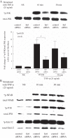
Fucosyltransferase 1 (fut1) siRNA inhibits phosphorylated JNK signaling in rheumatoid arthritis (RA) synovial fibroblasts. (A) Western blots were performed to determine whether TNF-α stimulates the phosphorylation of JNK. Phosphorylation of JNK signaling in TNF-α-stimulated fut1 siRNA-transfected RA synovial fibroblasts was significantly decreased at 10 minutes compared to control siRNA-transfected RA synovial fibroblasts. (B) Fut1 siRNA does not inhibit phosphorylation of NFκB, P38, and Erk1/2 in RA synovial fibroblasts. Means are presented with standard errror. *P <0.05 was significant; *p indicates phosphorylated signaling proteins (n = number of RA patient synovial fibroblasts).

Similar articles

Cited by

References

    1. Koch AE, Kunkel SL, Burrows JC, Evanoff HL, Haines GK, Pope RM, Strieter RM. Synovial tissue macrophage as a source of the chemotactic cytokine IL-8. J Immunol. 1991;16:2187–2195. - PubMed
    1. Ritchlin C. Fibroblast biology. Effector signals released by the synovial fibroblast in arthritis. Arthritis Res. 2000;16:356–360. doi: 10.1186/ar112. - DOI - PMC - PubMed
    1. Kunkel SL, Lukacs N, Kasama T, Strieter RM. The role of chemokines in inflammatory joint disease. J Leukoc Biol. 1996;16:6–12. - PubMed
    1. Choy EH, Panayi GS. Cytokine pathways and joint inflammation in rheumatoid arthritis. N Engl J Med. 2001;16:907–916. doi: 10.1056/NEJM200103223441207. - DOI - PubMed
    1. Koch AE. Chemokines and their receptors in rheumatoid arthritis: future targets? Arthritis Rheum. 2005;16:710–721. doi: 10.1002/art.20932. - DOI - PubMed

Publication types

MeSH terms

Substances