. 2014 Jan 28;2(1):1.
doi: 10.1186/2049-2618-2-1.

Microbes in the neonatal intensive care unit resemble those found in the gut of premature infants

Affiliations

Microbes in the neonatal intensive care unit resemble those found in the gut of premature infants

Brandon Brooks et al. Microbiome. .

Abstract

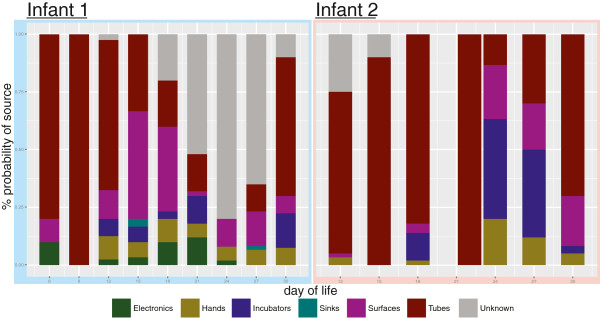
Background: The source inoculum of gastrointestinal tract (GIT) microbes is largely influenced by delivery mode in full-term infants, but these influences may be decoupled in very low birth weight (VLBW, <1,500 g) neonates via conventional broad-spectrum antibiotic treatment. We hypothesize the built environment (BE), specifically room surfaces frequently touched by humans, is a predominant source of colonizing microbes in the gut of premature VLBW infants. Here, we present the first matched fecal-BE time series analysis of two preterm VLBW neonates housed in a neonatal intensive care unit (NICU) over the first month of life.

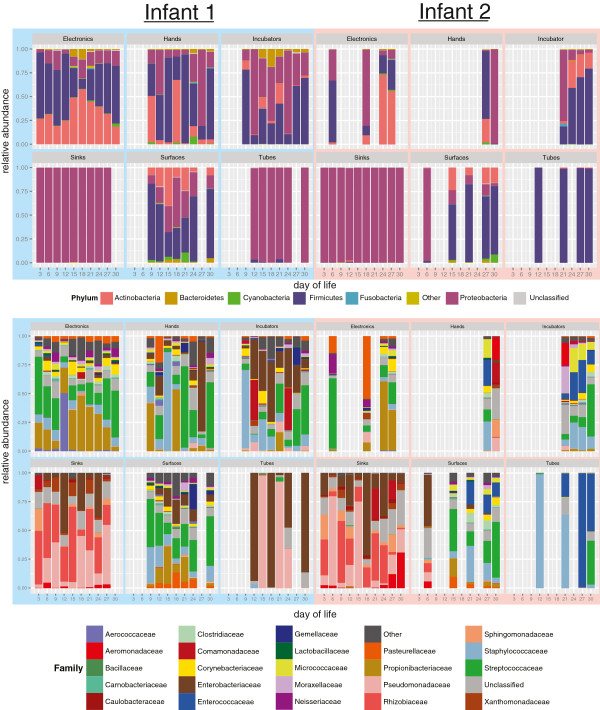
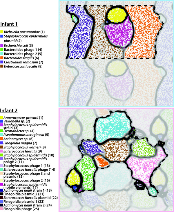
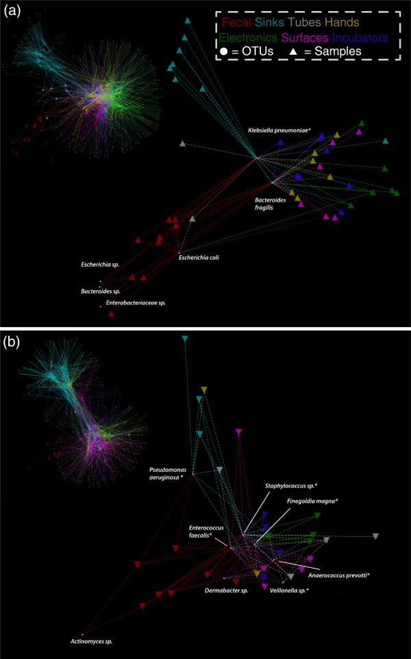
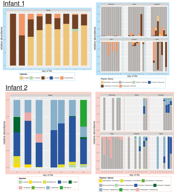
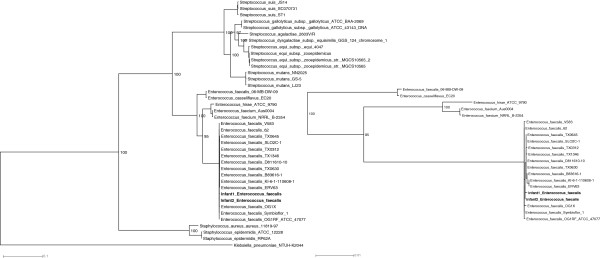
Results: Fresh fecal samples were collected every 3 days and metagenomes sequenced on an Illumina HiSeq2000 device. For each fecal sample, approximately 33 swabs were collected from each NICU room from 6 specified areas: sink, feeding and intubation tubing, hands of healthcare providers and parents, general surfaces, and nurse station electronics (keyboard, mouse, and cell phone). Swabs were processed using a recently developed 'expectation maximization iterative reconstruction of genes from the environment' (EMIRGE) amplicon pipeline in which full-length 16S rRNA amplicons were sheared and sequenced using an Illumina platform, and short reads reassembled into full-length genes. Over 24,000 full-length 16S rRNA sequences were produced, generating an average of approximately 12,000 operational taxonomic units (OTUs) (clustered at 97% nucleotide identity) per room-infant pair. Dominant gut taxa, including Staphylococcus epidermidis, Klebsiella pneumoniae, Bacteroides fragilis, and Escherichia coli, were widely distributed throughout the room environment with many gut colonizers detected in more than half of samples. Reconstructed genomes from infant gut colonizers revealed a suite of genes that confer resistance to antibiotics (for example, tetracycline, fluoroquinolone, and aminoglycoside) and sterilizing agents, which likely offer a competitive advantage in the NICU environment.

Conclusions: We have developed a high-throughput culture-independent approach that integrates room surveys based on full-length 16S rRNA gene sequences with metagenomic analysis of fecal samples collected from infants in the room. The approach enabled identification of discrete ICU reservoirs of microbes that also colonized the infant gut and provided evidence for the presence of certain organisms in the room prior to their detection in the gut.


Figures

Figure 1
Figure 1
Taxonomic classification of neonatal intensive care unit (NICU) room microbes for infants 1 and 2. Phylum-level (top) and family-level (bottom) classifications were assigned using the Ribosomal Database Project (RDP) classifier on assembled full-length 16S rRNA genes. Day of life (DOL) is plotted on the X axis and relative abundance, generated by ‘expectation maximization iterative reconstruction of genes from the environment’ (EMIRGE), is plotted on the Y axis.
Figure 2
Figure 2
Principle coordinates analysis (PCoA) based on UniFrac scores of room and gut microbes. Analysis reveals four discernible ecosystem clusters: skin associated communities, sinks, tubes, and feces.
Figure 3
Figure 3
Time-series coverage emergent self-organizing maps (ESOMs) reveal discrete genome bins for each infant’s dataset. The underlying ESOMs are shown in a tiled display with each data point colored by its taxonomic assignment. Labels to the left are colored to match their respective data points and numbers in parentheses correspond to the bin numbers in Table 4.
Figure 4
Figure 4
Spring-weighted edge-embedded network plots of room and fecal operational taxonomic units (OTUs). Found in two or more samples (infant 1 (a), infant 2 (b)). Left, the entire network is displayed. To better visualize the distribution of gut colonizers across room samples, only room samples sharing fecal OTUs are shown in the excerpt (right). Triangles represent samples and circles represent OTUs. The spring weight is derived from ‘expectation maximization iterative reconstruction of genes from the environment’ (EMIRGE) generated abundances and edges are colored by environment type. Each OTU has a taxonomic label and asterisks indicate OTUs detected in room samples before detection in the gut.
Figure 5
Figure 5
Community composition of gut colonizing microbes and room microbes through the first month of life. Time-series characterization of the fecal microbial community (left) and fecal microbes concurrently collected from the room (right) display discrete reservoirs of gut colonizers in the neonatal intensive care unit.
Figure 6
Figure 6
The most probable source of gut colonizing microbes. This was generated using the source-sink characterization software, SourceTracker. Neonatal intensive care unit room sequences were designated as putative sources and fecal sequences sinks.
Figure 7
Figure 7
Enterococcus faecalis phylogeny using 32 concatenated ribosomal proteins reveals closely related strains. The maximum likelihood phylogeny of E. faecalis strains was based on a concatenation of single-copy, highly conserved ribosomal proteins from our data set and available reference genomes. Bootstrap values greater than 50 are shown. An excerpt of the E. faecalis clade is shown to the right.

References

    1. Klepeis NE, Nelson WC, Ott WR, Robinson JP, Tsang AM, Switzer P, Behar JV, Hern SC, Engelmann WH. The National Human Activity Pattern Survey (NHAPS): a resource for assessing exposure to environmental pollutants. J Expo Anal Environ Epidemiol. 2001;11:231–252. doi: 10.1038/sj.jea.7500165. - DOI - PubMed
    1. Tringe SG, Zhang T, Liu X, Yu Y, Lee WH, Yap J, Yao F, Suan ST, Ing SK, Haynes M, Rohwer F, Wei CL, Tan P, Bristow J, Rubin EM, Ruan Y. The airborne metagenome in an indoor urban environment. PLoS One. 2008;3:e1862. doi: 10.1371/journal.pone.0001862. - DOI - PMC - PubMed
    1. Rintala H, Pitkäranta M, Toivola M, Paulin L, Nevalainen A. Diversity and seasonal dynamics of bacterial community in indoor environment. BMC Microbiol. 2008;8:56. doi: 10.1186/1471-2180-8-56. - DOI - PMC - PubMed
    1. Kelley ST, Gilbert JA. Studying the microbiology of the indoor environment. Genome Biol. 2013;14:202. doi: 10.1186/gb-2013-14-2-202. - DOI - PMC - PubMed
    1. La Duc MT, Dekas A, Osman S, Moissl C, Newcombe D, Venkateswaran K. Isolation and characterization of bacteria capable of tolerating the extreme conditions of clean room environments. Appl Environ Microbiol. 2007;73:2600–2611. doi: 10.1128/AEM.03007-06. - DOI - PMC - PubMed