. 2014 Jan 27:12:5.
doi: 10.1186/1741-7007-12-5.

Host insulin stimulates Echinococcus multilocularis insulin signalling pathways and larval development

Affiliations

Host insulin stimulates Echinococcus multilocularis insulin signalling pathways and larval development

Sarah Hemer et al. BMC Biol. .

Abstract

Background: The metacestode of the tapeworm Echinococcus multilocularis is the causative agent of alveolar echinococcosis, a lethal zoonosis. Infections are initiated through establishment of parasite larvae within the intermediate host's liver, where high concentrations of insulin are present, followed by tumour-like growth of the metacestode in host organs. The molecular mechanisms determining the organ tropism of E. multilocularis or the influences of host hormones on parasite proliferation are poorly understood.

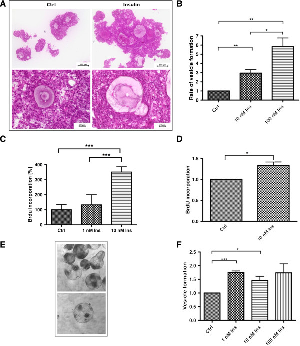
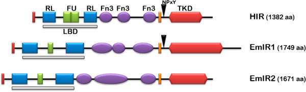
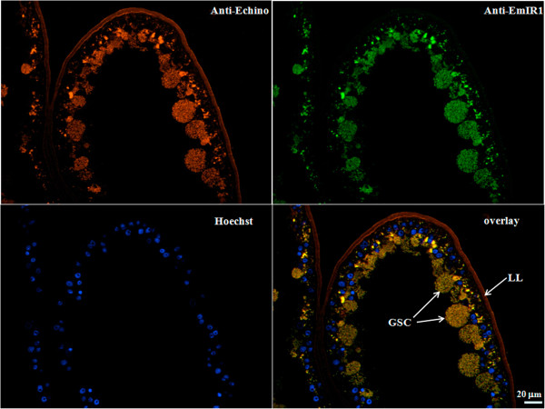
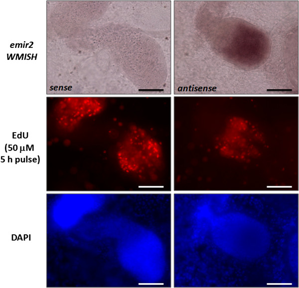
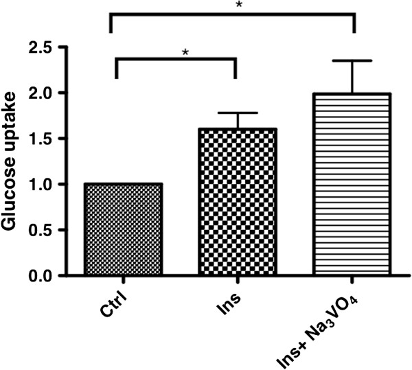
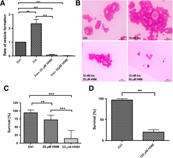
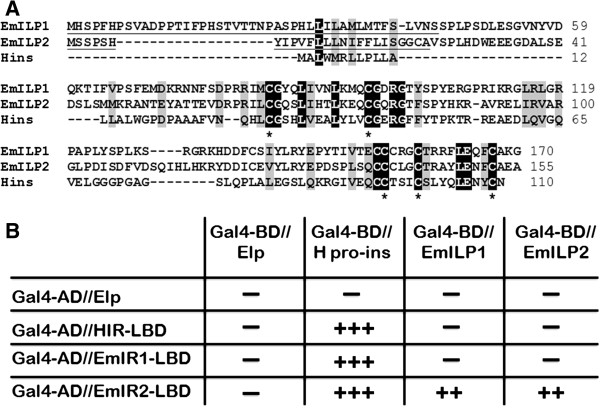
Results: Using in vitro cultivation systems for parasite larvae we show that physiological concentrations (10 nM) of human insulin significantly stimulate the formation of metacestode larvae from parasite stem cells and promote asexual growth of the metacestode. Addition of human insulin to parasite larvae led to increased glucose uptake and enhanced phosphorylation of Echinococcus insulin signalling components, including an insulin receptor-like kinase, EmIR1, for which we demonstrate predominant expression in the parasite's glycogen storage cells. We also characterized a second insulin receptor family member, EmIR2, and demonstrated interaction of its ligand binding domain with human insulin in the yeast two-hybrid system. Addition of an insulin receptor inhibitor resulted in metacestode killing, prevented metacestode development from parasite stem cells, and impaired the activation of insulin signalling pathways through host insulin.

Conclusions: Our data indicate that host insulin acts as a stimulant for parasite development within the host liver and that E. multilocularis senses the host hormone through an evolutionarily conserved insulin signalling pathway. Hormonal host-parasite cross-communication, facilitated by the relatively close phylogenetic relationship between E. multilocularis and its mammalian hosts, thus appears to be important in the pathology of alveolar echinococcosis. This contributes to a closer understanding of organ tropism and parasite persistence in larval cestode infections. Furthermore, our data show that Echinococcus insulin signalling pathways are promising targets for the development of novel drugs.


Figures

Figure 1
Figure 1
Effects of insulin on E. multilocularis larval development. A) Morphology of primary cell aggregates. Primary cells were isolated from axenic metacestode vesicles and cultivated in 2% FCS/(D)MEM supplemented with or without 10 nM human insulin for one week. The aggregates were fixed and embedded in Technovit 8100. Sections (4 μm) were stained with haematoxylin/eosin. B) Formation of metacestode vesicles. Primary cells were cultivated in conditioned medium supplemented with human insulin for three weeks and mature metacestode vesicles were counted. Control was set to 1 and results were normalised against the control. (*) P values below 0.05, (**) very significant for P between 0.001 and 0.01, (***) extremely significant for P <0.001. C) BrdU uptake by parasite cell cultures. Primary cells were isolated and incubated for 24 hours with insulin. BrdU was added for four hours and BrdU uptake was measured with the colorimetric BrdU ELISA kit (Roche, Mannheim, Germany). Asterisks mark significant values. D) BrdU uptake by mature metacestode vesicles. Metacestode vesicles were incubated for two days in the presence or absence of insulin and BrdU. BrdU uptake was measured after chromosomal DNA isolation with the colorimetric BrdU ELISA kit (Roche). E) Re-differentiation and microcyst formation of E. multilocularis protoscoleces. Examples of microcysts forming in in vitro protoscolex cultures. F) Microcyst formation of in vitro cultivated protoscoleces incubated with insulin for three weeks. Control was set to 1 and results were normalised against the control. (*) P values below 0.05, (**) very significant for P between 0.001 and 0.01, (***) extremely significant for P <0.001. BrdU, bromodeoxyuridine.
Figure 2
Figure 2
Domain structure of E. multilocularis insulin receptors. Schematic representation of the domain structures of the human insulin receptor (HIR), EmIR1 and EmIR2 according to SMART analyses (Letunic et al., 2012). Displayed are the location and size of the following predicted domains: TKD, tyrosine kinase domain; LBD, ligand binding domain; RL, receptor-L-domain; FU, furin-rich repeat; Fn3, fibronectin 3 domain. The presence of NPXY-motifs for binding of IRS is indicated by an arrowhead. Red bars at the N-terminus represent signal peptides, orange bars represent transmembrane domains. IRS, insulin receptor substrate; SMART, Simple Modular Architecture Research Tool.
Figure 3
Figure 3
Expression of EmIR1 and EmIR2 in E. multilocularis larval stages. A) Semi-quantitative RT-PCR of emir1 and emir2 expression. Serial 1/10 dilutions of cDNA from metacestode vesicles (MC), primary cells (PC) as well as dormant (PS-) and low pH/pepsin-activated protoscoleces (PS+) were subjected to gene-specific PCR using intron-flanking primers. PCR products were separated on a 1% agarose gel and stained with ethidium bromide. The constitutively expressed gene elp was used as control. B) Western blot and immunoprecipitation employing the EmIR1 anti-serum. EmIR1 was immunoprecipitated from metacestode vesicles and treated with β-mercaptoethanol (beta-MeOH) at concentrations of 0%, 1% and 10%. Probes were then separated on a 12.5% polyacrylamide gel and developed using the anti-EmIR1 antiserum. ‘pro’ and ‘beta’ indicate the pro-form and the β-subunit of EmIR, respectively. C) Immunoprecipitation and Western blot using the anti-EmIR2 serum. EmIR2 was immunoprecipitated from protoscolex preparations, samples were then supplemented with 1% or 10% of β-mercaptoethanol (β-ME) and separated on a 10% SDS gel. Western blot was carried out using the anti-EmIR2 antiserum. D) Immunodetection of EmIR1 and EmIR2 in different larval stages using immune sera. Parasite larvae were lysed, protein preparations were then separated by SDS-PAGE, blotted onto a membrane and detected by the antisera. The purified anti-EmIR2 immune serum recognized the EmIR2 β-subunit at 87 kDa and a second band at 60 kDa. The EmIR1 β-subunit was detected at 150 kDa using the anti-EmIR1 immune serum. Actin was used as a loading control. Mc, metacestode vesicles; Pc, primary cells; Ps-, dormant protoscoleces; Ps+, activated protoscoleces.
Figure 4
Figure 4
EmIR1 immunohistochemistry on metacestode vesicles. Cryosections of in vitro cultivated metacestode vesicles were probed with the anti-EmIR1 antiserum (anti-EmIR1) and detected with a FITC-coupled anti-rabbit-antibody. Nuclei were visualized by Hoechst-staining (Hoechst). Parasite surface structures were visualized using a general anti-Echinococcus metacestode antibody (Anti-Echi; Ingold et al., 2001 [50]). LL, laminated layer; GSC, glycogen storing cells. FITC, fluorescein isothiocyanate.
Figure 5
Figure 5
EmIR1 electron microscopic analyses. The anti-EmIR1 antiserum and gold-coupled anti-rabbit antibodies were used to detect EmIR1 in the metacestode germinal layer. A) Overview showing the location of the laminated layer, the germinal layer, undifferentiated (stem) cells and glycogen storage cells. B) Glycogen storage cell showing massive anti-EmIR1 staining (arrows). Scale bar in larger image represents 0.6 μm, scale bar in insert represents 3.6 μm.
Figure 6
Figure 6
Anti-EmIR2 immunohistochemistry. Sections of E. multilocularis protoscolex preparations (left panel) and primary cell culture aggregates (right panel) have been probed with the anti-EmIR2 antiserum. Brown color indicates positive signals. Cav, central cavity; S, suckers; R, rostellum; CC, calcareous corpuscles.
Figure 7
Figure 7
Whole mount in situ hybridisation (WMISH) detection of emir2. Germinal layer with developing protoscoleces stained for emir2 WMISH (upper panel), EdU incorporation (mid panel) and DAPI (lower panel). Sense (left) and antisense (right) probes have been used as indicated. Bar represents 50 μm. DAPI, 4',6-diamidino-2-phenylindole; EdU, 5-ethylnyl-2’-deoxyuridine.
Figure 8
Figure 8
Effects of insulin on metacestode glucose uptake. Uptake of C14-D-glucose by metacestode vesicles in the presence of 10 μM insulin alone,and insulin together with Na3VO4. Control was set to 1 and results were normalised against the control. (*) P values below 0.05, (**) very significant for P between 0.001 and 0.01, (***) extremely significant for P <0.001.
Figure 9
Figure 9
Effects of insulin on the phosphorylation of metacestode vesicle proteins. A) Detection of EmIR1 in vesicle membrane fractions (MF). Vesicles from in vitro culture were homogenized and the MF was isolated. Western blot detection was carried out on MF and whole vesicle preparations (Ves) using the anti-EmIR1 antiserum. ‘Pro’ and ‘β’ mark the receptor pro-form and β-subunits. B) Phosphorylation of EmIR1 in response to insulin. Metacestode MF was stimulated for 10 minutes with either 100 nM insulin or IGF-I, followed by 30 minutes incubation with 100 μM HNMPA(AM)3 or control DMSO. Phosphorylation of membrane proteins was carried out for 40 minutes in the presence of [32P] γ-ATP. Proteins of the MF were precipitated using the anti-EmIR1 antiserum. Proteins were then separated by 8% SDS-PAGE and phosphorylation was detected by autoradiography. Bands are visible at the size of the EmIR1 β-subunit and below. C) Phosphorylation of EmIR1 after insulin stimulation of metacestode vesicles. Vesicles were stimulated (+) or not (−) with 100 nM insulin for 10 minutes. Following solubilisation of membrane proteins, the EmIR1 β-subunit was precipitated using the anti-EmIR1 antiserum and separated by SDS-PAGE. Western blot detection was carried out using the anti-EmIR1 antiserum (lower panel) or an anti-phospho-tyrosine antibody (upper panel). D) Phosphorylation of Echinococcus PI3K/Akt pathway components in response to insulin. Vesicles were stimulated with 10 nM insulin for the times indicated above. Vesicle lysates were subsequently separated by SDS-PAGE and probed using antibodies against the phosphorylated Akt substrate motif or phospho 4E-BP as indicated. β-Actin was used as loading control. E) Inhibition of 4E-BP phosphorylation through HNMPA(AM)3. Vesicles were incubated for two hours with 100 μM HNMPA(AM)3 (HNM+) or the PI3K inhibitor LY294002 (LY+) before stimulation with 10 nM insulin. Crude lysates were probed with the anti-phospho 4E-BP antibody. β-Actin was used as loading control. DMSO, dimethyl sulphoxide; HNMPA, 2-hydroxynaphthalen-1-yl-methylphosphonic acid.
Figure 10
Figure 10
Effects of HNMPA(AM)3 on parasite larvae. A) Primary cells were isolated from axenic metacestode vesicles and cultivated in 2% FCS/(D)MEM supplemented with 10 nM human insulin, with or without HNMPA(AM)3. After three weeks of incubation, mature vesicles were counted. Insulin was added to the cultures in order to obtain vesicle formation within three weeks. B) Formation of primary cell aggregates in the presence of HNMPA(AM)3. Primary cells were incubated as in (A) for seven days before aggregates were fixed, embedded in Technovit 8100 and 4 μm sections stained with haematoxylin/eosin. Note the profound effect of HNMPA(AM)3 on parasite aggregates already after seven days. Ctrl, DMSO control. C) Protoscoleces were treated with HNMPA(AM)3 for two weeks under axenic conditions. Protoscolex viability was analysed by counter-staining with methylene blue. D) Metacestode vesicles were treated for one week with 100 μM HNMPA(AM)3 under axenic conditions. Survival was assessed by counting physically damaged vesicles. Vesicles were incubated in the presence of conditioned medium for optimal maintenance and survival conditions. (*) P values below 0.05, (**) very significant for P between 0.001 and 0.01, (***) extremely significant for P <0.001. DMSO, dimethyl sulphoxide; HNMPA, 2-hydroxynaphthalen-1-yl-methylphosphonic acid.
Figure 11
Figure 11
E. multilocularis insulin-like peptides and yeast two-hybrid experiments. A) Amino acid sequence comparison between the E. multilocularis insulin-like peptides EmILP1 and EmILP2 and human insulin (Hins). Highly conserved residues are printed in white on black background, residues with similar biochemical features are printed in black on grey background. Underlined sequences indicate predicted signal peptides. Asterisks indicate cysteine residues important for disulphide-bridge formation. B) Yeast two-hybrid experiments. Translational fusions were generated for the Gal4 activation domain (Gal4-AD) and the LBDs of the human insulin receptor (HIR-LBD) as well as the E. multilocularis receptors EmIR1 (EmIR1-LBD) and EmIR2 (EmIR2-LBD). The Gal4 DNA binding domain (Gal4-BD) was fused to human pro-insulin (H pro-ins) as well as to EmILP1 and EmILP2. Yeast strains were double transformed with the plasmid constructs as indicated and growth under different stringency conditions [10] was assessed. ‘-‘ indicates no growth, ‘++’ growth under medium stringency conditions, ‘+++’ growth under high stringency conditions. LBDs, ligand binding domains.

Similar articles

Cited by

References

    1. Kern P. Clinical features and treatment of alveolar echinococcosis. Curr Opin Infect Dis. 2010;12:505–512. doi: 10.1097/QCO.0b013e32833d7516. - DOI - PubMed
    1. Brehm K. Echinococcus multilocularis as an experimental model in stem cell research and molecular host-parasite interaction. Parasitology. 2010;12:537–555. doi: 10.1017/S0031182009991727. - DOI - PubMed
    1. Smyth JD, Howkins AB, Barton M. Factors controlling the differentiation of the hydatid organism, Echinococcus granulosus, into cystic or strobilar stages in vitro. Nature. 1966;12:1374–1377. doi: 10.1038/2111374a0. - DOI - PubMed
    1. Shojaee-Moradie F, Powrie JK, Sundermann E, Spring MW, Schüttler A, Sönksen PH, Brandenburg D, Jones RH. Novel hepatoselective insulin analog: studies with a covalently linked thyroxyl-insulin complex in humans. Diabetes Care. 2000;12:1124–1129. doi: 10.2337/diacare.23.8.1124. - DOI - PubMed
    1. Misbin RI, Merimee TJ, Lowenstein JM. Insulin removal by isolated perfused rat liver. Am J Physiol. 1976;12:171–177. - PubMed

Publication types

MeSH terms