. 2014 Jan 27:11:16.
doi: 10.1186/1742-2094-11-16.

Experimental mouse model of optic neuritis with inflammatory demyelination produced by passive transfer of neuromyelitis optica-immunoglobulin G

Affiliations

Experimental mouse model of optic neuritis with inflammatory demyelination produced by passive transfer of neuromyelitis optica-immunoglobulin G

Nithi Asavapanumas et al. J Neuroinflammation. .

Abstract

Background: Although optic neuritis (ON) is a defining feature of neuromyelitis optica (NMO), appropriate animal models of NMO ON are lacking. Most NMO patients are seropositive for immunoglobulin G autoantibodies (NMO-IgG) against the astrocyte water channel aquaporin-4 (AQP4).

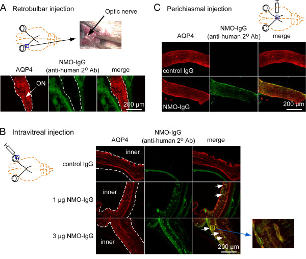
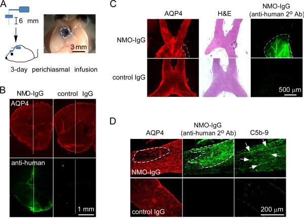
Methods: Several approaches were tested to develop a robust, passive-transfer mouse model of NMO ON, including NMO-IgG and complement delivery by: (i) retrobulbar infusion; (ii) intravitreal injection; (iii) a single intracranial injection near the optic chiasm; and (iv) 3-days continuous intracranial infusion near the optic chiasm.

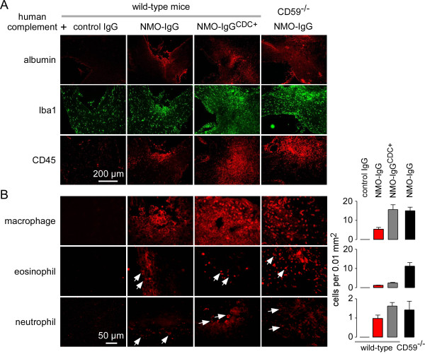
Results: Little ON or retinal pathology was seen using approaches (i) to (iii). Using approach (iv), however, optic nerves showed characteristic NMO pathology, with loss of AQP4 and glial fibrillary acidic protein immunoreactivity, granulocyte and macrophage infiltration, deposition of activated complement, demyelination and axonal injury. Even more extensive pathology was created in mice lacking complement inhibitor protein CD59, or using a genetically modified NMO-IgG with enhanced complement effector function, including significant loss of retinal ganglion cells. In control studies, optic nerve pathology was absent in treated AQP4-deficient mice, or in wild-type mice receiving control (non-NMO) IgG and complement.

Conclusion: Passive transfer of NMO-IgG and complement by continuous infusion near the optic chiasm in mice is sufficient to produce ON with characteristic NMO pathology. The mouse model of NMO ON should be useful in further studies of NMO pathogenesis mechanisms and therapeutics.


Figures

Figure 1
Figure 1
Passive transfer of neuromyelitis optica immunoglobulin G antibody and complement via retrobulbar infusion, intravitreal injection or single perichiasmal injection does not produce optic neuritis. (A) (Top) Lateral canthotomy was done in mice to expose the optic nerve (arrow). (Bottom) Absence of binding of neuromyelitis optica immunoglobulin G antibody (NMO-IgG) (as seen using an anti-human secondary antibody) to aquaporin-4 (AQP4) in the optic nerve (dashed line) 3 days after retrobulbar infusion of NMO-IgG and human complement (representative of three eyes from separate mice). (B) Binding of NMO-IgG to AQP4 in retina (dashed line) after intravitreal injection. No binding was seen with a control (non-NMO) IgG (n = 5 eyes). (Insert) Magnified view of NMO-IgG binding to AQP4 at perivascular end-feet of inner retinal Müller cells. (C) Binding of NMO-IgG (and not of control IgG) to AQP4 after single perichiasmal injection. Despite efficient binding of NMO-IgG to AQP4 in (B) and (C), no pathology was observed after 3 days (n = 5 eyes).
Figure 2
Figure 2
Continuous perichiasmal infusion of neuromyelitis optica immunoglobulin G antibody and complement leads to aquaporin-4 loss and complement activation in optic nerve. (A) (Left) Three-day continuous intracranial perichiasmal infusion of neuromyelitis optica immunoglobulin G antibody (NMO-IgG) and human complement was done by implantation of an osmostic pump. (Right) Area of diffusion in optic chiasm and brain of an infused blue dye (dashed line). (B) Immunofluorescence showing binding of NMO-IgG to aquaporin-4 (AQP4) in brain around the needle tract (white line) at 3-days after infusion of NMO-IgG or control IgG and complement. (C) AQP4 immunofluorescence, hematoxylin and eosin (H&E) staining and NMO-IgG immunofluorescence of the optic nerve. Dashed lines show lesion or area of NMO-IgG deposition. (D) Immunofluorescence showing AQP4 loss, NMO-IgG deposition and complement activation (C5b-9, arrows) in the optic nerve after infusion of NMO-IgG as in (A). White dashed lines demarcate the lesion.
Figure 3
Figure 3
Continuous perichiasmal infusion of neuromyelitis optica immunoglobulin G antibody and complement produces complement-dependent, neuromyelitis optica-like optic neuritis. (A) (Top) Micrographs showing aquaporin-4 (AQP4), glial fibrillary acidic protein (GFAP), myelin basic protein (MBP) and neurofilament (NF) immunoreactivity in the optic nerve after 3-days continuous perichiasmal infusion of complement with neuromyelitis optica immunoglobulin G antibody (NMO-IgG) or control IgG in wild-type mice and NMO-IgG in AQP4-/- mice. White dashed line demarcates area with loss of immunofluorescence. (Bottom) Higher magnification of yellow boxes. (B) (Top) AQP4, GFAP, MBP and NF immunofluorescence in the optic nerve after 3-days continuous perichiasmal infusion of (left) NMO-IgGCDC+ with or without complement in wild-type mice or (right) complement with control IgG or NMO-IgG in CD59-/- mice. White dashed line demarcates region with loss of immunofluorescence. (Bottom) Higher magnification of yellow boxes. (C) Hematoxylin and eosin staining of the optic nerve after 3-days continuous perichiasmal infusion in wild-type mice of control IgG or NMO-IgG or NMO-IgGCDC+, with or without complement. HC, human complement.
Figure 4
Figure 4
Inflammation in optic nerve lesions. (A) Immunostaining for albumin, ionized calcium-binding adaptor molecule-1 (Iba1); microglia and CD45 (leukocytes) in the optic nerve after 3-days continuous perichiasmal infusion of neuromyelitis optica immunoglobulin G antibody (NMO-IgG) and complement. (B) (Left) Immunostaining with markers for macrophages, neutrophils and eosinophils. (Right) Number of infiltrating cells of each per 0.01 mm2 (mean ± SEM, n = 3).
Figure 5
Figure 5
Continuous perichiasmal infusion of neuromyelitis optica immunoglobulin G antibody and complement produces retinal ganglion cells loss. (A) Retrograde labeling of retinal ganglion cells (RGCs) by injection of FluoroGold in the superior colliculus. After 7 days, neuromyelitis optica immunoglobulin G antibody (NMO-IgG) and complement were infused near the optic chiasm by minipump. Retinas were collected 7 days later. (B) Micrographs showing RGC loss. (C) Quantification of RGC number per 0.06 mm2 microscope field (mean ± SEM, n = 4, *P < 0.001). AQP4, aquaporin-4; HC, human complement; (ON, optic nerve).

Similar articles

Cited by

References

    1. Jarius S, Paul F, Franciotta D, Waters P, Zipp F, Hohlfeld R, Vincent A, Wildemann B. Mechanisms of disease: aquaporin-4 antibodies in neuromyelitis optica. Nat Clin Pract Neurol. 2008;4:202–214. - PubMed
    1. Jacob A, McKeon A, Nakashima I, Sato DK, Elsone L, Fujihara K, de Seze J. Current concept of neuromyelitis optica (NMO) and NMO spectrum disorders. J Neurol Neurosurg Psychiatry. 2013;84:922–930. doi: 10.1136/jnnp-2012-302310. - DOI - PubMed
    1. Wingerchuk DM, Lennon VA, Lucchinetti CF, Pittock SJ, Weinshenker BG. The spectrum of neuromyelitis optica. Lancet Neurol. 2007;6:805–815. doi: 10.1016/S1474-4422(07)70216-8. - DOI - PubMed
    1. Jarius S, Wildemann B. AQP4 antibodies in neuromyelitis optica: diagnostic and pathogenetic relevance. Nat Rev Neurol. 2010;6:383–392. doi: 10.1038/nrneurol.2010.72. - DOI - PubMed
    1. Lennon VA, Kryzer TJ, Pittock SJ, Verkman AS, Hinson SR. IgG marker of optic-spinal multiple sclerosis binds to the aquaporin-4 water channel. J Exp Med. 2005;202:473–477. doi: 10.1084/jem.20050304. - DOI - PMC - PubMed

Publication types

MeSH terms