Review
. 2014 Sep:38:105-16.
doi: 10.1016/j.yebeh.2013.12.022. Epub 2014 Jan 24.

Hippocampal granule cell pathology in epilepsy - a possible structural basis for comorbidities of epilepsy?

Affiliations
Review

Hippocampal granule cell pathology in epilepsy - a possible structural basis for comorbidities of epilepsy?

Michael S Hester et al. Epilepsy Behav. 2014 Sep.

Abstract

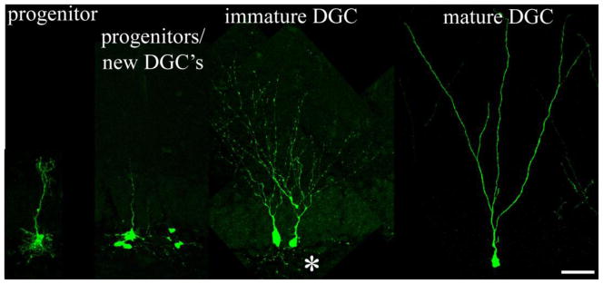
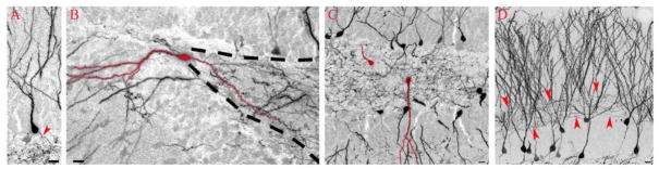
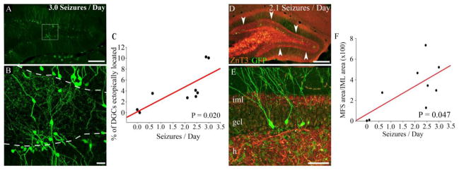
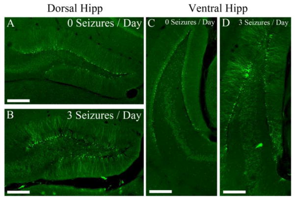
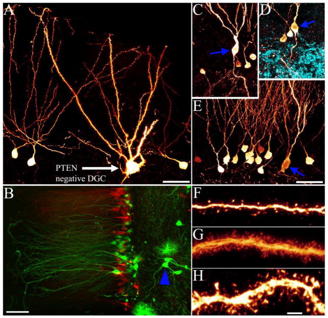
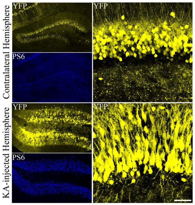
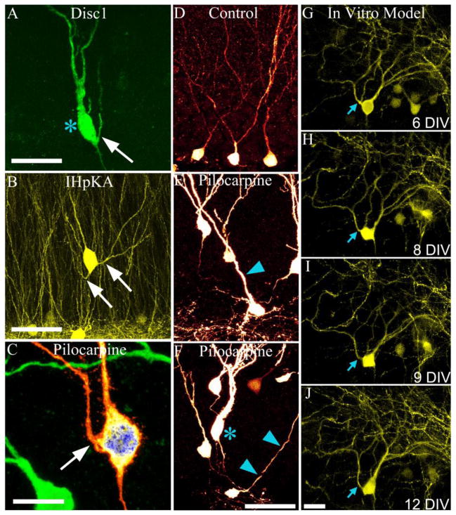
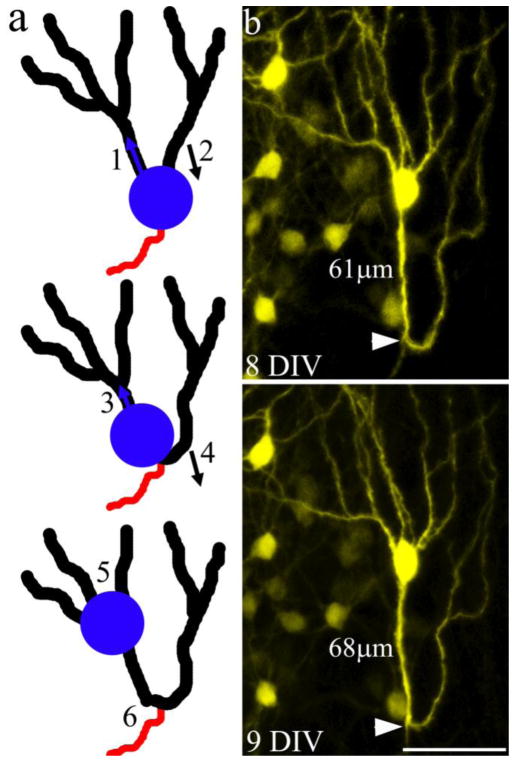
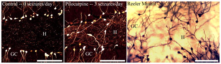
Temporal lobe epilepsy in both animals and humans is characterized by abnormally integrated hippocampal dentate granule cells. Among other abnormalities, these cells make axonal connections with inappropriate targets, grow dendrites in the wrong direction, and migrate to ectopic locations. These changes promote the formation of recurrent excitatory circuits, leading to the appealing hypothesis that these abnormal cells may by epileptogenic. While this hypothesis has been the subject of intense study, less attention has been paid to the possibility that abnormal granule cells in the epileptic brain may also contribute to comorbidities associated with the disease. Epilepsy is associated with a variety of general findings, such as memory disturbances and cognitive dysfunction, and is often comorbid with a number of other conditions, including schizophrenia and autism. Interestingly, recent studies implicate disruption of common genes and gene pathways in all three diseases. Moreover, while neuropsychiatric conditions are associated with changes in a variety of brain regions, granule cell abnormalities in temporal lobe epilepsy appear to be phenocopies of granule cell deficits produced by genetic mouse models of autism and schizophrenia, suggesting that granule cell dysmorphogenesis may be a common factor uniting these seemingly diverse diseases. Disruption of common signaling pathways regulating granule cell neurogenesis may begin to provide mechanistic insight into the cooccurrence of temporal lobe epilepsy and cognitive and behavioral disorders.

Keywords: Autism; Disc1; Epilepsy; Neurogenesis; Reelin; Schizophrenia; mTOR.


Conflict of interest statement

Conflict of Interests: The authors declare no competing financial interests

Figures

Figure 1
Figure 1
Photomontage of confocal maximum projections showing granule cell progenitors and granule cells at different maturational stages. To label granule cell progenitors, Gli1-CreERT2 mice were crossed with CAG-CAT-EGFP reporter mice. Gli1, a member of the sonic hedgehog family, drives Cre recombinase expression in granule cell progenitors. Activation of Cre recombinase by treating postnatal animals with tamoxifen leads to the persistent expression of green fluorescent protein (GFP) in recombined cells. Animals were killed and immunostained for GFP one week (progenitors), two weeks (immature), and two months (mature) after tamoxifen treatment. Mice were donated courtesy of Dr. Alexandra Joyner (Gli1-CreERT2; [207, 208]) and Dr. Jeffrey Robbins (CAG-CAT-EGFP reporter mice; [209]). Asterisk denotes basal dendrites on immature granule cells. Scale bar = 30 μm. Figure reprinted with permission from Danzer, 2008 [179] (Copyright © 2008 Sage Publications).
Figure 2
Figure 2
Normal and abnormal hippocampal dentate granule cells of pilocarpine-treated Gli1-CreERT2 x GFP-reporter mice. Typical granule cell morphology (A) consists of the cell body in the granule cell layer, apical dendrites extending towards the molecular layers, and a basal axon in the hilus (arrowhead). Common structural abnormalities develop in granule cells following status epilepticus, including cells with hilar basal dendrites (B), ectopically migrated cells (C), and axonal sprouting in the inner molecular layer (D, arrowheads). Scale bars = 10 μm.
Figure 3
Figure 3
Ectopically located newborn granule cells and the amount of mossy fiber sprouting correlate with seizure frequency. Newborn granule cells in the dorsal hippocampus of pilocarpine-treated Gli1-CreERT2 x GFP-reporter mice often migrate ectopically into the hilus (A,B). The percentage of newborn granule cells located in the hilus positively correlates to seizure frequency (C). Zinc transporter 3 (ZnT3) and GFP labeling show robust mossy fiber sprouting in the inner molecular layer (D,E). Arrowheads (D) highlight sprouted granule cell mossy fiber axons. The amount of mossy fiber sprouting positively correlates to seizure frequency (F). iml, inner molecular layer; gcl, granule cell layer; h, hilus. Scale bars: A and D 250 μm; B 20 μm; E 50 μm. Figure reprinted with permission from Hester and Danzer, 2013 [64].
Figure 4
Figure 4
Dorsal and ventral hippocampus are affected similarly following status epilepticus. Ectopic granule cells are largely absent from a Gli1-CreERT2 x GFP-reporter mouse that did not develop epilepsy following pilocarpine treatment (A,C) while an animal experiencing 3 seizures per day (B,D) exhibited frequent ectopic granule cells. Within animals, the number of abnormal granule cells is similar in dorsal (top) and ventral (bottom) hippocampus. Scale bars = 250 μm.
Figure 5
Figure 5
Hippocampal dentate granule cells from PTEN knockout mice (A,B,G) and mice rendered epileptic by inducing status epilepticus with pilocarpine (C–E, H). Note the hypertrophied soma of the PTEN knockout cell (A) relative to the hypertrophied somata of the granule cells from pilocarpine treated mice (C–E, blue arrowheads). PTEN deletion can also alter granule cell migration, leading to the appearance of ectopic cells in the dentate hilus (B, blue arrowhead; compare to figure 2, C for the pilocarpine model). Both PTEN deletion (G) and pilocarpine treatment (H) can lead to increases in dendritic spine density relative to control cells (F). Note: pilocarpine treatment can lead to increased spine density on some cells, and reduced density on others [39]. Scale bars = 30 μm (A), 100μm (B), 30 μm (C–E) and 10 μm (F–H). Panels C–E reprinted with permission from Murphy et al., 2011 [35]. Panels A, F and G reprinted with permission from Pun et al., 2012 [110].
Figure 6
Figure 6
Unilateral injection of kainic acid into the hippocampus increases phosphorylation of the downstream mTOR target S6 (blue) in the injected hemisphere, but not the contralateral (non-injected) hemisphere of thy1-YFP expressing (yellow) mice. Increased activation of the mTOR pathway is associated with a profound neuronal hypertrophy on the injected side. Scale bar = 40μm.
Figure 7
Figure 7
Disc1 knockout and epileptic mice exhibit similar granule cell abnormalities. A: Disc1 knockout granule cell with an elongated, enlarged soma (blue asterisk) and a recurrent basal dendrite (white arrow; used with permission from Duan et al., 2007 [129]). B: YFP-expressing ectopic granule cells in the dentate molecular layer with recurrent basal dendrites (white arrows) in the intrahippocampal kainic acid (IHpKA) model of epilepsy. C: GFP-expressing granule cell with a recurrent basal dendrite (white arrow) in the pilocarpine model of epilepsy. D–F: GFP-expressing control granule cells (D) and granule cells with misoriented dendrites from animals treated with pilocarpine (E,F, blue arrowheads). A granule cell with an enlarged and elongated soma (blue asterisk) is also shown. G–J: Time-lapse imaging of a granule cell in an organotypic hippocampal explant culture, which models epileptogenesis in vitro. Images over 6 days in vitro (DIV) show elongation of the soma and the shifting of an apical dendrite to the basal pole of the cell, creating a recurrent basal dendrite (blue arrows) (Images used with permission from Murphy and Danzer, 2011 [135]). Scale bars = 30 μm (A), 30 μm (B), 15 μm (C), 30 μm (D–F), 25 μm (G–J).
Figure 8
Figure 8
Model depicting granule cell dysmorphogenesis following somatic translocation into an apical dendrite. (a) As the soma moves into the initial segment of the left apical dendrite (1), the right apical dendrite is “left behind”, gradually shifting its position from the apical to the basal pole of the cell (2). Continuation of this process leads to a shortening of the initial dendritic segment on the left (3), and conversion of the right apical dendrite into a recurrent basal dendrite (4). In extreme cases, further migration of the soma leads to the absorption of branch points, converting these structures into primary dendrites (5), and the formation of dendritic loops projecting towards the hilus (6). (b) An example of a granule cell exhibiting a recurrent basal dendrite with a pronounced dendritic loop and an unusually large number of apical dendrites; the putative end product of the processes outlined in a. Note that the axon (white arrowhead) projects off the base of the dendritic loop, suggesting that this was the original location of the cell body. Scale bar = 50 μm. Images used with permission from Murphy and Danzer, 2011 [135].
Figure 9
Figure 9
Reeler mutant mice and epileptic mice exhibit similar granule cell abnormalities. Left panel: GFP-expressing granule cells from a Gli1-CreERT2::GFP reporter mouse that exhibited no epileptic seizures (control). Note the normal organization of the granule cell blades (GC) and the absence of ectopic cells in the hilus (H). Middle panel: Gli1-CreERT2::GFP reporter mouse treated with pilocarpine to induce epilepsy. In this animal, the granule cell layer is disorganized and numerous labeled cells are present in the hilus. Right panel: Golgi staining of granule cells in a reeler mutant mouse (used with permission from Drakew et al., 2002 [151]). Similar to the pilocarpine treated animal, the granule cell layer is disorganized and numerous ectopic cells are present in the hilus. Scale bars = 100 μm.

Similar articles

Cited by

References

    1. Bakker A, Kirwan CB, Miller M, Stark CE. Pattern separation in the human hippocampal CA3 and dentate gyrus. Science. 2008;319:1640–2. - PMC - PubMed
    1. Eichenbaum H, Sauvage M, Fortin N, Komorowski R, Lipton P. Towards a functional organization of episodic memory in the medial temporal lobe. Neurosci Biobehav Rev. 2012;36:1597–608. - PMC - PubMed
    1. Gilbert PE, Kesner RP, Lee I. Dissociating hippocampal subregions: double dissociation between dentate gyrus and CA1. Hippocampus. 2001;11:626–36. - PubMed
    1. Sahay A, Wilson DA, Hen R. Pattern separation: a common function for new neurons in hippocampus and olfactory bulb. Neuron. 2011;70:582–8. - PMC - PubMed
    1. Schacter DL, Addis DR, Buckner RL. Remembering the past to imagine the future: the prospective brain. Nat Rev Neurosci. 2007;8:657–61. - PubMed

Publication types