

The panorama of miRNA-mediated mechanisms in mammalian cells
- PMID: 24468964
- PMCID: PMC4031385
- DOI: 10.1007/s00018-013-1551-6
The panorama of miRNA-mediated mechanisms in mammalian cells
Abstract
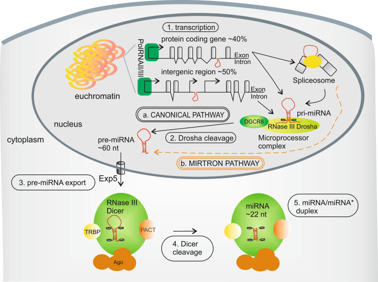
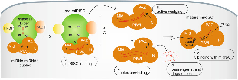
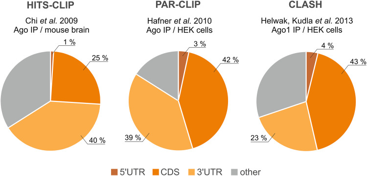
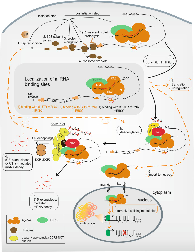
MicroRNAs comprise a large family of short, non-coding RNAs that are present in most eukaryotic organisms and are typically involved in downregulating the expression of protein-coding genes. The detailed mechanisms of miRNA functioning in animals and plants have been under investigation for more than decade. In mammalian cells, miRNA guides the effector complex miRISC to bind with partially complementary sequences, usually within the 3'UTR of mRNAs, and inhibit protein synthesis with or without transcript degradation. In addition to these main mechanisms, several other modes of miRNA-mediated gene expression regulation have been described, but their scale and importance remain a matter of debate. In this review, we briefly summarize the pathway of miRNA precursor processing during miRNA biogenesis and continue with the description of the miRISC assembly process. Then, we present the miRNA-mediated mechanisms of gene expression regulation in detail, and we gather information concerning the proteins involved in these processes. In addition, we briefly refer to the current applications of miRNA mechanisms in therapeutic strategies. Finally, we highlight some of the remaining controversies surrounding the regulation of mammalian gene expression by miRNAs.
Conflict of interest statement
The authors declare no conflict of interests.
Figures
Similar articles
-
Regulation of the MIR155 host gene in physiological and pathological processes.Gene. 2013 Dec 10;532(1):1-12. doi: 10.1016/j.gene.2012.12.009. Epub 2012 Dec 14. Gene. 2013. PMID: 23246696 Review.
-
RIP-Chip analysis supports different roles for AGO2 and GW182 proteins in recruiting and processing microRNA targets.BMC Bioinformatics. 2019 Apr 18;20(Suppl 4):120. doi: 10.1186/s12859-019-2683-y. BMC Bioinformatics. 2019. PMID: 30999843 Free PMC article.
-
Gene silencing in vitro and in vivo using intronic microRNAs.Methods Mol Biol. 2015;1218:321-40. doi: 10.1007/978-1-4939-1538-5_20. Methods Mol Biol. 2015. PMID: 25319661
-
Functional aspects of animal microRNAs.Cell Mol Life Sci. 2008 Feb;65(4):545-62. doi: 10.1007/s00018-007-7355-9. Cell Mol Life Sci. 2008. PMID: 17965831 Free PMC article. Review.
-
Conditions of appreciable influence of microRNA on a large number of target mRNAs.Mol Biosyst. 2009 Jun;5(6):638-43. doi: 10.1039/b808095j. Epub 2009 Apr 20. Mol Biosyst. 2009. PMID: 19462021
Cited by
-
miR-155: An Important Role in Inflammation Response.J Immunol Res. 2022 Apr 6;2022:7437281. doi: 10.1155/2022/7437281. eCollection 2022. J Immunol Res. 2022. PMID: 35434143 Free PMC article. Review.
-
MicroRNAs tend to synergistically control expression of genes encoding extensively-expressed proteins in humans.PeerJ. 2017 Aug 14;5:e3682. doi: 10.7717/peerj.3682. eCollection 2017. PeerJ. 2017. PMID: 28828274 Free PMC article.
-
miRTarBase 2025: updates to the collection of experimentally validated microRNA-target interactions.Nucleic Acids Res. 2025 Jan 6;53(D1):D147-D156. doi: 10.1093/nar/gkae1072. Nucleic Acids Res. 2025. PMID: 39578692 Free PMC article.
-
Interactions between cancer cells and normal cells via miRNAs in extracellular vesicles.Cell Mol Life Sci. 2015 May;72(10):1849-61. doi: 10.1007/s00018-014-1811-0. Epub 2015 Jan 7. Cell Mol Life Sci. 2015. PMID: 25563488 Free PMC article. Review.
-
The Interplay between MicroRNAs and Cellular Components of Tumour Microenvironment (TME) on Non-Small-Cell Lung Cancer (NSCLC) Progression.J Immunol Res. 2019 Feb 13;2019:3046379. doi: 10.1155/2019/3046379. eCollection 2019. J Immunol Res. 2019. PMID: 30944831 Free PMC article. Review.
References
Publication types
MeSH terms
Substances
LinkOut - more resources
Full Text Sources
Other Literature Sources
