. 2015 Jan 22;34(4):506-15.
doi: 10.1038/onc.2013.579. Epub 2014 Jan 27.

Progesterone receptor-B enhances estrogen responsiveness of breast cancer cells via scaffolding PELP1- and estrogen receptor-containing transcription complexes

Affiliations

Progesterone receptor-B enhances estrogen responsiveness of breast cancer cells via scaffolding PELP1- and estrogen receptor-containing transcription complexes

A R Daniel et al. Oncogene. .

Abstract

Progesterone and estrogen are important drivers of breast cancer proliferation. Herein, we probed estrogen receptor-α (ER) and progesterone receptor (PR) cross-talk in breast cancer models. Stable expression of PR-B in PR-low/ER+ MCF7 cells increased cellular sensitivity to estradiol and insulin-like growth factor 1 (IGF1), as measured in growth assays performed in the absence of exogenous progestin; similar results were obtained in PR-null/ER+ T47D cells stably expressing PR-B. Genome-wide microarray analyses revealed that unliganded PR-B induced robust expression of a subset of estradiol-responsive ER target genes, including cathepsin-D (CTSD). Estradiol-treated MCF7 cells stably expressing PR-B exhibited enhanced ER Ser167 phosphorylation and recruitment of ER, PR and the proline-, glutamate- and leucine-rich protein 1 (PELP1) to an estrogen response element in the CTSD distal promoter; this complex co-immunoprecipitated with IGF1 receptor (IGFR1) in whole-cell lysates. Importantly, ER/PR/PELP1 complexes were also detected in human breast cancer samples. Inhibition of IGF1R or phosphoinositide 3-kinase blocked PR-B-dependent CTSD mRNA upregulation in response to estradiol. Similarly, inhibition of IGF1R or PR significantly reduced ER recruitment to the CTSD promoter. Stable knockdown of endogenous PR or onapristone treatment of multiple unmodified breast cancer cell lines blocked estradiol-mediated CTSD induction, inhibited growth in soft agar and partially restored tamoxifen sensitivity of resistant cells. Further, combination treatment of breast cancer cells with both onapristone and IGF1R tyrosine kinase inhibitor AEW541 was more effective than either agent alone. In summary, unliganded PR-B enhanced proliferative responses to estradiol and IGF1 via scaffolding of ER-α/PELP1/IGF1R-containing complexes. Our data provide a strong rationale for targeting PR in combination with ER and IGF1R in patients with luminal breast cancer.


Figures

Figure 1
Figure 1
PR-B expression increases breast cancer cell growth in response to estradiol. (A) Western blots of PR-B and ER-alpha. Unmodified MCF7 and MCF7 cells expressing pSG5 or pSG5-PR-B were treated with estradiol (1nM; E2) for 24 h. (B) Soft agar colony formation of MCF7 pSG5 or pSG5-PR-B cells grown in ethanol (EtOH), estradiol (E2), or synthetic progestin, R5020 (14 days). (C) Soft agar colony formation assay in cells expressing vector or PR-B. Cells were treated with EtOH, estradiol (1nM; E2), IGF1 (5nM), both estradiol and IGF1, or R5020 (10nM) for 21 days. (D–E) Soft agar colony formation of MCF7L and BT474 cells grown in ethanol, estradiol, or the anti-progestin onapristone (1uM; Ona) for 7 days (±SEM, *p<0.05). (F) Western blots of PR, ER, and Actin (loading control) from BT474 and MCF7-L cells were treated for 24 hours with estradiol (1nM) and/or onapristone (1uM).
Figure 2
Figure 2
Gene expression profiling identified novel PR-dependent, estradiol-regulated genes. (A) Heat map of normalized gene expression levels in MCF7 cells stably expressing vector or PR-B. Cells were treated with EtOH or estradiol (6h). Blue lines (to the right of the heat map) indicate upregulated genes and black lines (to the right of the heat map) indicate downregulated genes between lanes 2 and 4. The Venn diagram shows the number of genes upregulated in response to estradiol (>2 fold, P <0.001) in MCF7 cells expressing vector only or PR-B. (B, D) SLC7A5 and MAOA mRNA expression. qRT-PCR of SLC7A5 and MAOA in MCF7 cells expressing vector or PR-B and treated with estradiol (6h) (upper panels). Relative SLC7A5 and MAOA mRNA expression in normal breast tissue and invasive breast carcinoma from the TCGA database (lower panels). (C, E) qRT-PCR to examine WISP2, PTGES, LXN, and TMPRSS2 in MCF7 cells expressing vector or PR-B and treated with estrogen (6h) (±SD or SEM, *p<0.05).
Figure 3
Figure 3
PR-B expression is required for estradiol induction of CTSD. (A–B) qRT-PCR of CTSD, TFF1, and SGK in pSG5 or pSG5-PR-B MCF7 cells treated (6h) with the indicated hormone (ie, estradiol or R5020) (±SD, *p<0.05). (C) TCGA database analysis of relative CTSD mRNA expression in normal breast tissue and invasive breast carcinoma.
Figure 4
Figure 4
PR is required for ER occupancy on the CTSD promoter. (A) qRT-PCR of CTSD in pSG5 or pSG5-PR-B T47D cells treated with ethanol or estradiol (24h). (B) qRT-PCR of CTSD mRNA in T47D cells stably expressing pSG5-PR-B or pSG5-DBM PR-B and treated with estradiol (24h). (Inset) Western blot of PR-B and ER in T47D cells stably expressing PR-B or DBM PR-B. (±SEM, *p<0.05). (C) pSG5-PR-B MCF7 cells treated with vehicle or estradiol (1h) were subjected to ChIP assays to examine ER recruitment to estradiol responsive regions in the CTSD distal promoter. Chromatin associated with ER immunocomplexes was subjected to qPCR, normalized to inputs, and expressed as estradiol-induced fold change over ethanol treatment. IgG was used as a negative control. Fold change values from two independent experimental repeats are presented. (D) MCF7 pSG5-PR-B cells were treated with ethanol or estradiol (40min). ChIP assays were performed using ER (right) or PR (left) antibodies compared to IgG negative controls. qPCR values normalized to input controls are shown, data is representative of 4–8 independent experiments. (E) ChIP assays to examine ER recruitment to the CTSD distal promoter were performed in MCF7L cells treated with ethanol or estradiol. Estradiol-induced ER recruitment was normalized to inputs and is expressed as fold blockade by onapristone. Values represent the average of three independent experimental repeats. (F) MCF7L cells were treated with estradiol, onapristone, or both (24h). CTSD mRNA expression was evaluated using qRT-PCR and normalized to a housekeeper gene (±SEM, *p<0.05).
Figure 5
Figure 5
PR-B coordinates ER signaling and transcriptional complexes. (A) Western blots of pSG5 or pSG5-PR MCF7 cells treated with estradiol or IGF1 (1h). (B) MCF7 (PR-null/ER-null subline C4-12) cells transiently transfected with ER and PR-B were starved and treated with estradiol or IGF-1 (10min). PR-containing complexes were isolated using PR-specific antibodies and protein G-agarose beads. Western blots were performed on immunocomplexes and whole cell lysates (N.B.; non-specfic band). (C) ChIP assays were performed in MCF7 pSG5-PRB cells treated with vehicle or estradiol (40min). CTSD distal promoter chromatin from immunocomplexes isolated by using antibodies specific for ER, PR, PELP1, or IGF1R was evaluated with qRT-PCR and normalized to inputs. Values are expressed as estradiol-induced fold change over vehicle. Data represent the average of 3–5 independent experimental repeats. (D) MCF7 cells were treated with vehicle, AEW541, or LY-294002 and vehicle or estradiol (24h). Cells were subjected qRT-PCR to asses CTSD and TFF1 mRNA levels normalized to housekeeper genes. Values are expressed as the average estradiol-induced fold change over vehicle. Data represent the average of 3 independent experimental repeats. (Inset) Western blots of MCF7L cells treated with vehicle or estradiol and vehicle or AEW541 (1h). (E) ChIP assay of ER recruitment to the CTSD distal and TFF1 promoters in MCF7L cell treated with vehicle, estradiol, AEW541, or both estradiol and AEW541 for 40 min. Values, expressed as a percent of input controls, are representative of 3 independent experiments.
Figure 6
Figure 6
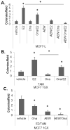
PR antagonist blocks estradiol-stimulated growth and sensitizes breast cancer cells to tamoxifen. (A) Soft agar colony formation of MCF7L cells grown in ethanol, estradiol, AEW, or onapristone (7 days). (B) Soft agar colony formation of MCF7 1GX cells treated with tamoxifen and either vehicle, estradiol, onapristone, or both (7 days). (C) Soft agar colony formation of MCF7 1GX cells treated with tamoxifen and estradiol and either vehicle, onapristone, AEW, or both (7 days) (±SEM, * p<0.05).
Figure 7
Figure 7
PR/ER/PELP1 containing complexes in human breast cancer samples. (A) Protein complexes were isolated from nine human tumor samples using PELP1-specific antibodies. Western blots were performed on immunocomplexes and input controls. Actin was used as a loading control. (B) PR-B is a component of a transcriptional complex where it coordinates estradiol-induced signaling to alter ER-mediated gene regulation.

Similar articles

Cited by

References

    1. Effects of chemotherapy and hormonal therapy for early breast cancer on recurrence and 15-year survival: an overview of the randomised trials. Lancet. 2005 May 14–20;365(9472):1687–717. - PubMed
    1. Davies C, Godwin J, Gray R, Clarke M, Cutter D, Darby S, et al. Relevance of breast cancer hormone receptors and other factors to the efficacy of adjuvant tamoxifen: patient-level meta-analysis of randomised trials. Lancet. 2011 Aug 27;378(9793):771–84. - PMC - PubMed
    1. Musgrove EA, Sutherland RL. Biological determinants of endocrine resistance in breast cancer. Nat Rev Cancer. 2009 Sep;9(9):631–43. - PubMed
    1. de Leeuw R, Neefjes J, Michalides R. A role for estrogen receptor phosphorylation in the resistance to tamoxifen. Int J Breast Cancer. 2011;2011:232435. - PMC - PubMed
    1. Vallabhaneni S, Nair BC, Cortez V, Challa R, Chakravarty D, Tekmal RR, et al. Significance of ER-Src axis in hormonal therapy resistance. Breast Cancer Res Treat. 2011 Nov;130(2):377–85. - PMC - PubMed

Publication types

MeSH terms