. 2015 Jan 29;34(5):546-57.
doi: 10.1038/onc.2013.582. Epub 2014 Jan 27.

Akt phosphorylates and activates HSF-1 independent of heat shock, leading to Slug overexpression and epithelial-mesenchymal transition (EMT) of HER2-overexpressing breast cancer cells

Affiliations

Akt phosphorylates and activates HSF-1 independent of heat shock, leading to Slug overexpression and epithelial-mesenchymal transition (EMT) of HER2-overexpressing breast cancer cells

R L Carpenter et al. Oncogene. .

Abstract

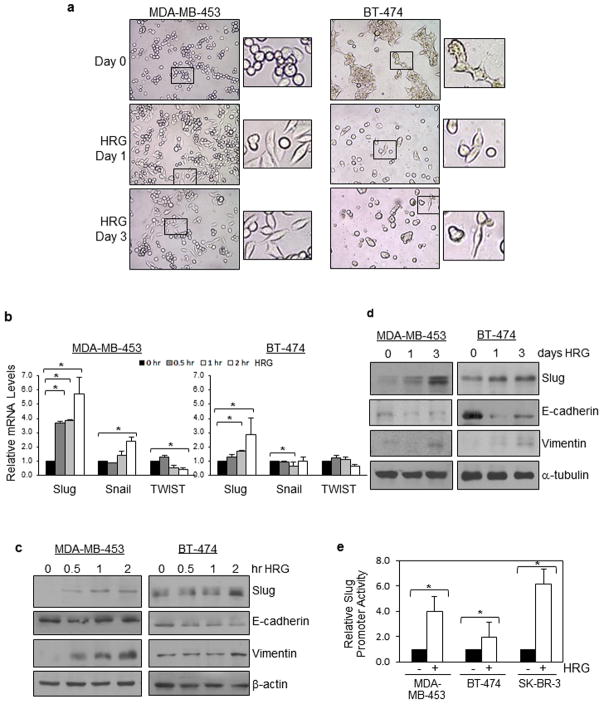
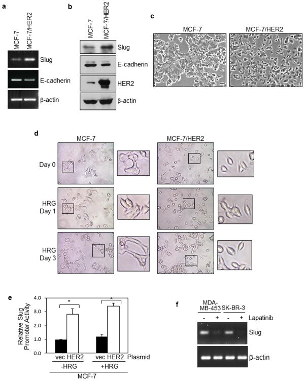
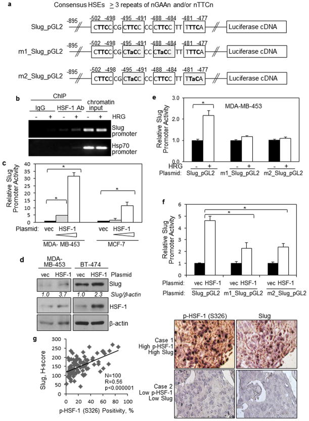
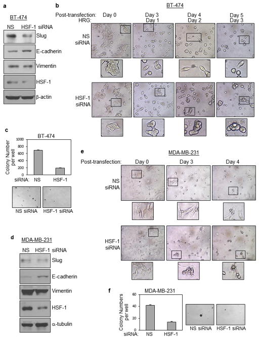
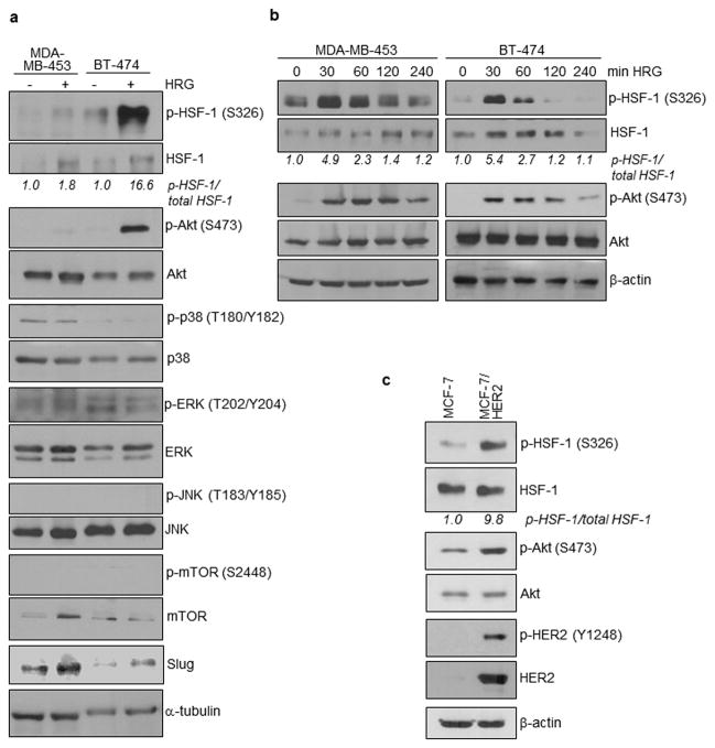
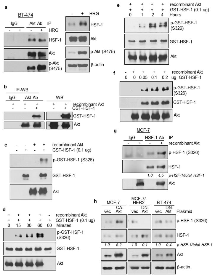
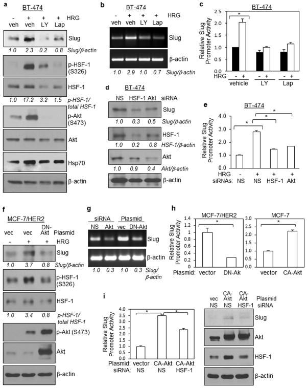
Epithelial-mesenchymal transition (EMT) is an essential step for tumor progression, although the mechanisms driving EMT are still not fully understood. In an effort to investigate these mechanisms, we observed that heregulin (HRG)-mediated activation of HER2, or HER2 overexpression, resulted in EMT, which is accompanied with increased expression of a known EMT regulator Slug, but not TWIST or Snail. We then investigated how HER2 induced Slug expression and found, for the first time, that there are four consensus HSF sequence-binding elements (HSEs), the binding sites for heat shock factor-1 (HSF-1), located in the Slug promoter. HSF-1 bound to and transactivated the Slug promoter independent of heat shock, leading to Slug expression in breast cancer cells. Mutation of the putative HSEs ablated Slug transcriptional activation induced by HRG or HSF-1 overexpression. Knockdown of HSF-1 expression by siRNA reduced Slug expression and HRG-induced EMT. The positive association between HSF-1 and Slug was confirmed by immunohistochemical staining of a cohort of 100 invasive breast carcinoma specimens. While investigating how HER2 activated HSF-1 independent of heat shock, we observed that HER2 activation resulted in concurrent phosphorylation of Akt and HSF-1. We then observed, also for the first time, that Akt directly interacted with HSF-1 and phosphorylated HSF-1 at S326. Inhibition of Akt using siRNA, dominant-negative Akt mutant, or small molecule inhibitors prevented HRG-induced HSF-1 activation and Slug expression. Conversely, constitutively active Akt induced HSF-1 phosphorylation and Slug expression. HSF-1 knockdown reduced the ability of Akt to induce Slug expression, indicating an essential role that HSF-1 plays in Akt-induced Slug upregulation. Altogether, our study uncovered the existence of a novel Akt-HSF-1 signaling axis that leads to Slug upregulation and EMT, and potentially contributes to progression of HER2-positive breast cancer.


Conflict of interest statement

Conflicts of Interest

The authors declare no conflicts of interest.

Figures

Figure 1
Figure 1. Heregulin induces EMT and Slug expression in HER2-amplified breast cancer cells
(a) Heregulin (HRG) induced EMT of HER2-amplified breast cancer cells. MDA-MB-453 and BT-474 were serum-starved overnight (day 0) and then treated with heregulin (100 ng/ml) for 1 or 3 days. Cultured cells were imaged using a phase-contract microscope. Representative images are shown. (b) Slug transcripts were induced by heregulin in HER2-amplified breast cancer cells. MDA-MB-453 and BT-474 were starved from serum overnight, treated with heregulin (100 ng/ml) for 0–120 min, and harvested for total RNA extraction and RT-qPCR. Levels of EMT regulators, Slug, Snail and TWIST were determined. * indicates p-values < 0.05. (c) (d) Slug protein expression was induced by heregulin in HER2-amplified breast cancer cells. MDA-MB-453 and BT-474 were starved from serum overnight, treated with heregulin (100 ng/ml) for 0–120 min (c) or for 1–3 days (d), and harvested for protein extraction and WB. Levels of Slug, E-cadherin (epithelial marker), and Vimentin (mesenchymal marker) were analyzed. (e) Slug promoter was significantly activated by heregulin in breast cancer cells with HER2 amplification. MDA-MB-453, BT-474 and SK-BR-3 cells were transfected with a firefly luciferase reporter under the control of the human Slug promoter, serum-starved for 16 hrs and treated with heregulin (100 ng/ml) for 2 hrs. Treated cells were lysed and subjected to luciferase assay. All cells were co-transfected with the Renilla luciferase expression vector, pRL-CMV, to control for transfection efficiency. The results were derived from at least three experiments. The student t-test was conducted to compute p-values. * indicates p-values < 0.05.
Figure 2
Figure 2. HER2 overexpression enhances Slug expression, leading to EMT in breast cancer cells
(a)(b) Ectopic HER2 expression induced Slug expression in breast cancer cells. MCF-7 (with normal HER2 expression level) and MCF-7/HER2 (MCF-7 cells stably expressing ectopic HER2) were analyzed for Slug and E-cadherin expression using RT-PCR (a) and WB (b). Enhanced HER2 expression in MCF-7/HER2 cells is indicated by WB. (c)(d) Ectopic HER2 expression induced EMT-like morphology changes in breast cancer cells. In panel c, both cell lines were cultured in normal growth condition with fetal calf serum. In panel d, cells were serum-starved and treated with heregulin (100 ng/ml) for 0–3 days. Representative images are shown. (e) Ectopic HER2 expression induced Slug promoter activity in breast cancer cells. MCF-7 cells (with normal HER2 levels) were transiently transfected with HER2 and the Slug luciferase reporter, serum-starved, and then stimulated with heregulin (100 ng/ml) for 2 hrs. All cells were co-transfected with the Renilla luciferase expression vector, pRL-CMV, to control for transfection efficiency. The results were derived from at least three experiments, and analyzed by the student t-test to compute p-values. * indicates p-values < 0.05. (f) Lapatinib, a small molecule HER2/EGFR inhibitor reduced Slug expression in HER2-amplified MDA-MB-453 and SK-BR-3 cells. Cells were pre-treated with lapatinib (5 uM) for 24 hrs and subjected to total RNA extraction and RT-PCR for Slug transcript levels.
Figure 3
Figure 3. HSF-1 binds to and transactivates the Slug gene promoter, leading to Slug expression in breast cancer cells
(a) Identification of four putative HSEs within the human Slug gene promoter. TFSearch, a web-based search engine for transcription factor-binding sites, was used to search for HSEs within the Slug promoter. Consensus HSEs are shown on the top. Structures of the wild-type Slug promoter reporter and two mutant reporters are shown. Each of the two mutant promoters contains mutations at two of the four putative sites, in order to destroy the three repeats required for binding to HSF-1 trimers. Clear boxes mark the putative HSEs. Lower case letters indicate mutated bases. (b) HSF-1 binds to the Slug promoter and the binding was enhanced by heregulin. Serum-starved BT-474 cells treated with and without heregulin (100 ng/ml) were used in the ChIP assay. HSF-1 antibody (Ab) was used to immunoprecipitate HSF-1 while IgG served as the negative controls. Chromatin input was used to control for loading. PCR was conducted to detect HSF-1 binding to Slug promoter and a known HSF-1 target gene, Hsp70. (c) Ectopic HSF-1 expression significantly induced Slug promoter activity. Cells were transfected with the control vector or the HSF-1 vector, and the Slug luciferase reporter for 48 hrs and subjected to luciferase assay. All cells were co-transfected with the Renilla luciferase expression vector, pRL-CMV, to control for transfection efficiency. The results represent means and standard deviations from at least three experiments, and were analyzed by the student t-test to compute p-values. * indicates p-values < 0.05. (d) Ectopic HSF-1 expression enhanced Slug expression. BT-474 and MDA-MB-453 cells transfected with the control vector or the HSF-1 vector were analyzed by WB to determine Slug and HSF-1 expression levels. (e)(f) Identified HSEs are important for heregulin- and HSF-1-mediated induction of Slug promoter activation. MDA-MB-453 cells transfected with the wild-type and mutant slug reporters were serum-starved and treated with heregulin for 2 hrs, and then subjected to luciferase assay. All cells were co-transfected with the Renilla luciferase reporter, pRL-CMV, to control for transfection efficiency. The results were derived from at least three experiments, and analyzed by the student t-test to compute p-values. * indicates p-values < 0.05. (g) Levels of p-HSF-1 (S326) were directly associated with those of Slug in invasive breast carcinoma specimens. IHC was conducted to analyze 100 invasive carcinomas. Linear regression was used to compute R and p values (R=0.56, p<0.000001). Right, representative images.
Figure 4
Figure 4. HSF-1 expression knockdown prevents heregulin-induced EMT and suppresses growth of breast cancer cells
(a) HSF-1 siRNA reduced expression of Slug in epithelial BT-474 cells. BT-474 cells transfected with non-specific (NS) siRNA or Slug siRNA were analyzed by WB. (b) HSF-1 knockdown has in part prevented heregulin-induced EMT. BT-474 cells transfected NS siRNA or Slug siRNA were serum-starved and treated with heregulin (100 ng/ml) for 0–3 days. Representative images are shown. (c) HSF-1 siRNA reduced the propensity of BT-474 cells to grow in an anchorage-independent fashion. BT-474 cells transfected NS siRNA or Slug siRNA were seeded into 6-well culture plates with agarose (2000 cells/well). After colonies were formed to the appropriate size, colonies were counted under a microscope. Data represent means and standard deviations of three independent experiments. The student t-test was performed to calculate p-values. * indicates p-values < 0.05. (d) HSF-1 siRNA reduced Slug expression in mesenchymal MDA-MB-231 cells. MDA-MB-231 cells transfected with NS siRNA or Slug siRNA were examined by WB. (e) HSF-1 knockdown did not result in MET of mesenchymal, post-EMT MDA-MB-231 cells, but induced significant cell death. MDA-MB-231 cells transfected NS siRNA or Slug siRNA imaged for 0–4 days post transfections. Representative images are shown. (f) HSF-1 siRNA reduced the ability of MDA-MB-231 cells to grow in an anchorage-independent fashion. MDA-MB-231 cells transfected NS siRNA or Slug siRNA were seeded into 6-well culture plates with agarose (2000 cells/well). After colonies have formed to the appropriate size, colonies were counted. Results represent means and standard deviations of three independent experiments, and were analyzed by the student t-test. * indicates p-values < 0.05.
Figure 5
Figure 5. Concurrent activation of Akt and HSF-1 by heregulin/HER2 in breast cancer cells
(a) Heregulin induced phosphorylation of both HSF-1 and Akt in HER2-amplified breast cancer cell lines. MDA-MB-453 and BT-474 cells were treated with and without heregulin (100 ng/ml) for 2 hrs and the whole cell lysates were analyzed by WB for levels of HER2 downstream kinases and Slug. (b) Kinetics for HSF-1 activation is in concordance with that for Akt. The two cell lines were treated with heregulin for 0–240 min and the whole cell lysates were analyzed by WB to determine levels of p-HSF-1 (S326) and p-Akt (S473). (c) Ectopic HER2 expression led to increased activation of both HSF-1 and Akt. MCF-7 and MCF-7/HER2 cell lines were examined using WB.
Figure 6
Figure 6. Akt directly interacts with and phosphorylates HSF-1 at S326
(a) Akt interacts with HSF-1 constitutively, independent of heregulin treatment. IP-WB was conducted using whole cell lysates extracted from BT-474 cells treated with and without heregulin. An Akt Ab was used to immunoprecipitate Akt whereas IgG was used as negative controls. WB results are shown in the right panel. (b) Recombinant Akt directly interacts with recombinant HSF-1. IP-WB was conducted. (c) Recombinant Akt directly phosphorylates recombinant HSF-1 protein at S326. Cell-free Akt kinase assay was conducted followed by WB. (d,e) HSF-1 protein was phosphorylated by Akt in a time-dependent fashion. (f) HSF-1 protein was phosphorylated by Akt in a dose-dependent fashion. (g) Cellular HSF-1 is directly phosphorylated by Akt at S326. HSF-1 immunoprecipitated from MCF-7 cells was subjected to the cell-free Akt kinase assay followed by WB. (h) Ectopic expression of constitutively activated Akt (CA-Akt) significantly enhanced HSF-1 phosphorylation, whereas ectopic expression of dominant-negative Akt (DN-Akt) substantially reduced HSF-1 phosphorylation. Transfected cells were lysed and subjected to WB.
Figure 7
Figure 7. Slug expression is suppressed by blocking the HER2-Akt-HSF-1 signaling axis; HSF-1 is essential for Akt-induced Slug expression
In luciferase assays (panels b, d and e), three independent experiments were performed to derive means and standard deviations. All cells were co-transfected with the Renilla luciferase reporter, pRL-CMV, to control for transfection efficiency. The results were analyzed by the student t-test to compute p-values. * indicates p-values < 0.05. (a)(b) Small molecule inhibitors to PI3K/Akt (LY294002; LY; 50 uM) and HER2 (Lapatinib; Lap; 25 uM) pre-treatment for 1 2 hrs suppressed heregulin-induced Slug expression in BT-474 cells. In panel a, WB was conducted. Hsp70 served as positive controls for HSF-1 activity. Heregulin exposure was for 1 hr at 100 ng/ml. In panel b, total RNA was analyzed by RT-PCR. Veh, vehicle (1% DMSO) (c) Both LY294002 and Lapatinib blocked heregulin induction of Slug promoter activity. BT-474 cells transfected with the Slug luciferase reporter were serum-starved, treated with vehicle or indicated inhibitor (50 uM LY294002 or 25 uM Lapatinib) for 2 hrs, and then treated with and without heregulin for 4 hrs. Harvested cells were lysed and subjected to luciferase assay. (d) HSF-1 and Akt siRNAs reduced Slug expression in BT-474 cells, as shown by WB. (e) HSF-1 and Akt siRNAs prevented heregulin-induced activation of Slug promoter in BT-474 cells. BT-474 cells co-transfected with siRNA and the Slug luciferase reporter were serum-starved and treated with heregulin for 4 hrs, and then subjected to luciferase assay. (f) DN-Akt blocked heregulin-induced Slug expression and HSF-1 activation in MCF-7/HER2 cells, as shown by WB. (g) Akt siRNA and DN-Akt inhibited Slug transcription in BT-474 cells as shown by RT-PCR. (h) DN-Akt reduced activity of the Slug promoter in MCF-7/HER2 cells while CA-Akt enhanced its activity in MCF-7 cells. Luciferase assay was conducted to measure Slug promoter activity. (i) HSF-1 is essential for CA-Akt-induced Slug expression. MCF-7 cells were used. Left, luciferase assay. Right, WB.

Similar articles

Cited by

References

    1. Thiery JP, Acloque H, Huang RY, Nieto MA. Epithelial-mesenchymal transitions in development and disease. Cell. 2009;139:871–890. - PubMed
    1. De Craene B, Berx G. Regulatory networks defining EMT during cancer initiation and progression. Nat Rev Cancer. 2013;13:97–110. - PubMed
    1. Rhim AD, Mirek ET, Aiello NM, Maitra A, Bailey JM, McAllister F, et al. EMT and dissemination precede pancreatic tumor formation. Cell. 2012;148:349–361. - PMC - PubMed
    1. Trimboli AJ, Fukino K, de Bruin A, Wei G, Shen L, Tanner SM, et al. Direct evidence for epithelial-mesenchymal transitions in breast cancer. Cancer Res. 2008;68:937–945. - PubMed
    1. Hajra KM, Chen DY, Fearon ER. The SLUG zinc-finger protein represses E-cadherin in breast cancer. Cancer Res. 2002;62:1613–1618. - PubMed

Publication types

MeSH terms