. 2014 Aug;62(2):63-71.
doi: 10.1016/j.vph.2014.01.005. Epub 2014 Jan 25.

Low anticoagulant heparin blocks thrombin-induced endothelial permeability in a PAR-dependent manner

Affiliations

Low anticoagulant heparin blocks thrombin-induced endothelial permeability in a PAR-dependent manner

Joyce N Gonzales et al. Vascul Pharmacol. 2014 Aug.

Abstract

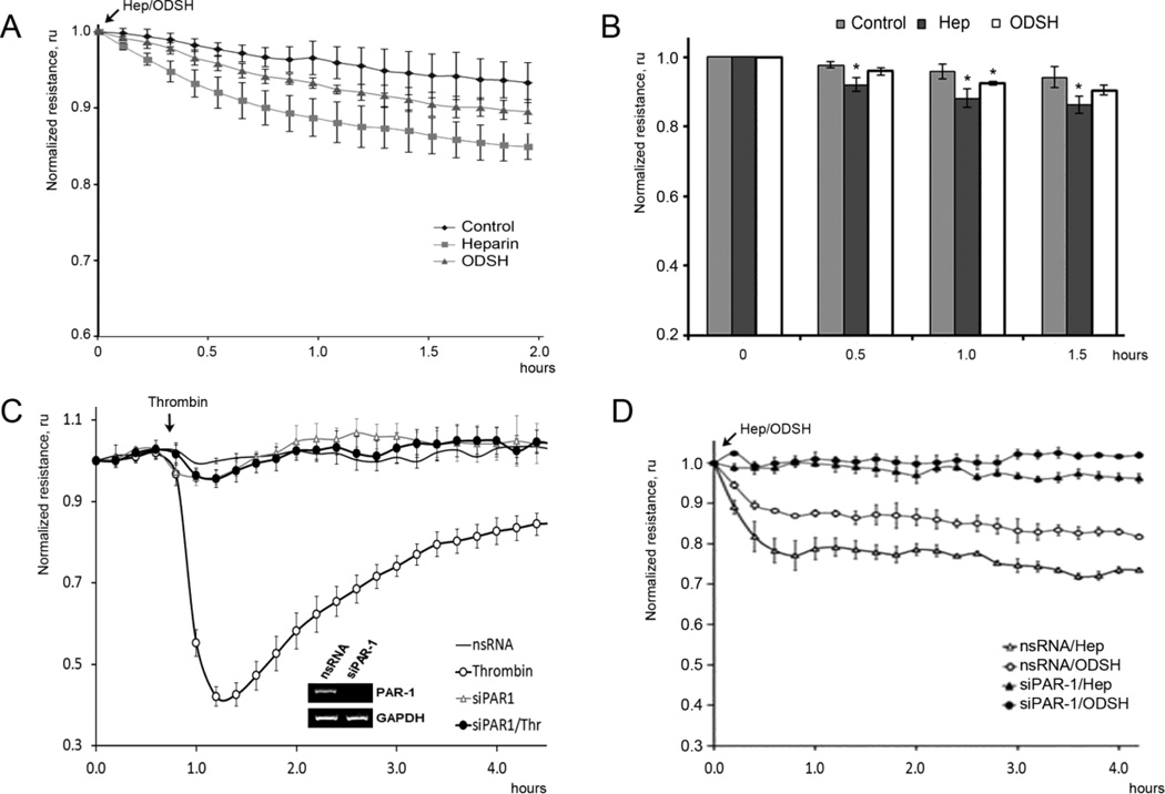
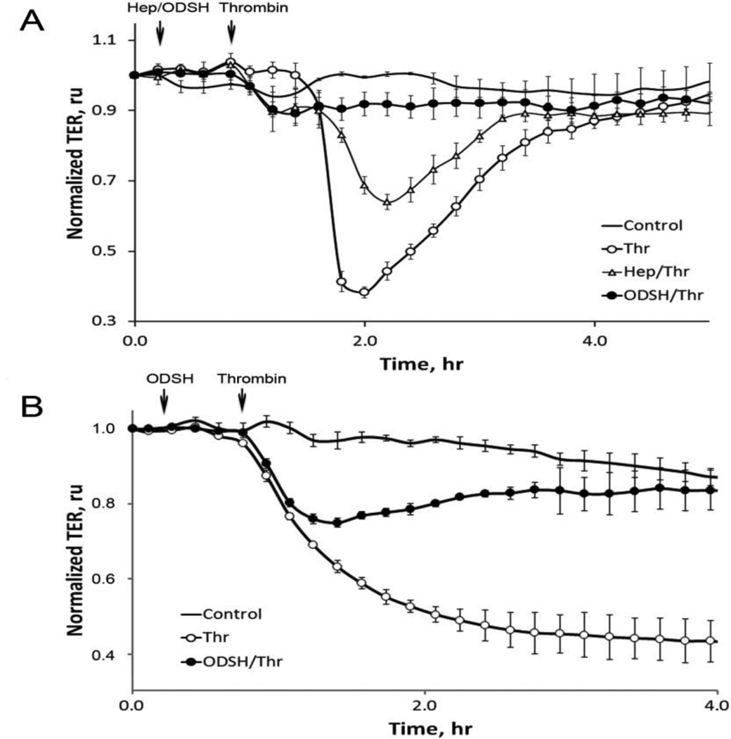
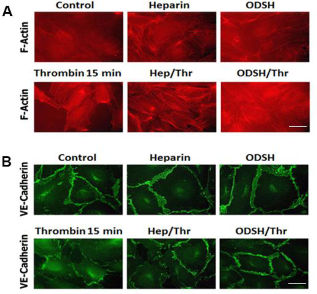
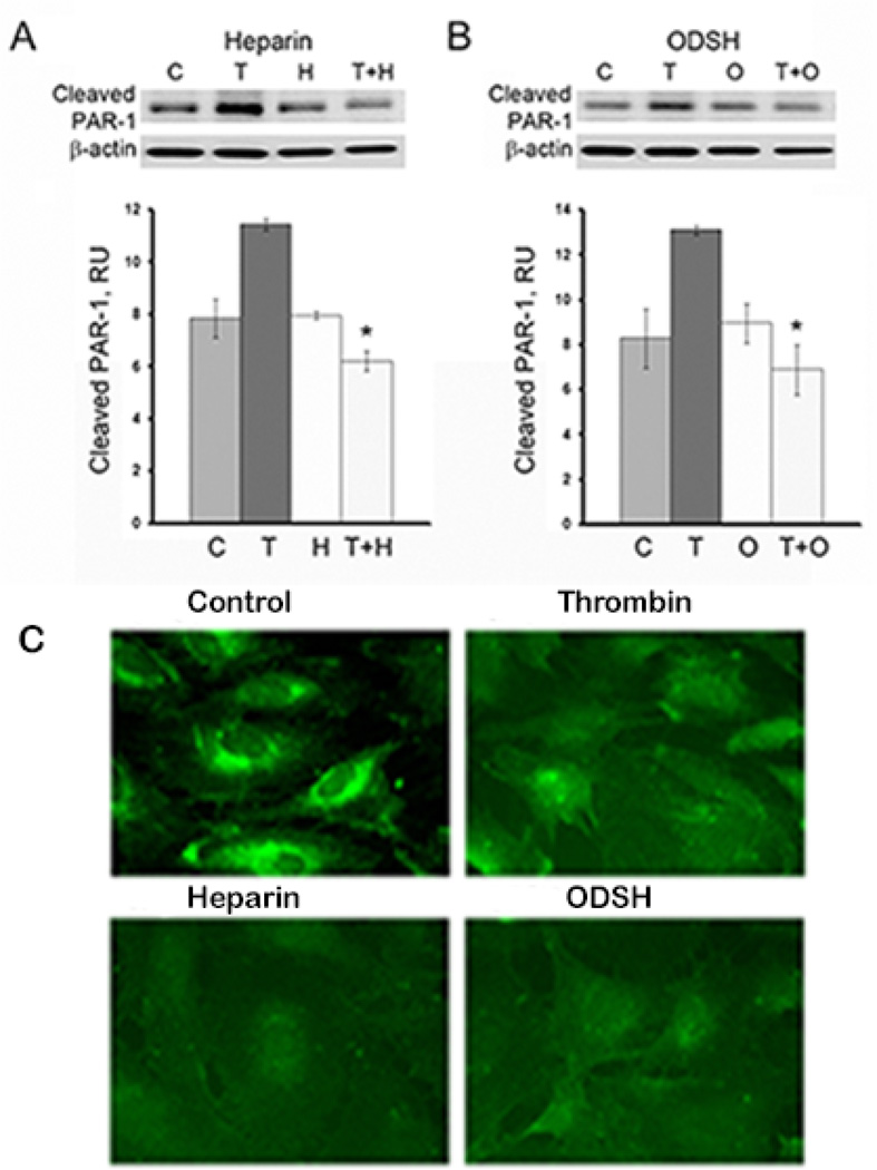
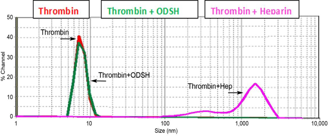
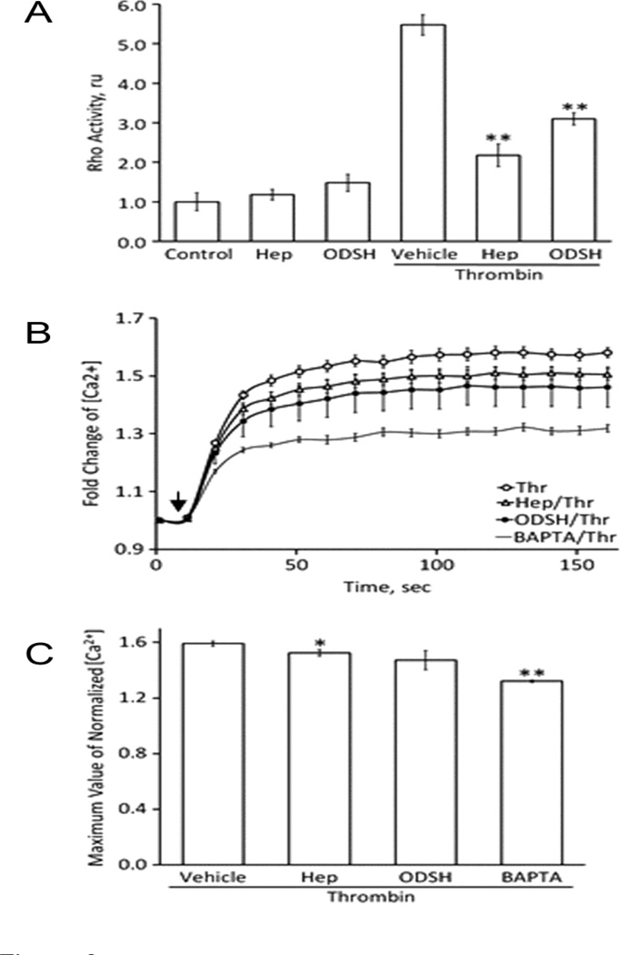
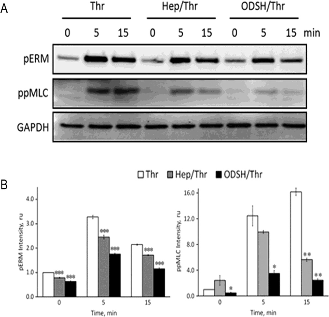
Acute lung injury and acute respiratory distress syndrome are accompanied by thrombin activation and fibrin deposition that enhance lung inflammation, activate endothelial cells and disrupt lung paracellular permeability. Heparin possesses anti-inflammatory properties but its clinical use is limited by hemorrhage and heparin induced thrombocytopenia. We studied the effects of heparin and low anticoagulant 2-O, 3-O desulfated heparin (ODSH) on thrombin-induced increases in paracellular permeability of cultured human pulmonary endothelial cells (ECs). Pretreatment with heparin or ODSH blocked thrombin-induced decrease in the EC transendothelial electrical resistance (TER), attenuated thrombin-stimulated paracellular gap formation and actin cytoskeletal rearrangement. Our data demonstrated that heparin and ODSH had inhibitory effects on thrombin-induced RhoA activation and intracellular calcium elevation. Thrombin-stimulated phosphorylation of the cytoskeletal regulatory proteins, myosin light chain and ezrin/radixin/moesin was also reduced. In these effects, low anticoagulant ODSH was more potent than heparin. Heparin or ODSH alone produced decreases in the EC TER that were abolished by siRNA-mediated depletion of the thrombin receptor, PAR-1. We also demonstrated that, in contrast to heparin, ODSH did not possess thrombin-binding activity. Results suggest that heparin and low anticoagulant ODSH can interfere with thrombin-activated signaling.

Keywords: Endothelium; Heparin; ODSH; PAR; Thrombin.


Conflict of interest statement

Conflict of Interest Statement

Dr. Kennedy Is the Scientific founder of ParinGenix, Inc. and technology inventor and consultant to ParinGenix, Inc.. He maintains founder’s stock in the company. He is the inventor of 2-O, 3-O desulfated heparin. He maintains no other conflict of interest.

All other authors declare no conflict of interest.

Figures

Figure 1
Figure 1. Effect of heparin and ODSH on the EC TER
Panels A, B. Heparin or ODSH increase the permeability of HPAEC. The cells were treated with heparin (50 µg/ml) or ODSH (50 µg/ml) and transendothelial resistance (TER) was recorded in real time in ECIS assay. Panel C. Depletion of PAR-1 with specific siRNA attenuated thrombin-induced barrier disruption in HPAEC. The cells grown in ECIS arrays were transfected with specific siRNA for PAR-1 or non-silencing (ns) RNA as described in Materials and Methods. 48 hours later, the cells were treated with thrombin (20 nM) and changes in the monolayer integrity were analyzed by ECIS assay. Depletion of PAR-1 was confirmed by RT-PCR. Panel D. Depletion of PAR-1 with specific siRNA attenuated heparin- or ODSH-induced TER decrease. The involvement of PAR-1 in heparin- or ODSH-induced TER decrease was examined in the same conditions used in panel A. TER values were normalized with the initial resistance and expressed as the mean of three or four individual experiments. Arrows indicate time points when reagents were added. Data is expressed as a mean of four individual experiments ± SE, and statistical significances compare to control were evaluated using Student’s t-test; * (p < 0.05).
Figure 2
Figure 2. Effect of heparin or ODSH on thrombin-induced endothelial permeability
Panel A. Heparin or ODSH prevents thrombin-induced TER loss in HPAEC. The cells grown in ECIS arrays were pretreated with heparin or ODSH for 40 min and then challenged with 20 nM thrombin as described in Materials and Methods. Arrows indicate time points when the effectors were added. TER values were normalized with the initial resistance and expressed as mean ± standard error (SE), relative units (ru) of four individual experiments. Panel B. ODSH prevents thrombin-induced TER loss in HLMVEC. The same experiment as in Panel A but another EC (HLMVEC) was used. Thrombin concentration – 0.5 nM.
Figure 3
Figure 3. Heparin or ODSH pretreatment prevents thrombin-induced actin cytoskeleton rearrangement and prevents thrombin-induced cell-cell contact loss in HPAEC
Panel A. The cells grown on coverslips were pretreated with heparin or ODSH (50 µg/ml, 40 min) and challenged with thrombin (20 nM, 15 min). F-actin visualization by Alexa-488-phalloidin staining was performed as described in Materials and Methods. Panel B. The cells grown on coverslips were pretreated with heparin or ODSH (50 µg/ml, 40 min) then challenged with thrombin (20 nM, 15 min). Visualization of adherens junctions by VE-cadherin immunostaining was performed as described in Materials and Methods. Scale bars represent 20microns.
Figure 4
Figure 4. Heparin and ODSH protect PAR-1 from cleavage by thrombin
Panels A, B. HPAEC were pre-treated with heparin (A) or ODSH (B) (50 µg/ml, 40 min), washed with fresh serum-free cell culture medium to remove unbound heparins, then challenged with thrombin (8 nM, 10 min) and analyzed by Western blotting using antibody specifically recognizing thrombin-cleaved PAR-1. Statistical analyses of the experimental data expressed in relative units are presented as a mean of three individual experiments ± SE. *P < 0.01 versus cells treated with thrombin alone. Panel C. Heparin and ODSH affect cell surface expression of PAR-1. Exposure to thrombin, heparin or ODSH alters cell surface expression of PAR-1 as revealed by immunofluorescence. Cells were incubated with thrombin (20 ng/ml) or heparin (50 µg/ml) or ODSH (50 µg/ml) for 30 min prior to immunostaining with anti-PAR-1 antibody. Images of control, thrombin-, heparin- and ODSH-treated EC are shown. Results are representative of a minimum of 3 individual experiments.
Figure 5
Figure 5. ODSH has low binding affinity for thrombin
The particle size of the macromolecular complexes was analyzed using the Microtac Nanotrac ULTRA dynamic light scattering instrument. The instrument detects particle size distributions in low concentration and is sensitive to size ranges below 10 nm. Thrombin was diluted in 300 µl of PBS and particle size was measured (red line indicated by arrow). After that, a single dose of ODSH (5 µl/ml, 50 mg/ml) (green line indicated by arrow) or heparin (5 µl/ml, 25 mg/ml) (pink line indicated by arrow) was added into the thrombin solution, incubated for 5 min at RT and resulted complex sizes were measured. Measurements were repeated at least two times in two separate experiments.
Figure 6
Figure 6. Thrombin-induced increases in RhoA activity and intracellular calcium can be attenuated by pretreatment with heparin or ODSH
Panel A. HPAEC monolayers were pretreated with heparin (50 µg/ml) or ODSH (50 µg/ml) for 40 min before challenging with thrombin (20 nM). The cells were harvested 5 minutes after thrombin addition and used for RhoA activation G-LISA assay. RhoA activation induced by thrombin was significantly inhibited in the heparin- or ODSH-pretreated EC. **P<0.001 (Hep- or ODSH-pretreatment vs. thrombin treatment alone). Panel B. The increase of intracellular calcium in the response of thrombin treatment (20 nM) was recorded for 160 seconds after thrombin addition (indicated as arrow). BAPTA-AM (10 µM), cell-permeable calcium chelator. Panel C. Statistical analysis of the results shown in Panel B. Data is expressed as a mean of six individual experiments ± SE. *P<0.05 and **P<0.01 (Hep- or ODSH-pretreatment vs. thrombin alone treatment.
Figure 7
Figure 7. Heparin or ODSH pretreatment down-regulates thrombin-induced cell signaling cascades
Panel A. HPAEC monolayers were pretreated with heparin (50 µg/ml) or ODSH (50 µg/ml) for 40 min before challenging with thrombin (20 nM). The cells were harvested at indicated time points and analyzed by Western blotting for ezrin/radixin/moesin phosphorylation (pERM) and MLC di-phosphorylation (ppMLC). GAPDH immunostaining was used as a loading control. Panel B. Statistical analysis of the results shown in panel A. Experimental data is expressed in relative units and presented as a mean of four individual experiments ± SE. *P < 0.05 and **P < 0.01, and ***p<0.001 versus cells treated with thrombin alone.

References

    1. Bao Y, Geng Y, Jing H. Effect of hirudin on the levels of acute lung injury rat tumor necrosis factor-α and matrix metalloproteinase-12. Mol. Med. Rep. 2012;5:873–875. - PubMed
    1. Bernard GR, Vincent JL, Laterre PF, LaRosa SP, Dhainaut JF, Lopez-Rodriguez A, Steingrub JS, Garber GE, Helterbrand JD, Ely EW, Fisher CJ., Jr Efficacy and safety of recombinant human activated protein C for severe sepsis. N. Engl. J. Med. 2001;344:699–709. - PubMed
    1. Birukova AA, Birukov KG, Smurova K, Adyshev D, Kaibuchi K, Alieva I, Garcia JGN, Verin AD. Novel role of microtubules in thrombin-induced endothelial barrier dysfunction. FASEB. J. Off. Publ. Fed. Am. Soc. Exp. Biol. 2004a;18:1879–1890. - PubMed
    1. Birukova AA, Smurova K, Birukov KG, Kaibuchi K, Garcia JGN, Verin AD. Role of Rho GTPases in thrombin-induced lung vascular endothelial cells barrier dysfunction. Microvasc. Res. 2004b;67:64–77. - PubMed
    1. Bode W, Turk D, Karshikov A. The refined 1.9-A X-ray crystal structure of D-Phe-Pro-Arg chloromethylketone-inhibited human alpha-thrombin: structure analysis, overall structure, electrostatic properties, detailed active-site geometry, and structure-function relationships. Protein Sci. Publ. Protein Soc. 1992;1:426–471. - PMC - PubMed

Publication types

MeSH terms