. 2014 Feb;133(2):301-310.
doi: 10.1097/01.prs.0000436840.69752.7e.

Lymph node transplantation results in spontaneous lymphatic reconnection and restoration of lymphatic flow

Affiliations

Lymph node transplantation results in spontaneous lymphatic reconnection and restoration of lymphatic flow

Seth Z Aschen et al. Plast Reconstr Surg. 2014 Feb.

Abstract

Background: Although lymph node transplantation has been shown to improve lymphatic function, the mechanisms regulating lymphatic vessel reconnection and functional status of lymph nodes remains poorly understood.

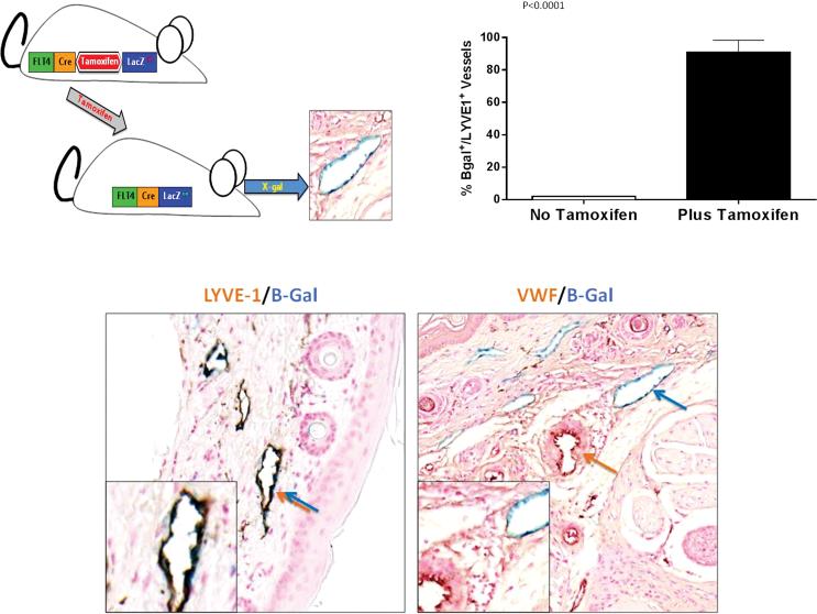
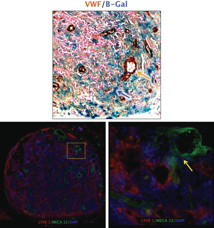
Methods: The authors developed and used LacZ lymphatic reporter mice to examine the lineage of lymphatic vessels infiltrating transferred lymph nodes. In addition, the authors analyzed lymphatic function, expression of vascular endothelial growth factor (VEGF)-C, maintenance of T- and B-cell zone, and anatomical localization of lymphatics and high endothelial venules.

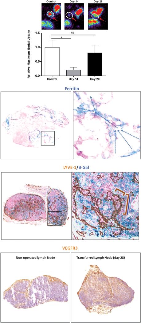
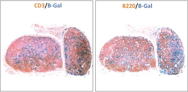
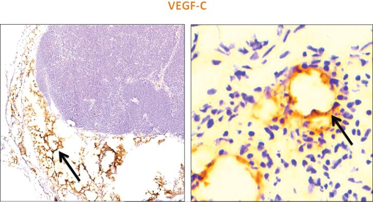
Results: Reporter mice were specific and highly sensitive in identifying lymphatic vessels. Lymph node transfer was associated with rapid return of lymphatic function and clearance of technetium-99 secondary to a massive infiltration of recipient mouse lymphatics and putative connections to donor lymphatics. T- and B-cell populations in the lymph node were maintained. These changes correlated with marked increases in the expression of VEGF-C in the perinodal fat and infiltrating lymphatics. Newly formed lymphatic channels in transferred lymph nodes were in close anatomical proximity to high endothelial venules.

Conclusions: Transferred lymph nodes have rapid infiltration of functional host lymphatic vessels and maintain T- and B-cell populations. This process correlates with increased endogenous expression of VEGF-C in the perinodal fat and infiltrating lymphatics. Anatomical proximity of newly formed lymphatics and high endothelial venules supports the hypothesis that lymph node transfer can improve lymphedema by exchanges with the systemic circulation.


Figures

Figure 1
Figure 1. FLT-4-LacZ mice can be used as an inducible lymphatic reporter mouse
(A) Schematic figure of FLT-4Cre/LacZ LoxP mice. Treatment with tamoxifen activates LacZ expression and enables visualization of lymphatic vessels with B-gal. (B) High level activation of LacZ expression in LYVE-1 positive lymphatics is inducible with tamoxifen. (C) Representative high power (40×) images of skin sections double stained for B-galactosidase (blue) and LYVE-1 (brown stain, left panel) or Von-Willebrand Factor (VWF; brown stain, right panel) demonstrate specificity of Lac-Z expression only in lymphatics vessels (blue/brown double arrow on the left). Notice no overlap with VWF (separate brown and blue arrows) on the right panel.
Figure 2
Figure 2. Lymph node transplantation restores lymphatic transport capacity and function by promoting infiltration of lymphatics from the recipient mouse
(A) Representative heat maps (top) and quantification of relative Tc99 uptake in axillary lymph nodes after injection in the distal upper extremity of control mice (axillary incision without lymphadenectomy), and experimental mice 14 and 28 days after lymph node transplantation. Note recovery of Tc99 uptake by day 28. (B) Representative low (left panel; 2.5×) and high (right panel; 40×) powered images of ferritin staining in lymph nodes harvested 28 days after transplantation (arrows denote direction of lymphatic flow from lymphatic sinuses toward the cortex). (C) Representative low (left panel; 2.5×) and high powered (right panel; 40× zoom of boxed region in left panel) images of transplanted lymph nodes double stained with the lymphatic specific marker LYVE-1 (brown stain) and B-gal (blue stain). Note connection of double stained vessels (overlapping blue/brown brackets) with single stained brown vessel (brown bracket) suggesting connection of recipient and donor lymphatics within the lymph node. (D) Representative low power (2.5×) images of control (sham-operated; left panel) and transplanted lymph node (right panel) stained for VEGF-R3 (brown stain) demonstrating massive infiltration of VEGF-R3 positive cells in transplanted node.
Figure 3
Figure 3. Transplanted lymph nodes retain T and B cells
Representative low power (2.5×) images of transplanted lymph nodes double stained with B-galactosidase (blue) and CD3 (brown staining of T cells; left panel) or B220 (brown staining of B cells; right panel). Note that transplanted lymph nodes retain both cell populations in addition to maintenance of lymph node cellularity.
Figure 4
Figure 4. Lymphatic regeneration correlates with expression of VEGF-C
Representative low (left panel; 5×) and high power (right panel; 40×) images of transplanted lymph nodes stained for VEGF-C (brown staining). Note high-level expression in perinodal fat and in infiltrating lymphatic vessels (black arrows).
Figure 5
Figure 5. Lymphatic vessels in lymph nodes are in close proximity to high endothelial venules (HEVs)
(A) Representative high power (40×) images of transplanted lymph nodes double stained with B-galactosidase (blue) and Von Willebrand factor (VWF; brown). Note close proximity of lymphatic vessels shown in blue and blood vessels stained brown. (B) Representative low-power (2.5×) florescent images of transplanted lymph nodes double stained with LYVE-1 (red) and HEV marker MECA-32 (green). Note large numbers of lymphatic channels (C) Representative high-power (40×) image of yellow-boxed area shown in B. Note close proximity of lymphatics and HEVs (yellow arrow).

References

    1. Petrek JA, Heelan MC. Incidence of breast carcinoma-related lymphedema. Cancer. 1998;83:2776–2781. - PubMed
    1. Cheng MH, Chen SC, Henry SL, et al. Vascularized groin lymph node flap transfer for postmastectomy upper limb lymphedema: flap anatomy, recipient sites, and outcomes. Plast Reconstr Surg. 2013;131:1286–1298. - PubMed
    1. Becker C, Assouad J, Riquet M, et al. Postmastectomy lymphedema: long-term results following microsurgical lymph node transplantation. Ann Surg. 2006;243:313–315. - PMC - PubMed
    1. Tobbia D, Semple J, Baker A, et al. Experimental assessment of autologous lymph node transplantation as treatment of postsurgical lymphedema. Plast Reconstr Surg. 2009;124:777–786. - PubMed
    1. Saaristo AM, Niemi TS, Viitanen TP, et al. Microvascular breast reconstruction and lymph node transfer for postmastectomy lymphedema patients. Ann Surg. 2012;255:468–473. - PubMed

Publication types

LinkOut - more resources