

Review
doi: 10.1002/embr.201338166.
Epub 2014 Jan 27.
RBR E3-ligases at work
Affiliations
- PMID: 24469331
- PMCID: PMC3989860
- DOI: 10.1002/embr.201338166
Item in Clipboard
Review
RBR E3-ligases at work
EMBO Rep.
2014 Feb.
Abstract
The RING-in-between-RING (RBR) E3s are a curious family of ubiquitin E3-ligases, whose mechanism of action is unusual in several ways. Their activities are auto-inhibited, causing a requirement for activation by protein-protein interactions or posttranslational modifications. They catalyse ubiquitin conjugation by a concerted RING/HECT-like mechanism in which the RING1 domain facilitates E2-discharge to directly form a thioester intermediate with a cysteine in RING2. This short-lived, HECT-like intermediate then modifies the target. Uniquely, the RBR ligase HOIP makes use of this mechanism to target the ubiquitin amino-terminus, by presenting the target ubiquitin for modification using its distinctive LDD region.
Figures
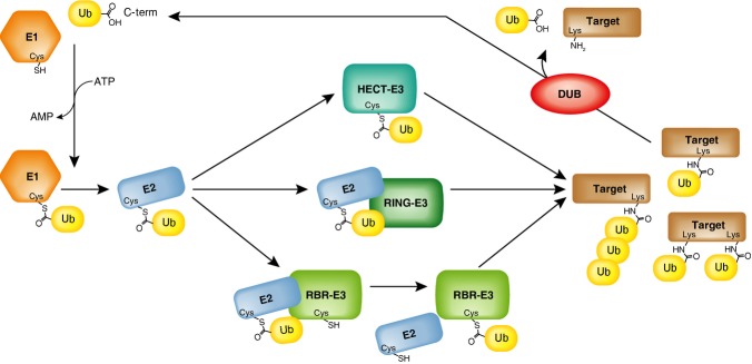
RBR E3-ligases have a unique mode of transferring ubiquitin to a target. The ubiquitin (Ub) C-terminus is activated in an ATP-dependent manner by an E1 activating enzyme, and is subsequently transferred to form a thioester intermediate on an E2 conjugase. The final transfer of ubiquitin onto its target is mediated by E3-ligases that either form a thioester intermediate with the ubiquitin (HECT E3-ligases), mediate a direct transfer of the ubiquitin from the E2 onto its target (RING E3-ligases), or function as RING/HECT-type hybrids (RBR E3-ligases). Through this cascade of E1, E2 and E3 enzymes, the ubiquitination machinery mediates the formation of mono-ubiquitination, multi-mono-ubiquitination, or ubiquitin chain formation on its targets. The ubiquitin signal can be removed by de-ubiquitination enzymes (DUBs).
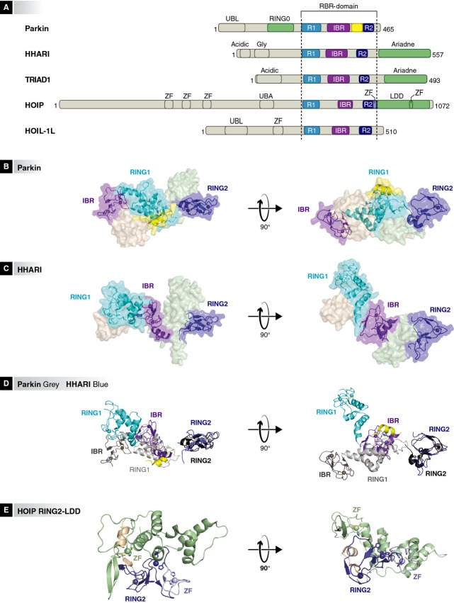
Domain arrangements in RBR E3-ligases. The domain borders are drawn to scale according to Uniprot definitions (http://www.uniprot.org ). Ubiquitin like domain (UBL), acidic region (Acidic), glycine-rich region (Gly), zinc finger (ZF), ubiquitin-associated domain (UBA). The RBRs are represented in RING1:cyan (R1), purple (IBR) and blue (R2). The Parkin RING0, C-terminal Ariadne domain and linear ubiquitin determining domain (LDD) are represented in pale green and the Parkin linker/tether helix (also called PUB or REP) is yellow. B) Crystal structure of full-length Parkin (surface representation) (PDB 4k95). The RBR (cartoon) is autoinhibited by the N-terminal regions of Parkin. The colors correspond to the colors in the schematic representation in Figure 2A. C) Surface representation of the crystal structure of full length HHARI (PDB 4kbl). The RBR is shown as cartoon. D) A cartoon of superposed RBR domains of Parkin and HHARI (SSM (WinCoot 0.7.1) superposition of RING2 domain of HHARI on the RING2 domain of Parkin). Parkin RING1:light grey80, IBR:dark grey40, PUB domain: yellow, RING2:black. HHARI RING1:cyan, IBR:purple, RING2:blue. E) Crystal structure of HOIP RING2-LDD (PDB 4ljo).
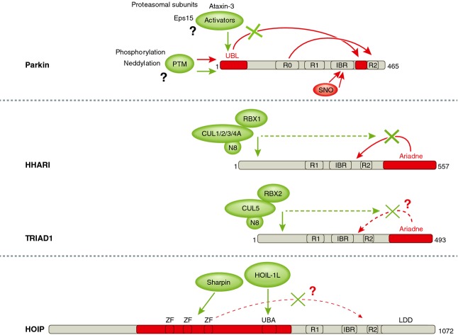
Regulation of the E3-ligase activity of Parkin, HHARI, TRIAD1 and HOIP. Schematic representation of the RBR E3-ligases. The domains involved in autoinhibitory interactions are shown in red, whereas activators are shown in green. Autoinhibitory interactions between domains are depicted by the red arrows, and green arrows indicate interactions and domains that are influenced by the activators. Dotted lines are used for interactions that have not been fully characterized yet. See text for details. IBR = in between ring; LDD = linear-ubiquitin-chain determining domain; N8 = Nedd8; PTM = post-translational modification; SNO = s-nitrosylation; R = RING; UBA = ubiquitin associated domain; ZF = zinc finger.
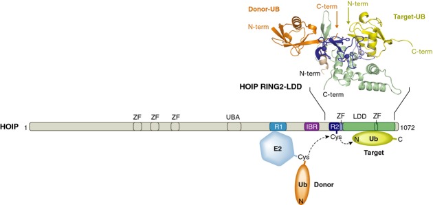
Linear ubiquitin chain formation by HOIP. The positioning of the target ubiquitin (yellow) by the HOIP LDD (green) is critical for the transfer of the donor-ubiquitin (orange) from the E2 (blue), via a cysteine on RING2 onto the N-terminus of the target ubiquitin. The crystal structure of HOIP RING2-LDD in complex with a donor and a target ubiquitin (PDB 4ljo) shows a snapshot of the orientation of the proteins just before the two ubiquitins are linked together by HOIP. IBR = in between ring; LDD = linear-ubiquitin-chain determining domain; R = RING; UBA = ubiquitin associated domain; ZF = zinc finger.
References
-
- Morett E, Bork P. A novel transactivation domain in parkin. Trends Biochem Sci. 1999;24:229–231. - PubMed
-
- Marin I, Ferrus A. Comparative genomics of the RBR family, including the Parkinson's disease-related gene parkin and the genes of the ariadne subfamily. Mol Biol Evol. 2002;19:2039–2050. - PubMed
-
- Marin I, Lucas JI, Gradilla AC, Ferrus A. Parkin and relatives: the RBR family of ubiquitin ligases. Physiol Genomics. 2004;17:253–263. - PubMed
Publication types
MeSH terms
Substances
LinkOut - more resources
Full Text Sources
Other Literature Sources
