. 2014 Apr;34(7):1349-62.
doi: 10.1128/MCB.01052-13. Epub 2014 Jan 27.

TRF2-tethered TIN2 can mediate telomere protection by TPP1/POT1

Affiliations

TRF2-tethered TIN2 can mediate telomere protection by TPP1/POT1

David Frescas et al. Mol Cell Biol. 2014 Apr.

Abstract

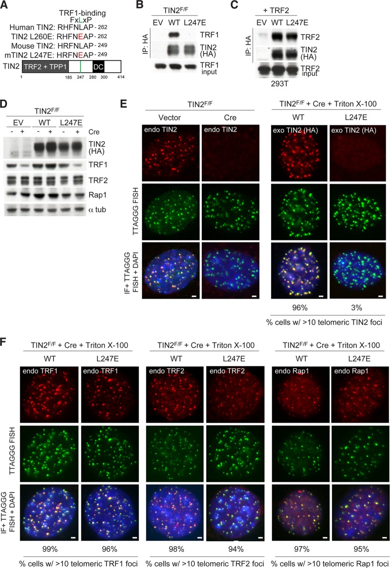
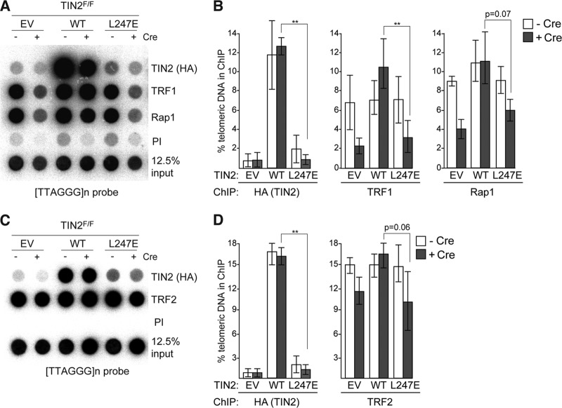
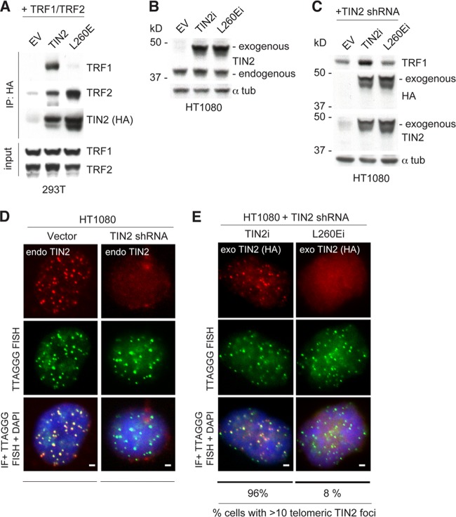
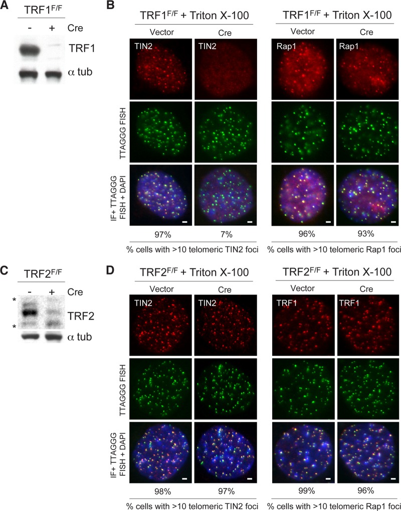
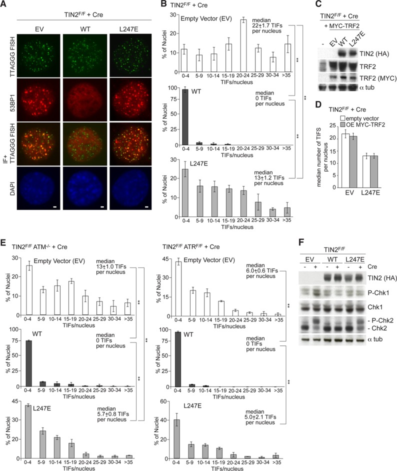
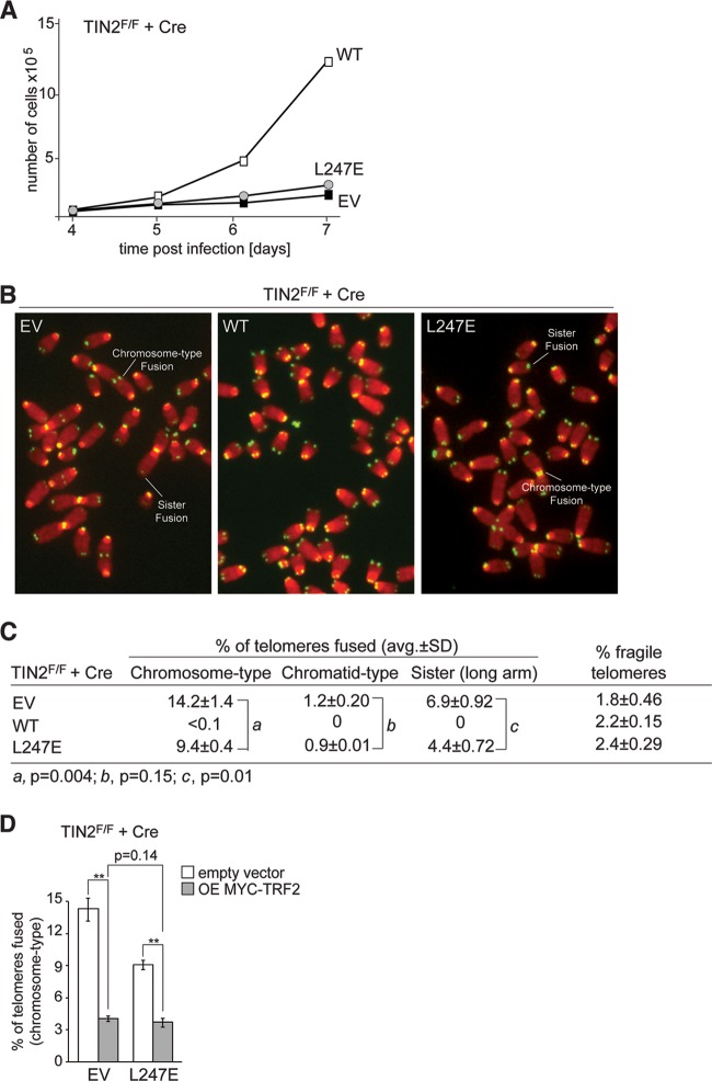
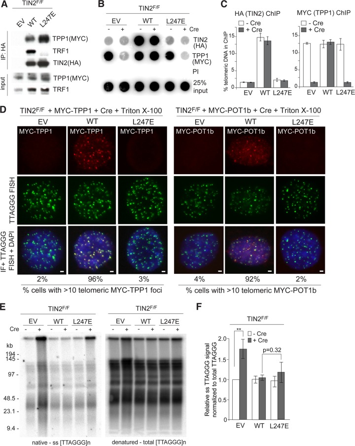
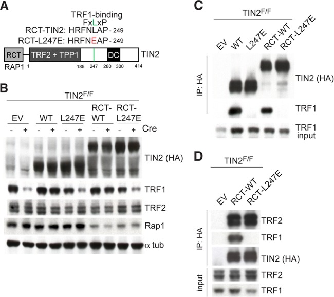
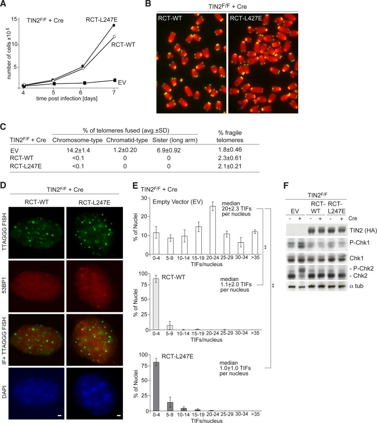
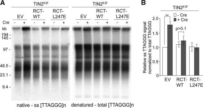
The shelterin protein TIN2 is required for the telomeric accumulation of TPP1/POT1 heterodimers and for the protection of telomeres by the POT1 proteins (POT1a and POT1b in the mouse). TIN2 also binds to TRF1 and TRF2, improving the telomeric localization of TRF2 and its function. Here, we ask whether TIN2 needs to interact with both TRF1 and TRF2 to mediate the telomere protection afforded by TRF2 and POT1a/b. Using a TIN2 allele deficient in TRF1 binding (TIN2-L247E), we demonstrate that TRF1 is required for optimal recruitment of TIN2 to telomeres and document phenotypes associated with the TIN2-L247E allele that are explained by insufficient TIN2 loading onto telomeres. To bypass the requirement for TRF1-dependent recruitment, we fused TIN2-L247E to the TRF2-interacting (RCT) domain of Rap1. The RCT-TIN2-L247E fusion showed improved telomeric localization and was fully functional in terms of chromosome end protection by TRF2, TPP1/POT1a, and TPP1/POT1b. These data indicate that when sufficient TIN2 is loaded onto telomeres, its interaction with TRF1 is not required to mediate the function of TRF2 and the TPP1/POT1 heterodimers. We therefore conclude that shelterin can protect chromosome ends as a TRF2-tethered TIN2/TPP1/POT1 complex that lacks a physical connection to TRF1.


Figures

FIG 1
FIG 1
TIN2 allele lacking TRF1 binding is lost at telomeres. (A) Schematic of the mouse TIN2 protein indicating regions of shelterin binding and location of mutations identified in dyskeratosis congenita (DC) patients. Homology between human and mouse TIN2 is shown surrounding the FXLXP TRF1 binding motif. Amino acids in red indicate mutations introduced to generate TRF1 binding mutants. (B) Cell extracts from TIN2F/F MEFs exogenously expressing the indicated FLAG-HA2 TIN2 alleles were immunoprecipitated (IP) with anti-HA resin, and immunocomplexes were probed for the indicated proteins. (C) Cell extracts from 293T cells coexpressing the indicated FLAG-HA2 TIN2 alleles and TRF2 were immunoprecipitated with anti-HA resin, and immunocomplexes were probed for the indicated proteins. (D) Immunoblotting of cell extracts from TIN2F/F MEFs expressing TIN2 alleles (as in panel B) or empty vector (EV) with or without Cre treatment (72 h). (E) Left, loss of TIN2 as observed by IF-FISH from TIN2F/F MEFs with or without Cre treatment (72 h). Right, IF of FLAG-HA2 TIN2 alleles (anti-HA antibody, red) and telomeric FISH (green) following Triton X-100 extraction of soluble proteins in TIN2−/− MEFs (72 h post-Cre). (F) Left, IF of TRF2 (anti-TRF1 antibody, red) in cells expressing FLAG-HA2 TIN2 alleles and telomeric FISH (green) following Triton X-100 extraction of soluble proteins in TIN2−/− MEFs (72 h post-Cre). Middle, IF of TRF2 (anti-TRF2 antibody, red) in cells expressing FLAG-HA2 TIN2 alleles and telomeric FISH (green) following Triton X-100 extraction of soluble proteins in TIN2−/− MEFs (72 h post-Cre). Right, IF of Rap1 (anti-Rap1 antibody, red) in cells expressing FLAG-HA2 TIN2 alleles and telomeric FISH (green) following Triton X-100 extraction of soluble proteins in TIN2−/− MEFs (72 h post-Cre). DNA is stained with DAPI (blue). At least 200 cells were used for quantification of TIN2 and Rap1 foci. Bars, 1.5 μm.
FIG 2
FIG 2
TIN2-L247E protein is absent from telomeres by ChIP. (A) Telomeric DNA ChIP for FLAG-HA2 TIN2 alleles (anti-HA), TRF1, or Rap1 in TIN2F/F MEFs with or without Cre treatment (72 h). (B) Quantification of the ChIP signals from three identical experiments, as described for panel A. ChIP signals were normalized to the input, and the background (PI) was subtracted. **, P < 0.05 (paired Student's t test). (C) Telomeric DNA ChIP for TRF2 in TIN2F/F MEFs expressing FLAG-HA2 TIN2 alleles (anti-HA) with or without Cre treatment (72 h). (D) Quantification of the ChIP signals from three identical experiments, as described for panel C. ChIP signals were normalized to the input, and the background (PI) was subtracted. **, P < 0.05 (paired Student's t test).
FIG 3
FIG 3
TIN2-L260E affects TRF1 accumulation at human telomeres. (A) Cell extracts from 293T cells exogenously expressing the indicated human FLAG-HA2 TIN2 alleles (WT and L260E) and MYC-tagged TRF1 and MYC-tagged TRF2 were immunoprecipitated (IP) with anti-HA resin, and immunocomplexes were probed for the indicated proteins. (B) FLAG-HA2-tagged TIN2 alleles were introduced into HT1080 human fibrosarcoma cells. Immunoblotting for these proteins was conducted from whole-cell extracts. Endogenous and exogenous TIN2 proteins were identified using an anti-TIN2 antibody. (C) Protein levels of FLAG-HA2-tagged shRNA-insensitive proteins (indicated as TIN2i and L247Ei) and TRF1 in HT1080 human fibroblasts transduced with a TIN2 shRNA. Immunoblotting for these proteins was conducted in whole-cell extracts and detected by anti-HA and anti-TIN2 antibodies. (D) Loss of TIN2 as observed by IF-FISH from HT1080 cells transduced with a TIN2 shRNA, as described for panel C. DNA is stained with DAPI (blue). (E) IF of FLAG-HA2 TIN2i alleles (anti-HA antibody, red) and telomeric FISH (green) following Triton X-100 extraction of soluble proteins in HT1080 cells, as described for panel C. DNA is stained with DAPI (blue). At least 200 cells were used for quantification of TIN2 foci. Bars, 1.5 μm.
FIG 4
FIG 4
Accumulation of TIN2 at telomeres is reduced in TRF1-deficient MEFs. (A) An anti-TRF1 antibody was used to detect TRF1 protein in immunoblots in TRF1F/F MEFs with or without Cre treatment (72 h). (B) Endogenous TIN2 (left) and endogenous Rap1 (right) were detected by IF in combination with telomeric FISH (green) with or without Cre treatment (72 h). Triton X-100 was used to extract soluble proteins. DNA is stained with DAPI (blue). Bars, 1.5 μm. (C) An anti-TRF2 antibody was used to detect TRF2 protein in immunoblots in TRF2F/F MEFs with or without Cre treatment (72 h). *, unspecific bands. (D) Endogenous TIN2 (left) and endogenous TRF1 (right) were detected by IF in combination with telomeric FISH (green) with or without Cre treatment (72 h). Triton X-100 was used to extract soluble proteins. DNA is stained with DAPI (blue). Bars, 1.5 μm.
FIG 5
FIG 5
DNA damage response in TIN2-L247E-expressing cells. (A) IF of 53BP1 (red) and telomeric FISH (green) in TIN2−/− MEFs (72 h post-Cre) expressing empty vector (EV) or the indicated FLAG-HA2 TIN2 alleles. DNA is stained with DAPI (blue). Bars, 1.5 μm. (B) Quantification of TIFs in TIN2F/F MEFs transduced with an empty vector (top), wild-type TIN2 (middle), or L247E (bottom), as scored for 53BP1 TIFs per nucleus (n > 200) after Cre (72 h). Averages and median values are from three independent experiments and standard deviations (SDs). **, P < 0.05 (paired Student's t test). (C) An anti-TRF2 antibody and an anti-MYC antibody were used to detect exogenously expressed MYC-tagged TRF2 proteins in immunoblots in TIN2F/F MEFs expressing FLAG-HA2 TIN2 alleles (anti-HA) and MYC-TRF2. (D) Quantification of a median number of TIFs per nucleus in TIN2-deleted cells expressing an EV or TIN2-L247E with or without exogenously expressed TRF2. Median values are from three independent experiments and SDs. (E) Left, quantification of TIFs in TIN2F/F ATM−/− MEFs transduced with an empty vector (top), wild-type TIN2 (middle), or L247E (bottom), as scored for 53BP1 TIFs per nucleus (n > 100) after Cre (72 h post-Cre). Averages and median values are from three independent experiments and SDs. **, P < 0.05 (paired Student's t test). Right, quantification of TIFs in TIN2F/F ATRF/F MEFs transduced with an empty vector (top), wild-type TIN2 (middle), or L247E (bottom), as scored for 53BP1 TIFs per nucleus (n > 100) after Cre (72 h post-Cre). Averages and median values are from three independent experiments and SDs. **, P < 0.05 (paired Student's t test). Endoreduplicated (tetraploid) cells were excluded from the analysis. (F) Immunoblots for Chk1 and Chk2 phosphorylation in TIN2F/F MEFs expressing the indicated FLAG-HA2 TIN2 alleles and treated with Cre (72 h) as indicated.
FIG 6
FIG 6
Growth arrest and telomere fusions in cells expressing TIN2-L247E. (A) Growth curve of SV40-LT-immortalized TIN2−/− MEFs expressing an empty vector (EV) or the indicated FLAG-HA2 TIN2 alleles. (B) Examples of telomere fusions in metaphases of TIN2-deficient cells expressing the indicated TIN2 alleles or no TIN2 (EV) (72 h post-Cre). Green, TelC PNA probe; red, DAPI DNA stain. (C) Summary of telomere phenotypes in TIN2-deficient cells expressing TIN2 alleles, as determined by FISH, as shown in panel B. Values are averages and SDs from 3 independent experiments. Chromosome-type and chromatid-type telomere fusions were scored on 3,000 to 5,000 telomeres/experiment. Long-arm sister telomere fusions and fragile telomeres were scored on 2,000 to 3,000 telomeres/experiment. (D) Quantification of chromosome-type fusions in TIN2-deleted cells expressing an EV or TIN2-L247E with or without exogenously expressed TRF2. Averages and SDs are from three independent experiments (2,000 to 3,000 telomeres/experiment). **, P < 0.05 (paired Student t test).
FIG 7
FIG 7
Unaltered telomeric overhangs despite diminished TPP1 and POT1b at telomeres. (A) TIN2F/F MEFs exogenously expressing MYC-TPP1 were infected with empty vector (EV) or the indicated FLAG-HA2 TIN2 alleles. Cell extracts were immunoprecipitated (IP) with anti-HA resin, and immunocomplexes were probed for the indicated proteins. (B) Telomeric DNA ChIP for FLAG-HA2 TIN2 alleles (anti-HA) and MYC-TPP1 (anti-MYC) in TIN2F/F MEFs with or without Cre treatment (72 h). (C) Quantification of the ChIP signals from two identical experiments, as described for panel B. ChIP signals were normalized to the input, and the background (PI) was subtracted. Error bars represent standard errors of the means. (D) TIN2F/F MEFs exogenously expressing MYC-tagged TPP1 (left) or MYC-tagged POT1b (right) were infected with EV or the indicated FLAG-HA2 TIN2 alleles. Indirect immunofluorescence (IF) of MYC-tagged proteins (anti-MYC antibody, red) and telomeric FISH (green) was conducted following Triton X-100 extraction of soluble proteins in TIN2−/− MEFs (72 h post-Cre). DNA is stained with DAPI (blue). Bars, 1.5 μm. (E) In-gel hybridization assay for single-stranded (ss) telomeric DNA after TIN2 deletion in cells expressing EV, TIN2, or L247E-TIN2. Left, TelC signals under the native condition; right, same gel rehybridized after in situ denaturation of the DNA. (F) Quantification of overhang signals from three independent experiments, as described for panel E, were normalized to the total telomeric signals and compared to TIN2F/F MEFs without Cre. **, P < 0.05 (paired Student's t test).
FIG 8
FIG 8
Fusion of RCT domain of Rap1 to TIN2 alleles. (A) Schematic of the Rap1 RCT-domain TIN2 fusion protein indicating regions of shelterin binding (TRF1, TRF2, and TPP1) and location of mutations identified in DC patients. The RCT domain of mouse Rap1 (amino acids [aa] 295 to 393) was fused to the N terminus (aa 2) of TIN2 and L247E to generate RCT-TIN2 alleles. All alleles have an N-terminal FLAG-HA2 tag. The residue in red indicates the mutation introduced to generate the TRF1 binding mutant. (B) The indicated FLAG-HA2-tagged proteins were introduced into TIN2F/F MEFs. Immunoblotting for the indicated proteins was conducted in cell extracts from MEFs with or without Cre treatment (72 h). (C) Extracts from TIN2F/F MEFs infected with empty vector (EV) or the indicated FLAG-HA2 TIN2 alleles following Cre treatment (72 h) were immunoprecipitated (IP) with anti-HA resin, and immunocomplexes were probed for the indicated proteins. (D) Extracts from TIN2F/F MEFs infected with EV or the indicated FLAG-HA2 RCT-TIN2 alleles following Cre treatment (72 h) were immunoprecipitated with anti-HA resin, and immunocomplexes were probed for the indicated proteins.
FIG 9
FIG 9
Fusion of the Rap1 RCT domain to TIN2-L247E restores telomere association. (A) Indirect immunofluorescence (IF) for the indicated FLAG-HA2 TIN2 alleles (anti-HA antibody, red) and telomeric FISH (green) in TIN2−/− MEFs (72 h post-Cre). Soluble proteins were extracted by Triton X-100 treatment prior to fixation. DNA is stained with DAPI (blue). Bars, 1.5 μm. (B) Telomeric DNA ChIP for the FLAG-HA2 TIN2 alleles (anti-HA), TRF1, and Rap1 in TIN2F/F MEFs with or without Cre treatment (72 h). (C) Quantification of the ChIP signals from three identical experiments, as performed for panel B. ChIP signals were normalized to the input, and the background (PI) was subtracted. **, P < 0.05 (paired Student's t test).
FIG 10
FIG 10
RCT-L247E rescues telomere deprotection phenotypes of TIN2 deletion. (A) Growth curve of SV40-LT-immortalized TIN2−/− MEFs expressing an empty vector (EV) or the indicated FLAG-HA2 RCT-TIN2 alleles. (B) Examples of metaphases in TIN2-deficient cells expressing RCT-TIN2 alleles by telomeric FISH. Green, TelC PNA probe; red, DAPI DNA stain. (C) Summary of telomere phenotype TIN2-deficient cells expressing RCT-TIN2 alleles, as determined by FISH, as shown in panel B. Values are averages and SDs from 3 independent experiments. Chromosome-type and chromatid-type telomere fusions were scored on 3,000 to 5,000 telomeres/experiment. Long-arm sister telomere fusions and fragile telomeres were scored on 2,000 to 3,000 telomeres/experiment. (D) IF for 53BP1 (red) and telomeric FISH (green) in TIN2−/− MEFs (72 h post-Cre) transduced with RCT-TIN2 or RCT-L247E. DNA is stained with DAPI (blue). Bars, 1.5 μm. (E) Quantification of 53BP1 TIFs, as determined for panel D (n > 100 nuclei) after Cre (72 h). Averages and median values are from three independent experiments and SDs. **, P < 0.05 (paired Student's t test). (F) Immunoblots for Chk1 and Chk2 phosphorylation in TIN2F/F MEFs expressing the indicated FLAG-HA2 TIN2 alleles and treated with Cre (72 h) as indicated.
FIG 11
FIG 11
RCT-TIN2-L427E expressing cells have telomeres with normal 3′ overhangs. (A) In-gel hybridization assay for ss telomeric DNA after TIN2 deletion in cells expressing empty vector, RCT-TIN2, or RCT-L247E fusion proteins. Left, TelC signals under the native condition; right, same gel rehybridized after in situ denaturation of the DNA. (B) Quantification of overhang signals from three identical experiments, as described for panel A, were normalized to the total telomeric signals and compared to TIN2F/F MEFs without Cre. **, P < 0.05 (paired Student's t test).

Similar articles

Cited by

References

    1. Palm W, de Lange T. 2008. How shelterin protects mammalian telomeres. Annu. Rev. Genet. 42:301–334. 10.1146/annurev.genet.41.110306.130350 - DOI - PubMed
    1. Houghtaling BR, Cuttonaro L, Chang W, Smith S. 2004. A dynamic molecular link between the telomere length regulator TRF1 and the chromosome end protector TRF2. Curr. Biol. 14:1621–1631. 10.1016/j.cub.2004.08.052 - DOI - PubMed
    1. Liu D, O'Connor MS, Qin J, Songyang Z. 2004. Telosome, a mammalian telomere-associated complex formed by multiple telomeric proteins. J. Biol. Chem. 279:51338–51342. 10.1074/jbc.M409293200 - DOI - PubMed
    1. Liu D, Safari A, O'Connor MS, Chan DW, Laegeler A, Qin J, Songyang Z. 2004. PTOP interacts with POT1 and regulates its localization to telomeres. Nat. Cell Biol. 6:673–680. 10.1038/ncb1142 - DOI - PubMed
    1. Ye JZ, Donigian JR, Van Overbeek M, Loayza D, Luo Y, Krutchinsky AN, Chait BT, de Lange T. 2004. TIN2 binds TRF1 and TRF2 simultaneously and stabilizes the TRF2 complex on telomeres. J. Biol. Chem. 279:47264–47271. 10.1074/jbc.M409047200 - DOI - PubMed

Publication types

MeSH terms