. 2014 Sep 1;135(5):1072-84.
doi: 10.1002/ijc.28741. Epub 2014 Feb 25.

17β-estradiol upregulates GREB1 and accelerates ovarian tumor progression in vivo

Affiliations

17β-estradiol upregulates GREB1 and accelerates ovarian tumor progression in vivo

Laura A Laviolette et al. Int J Cancer. .

Abstract

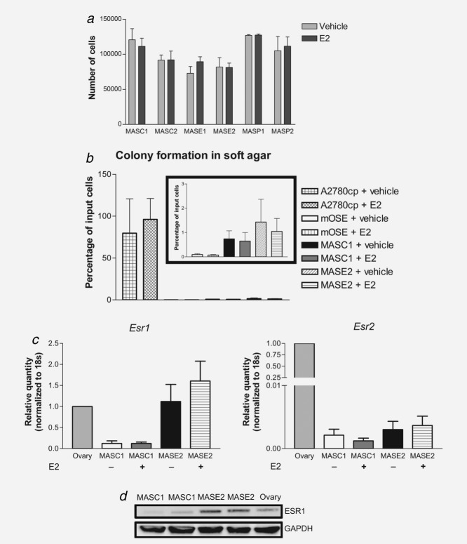
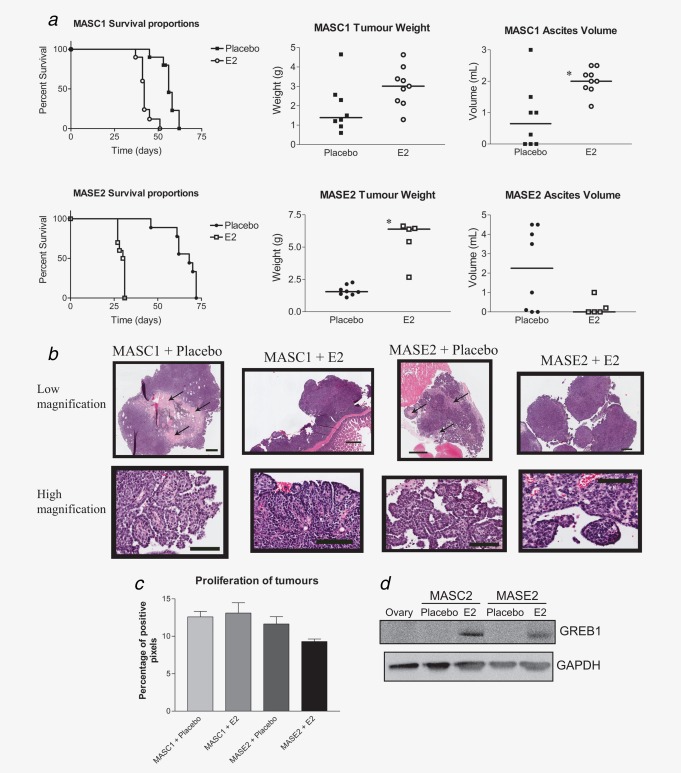
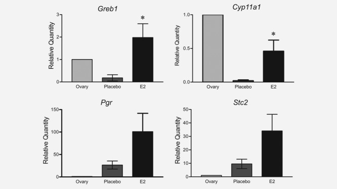
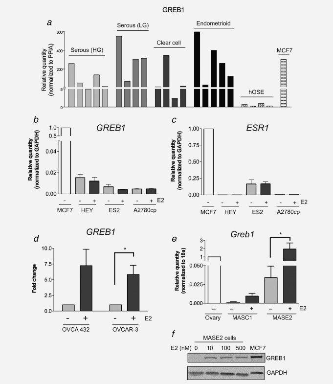
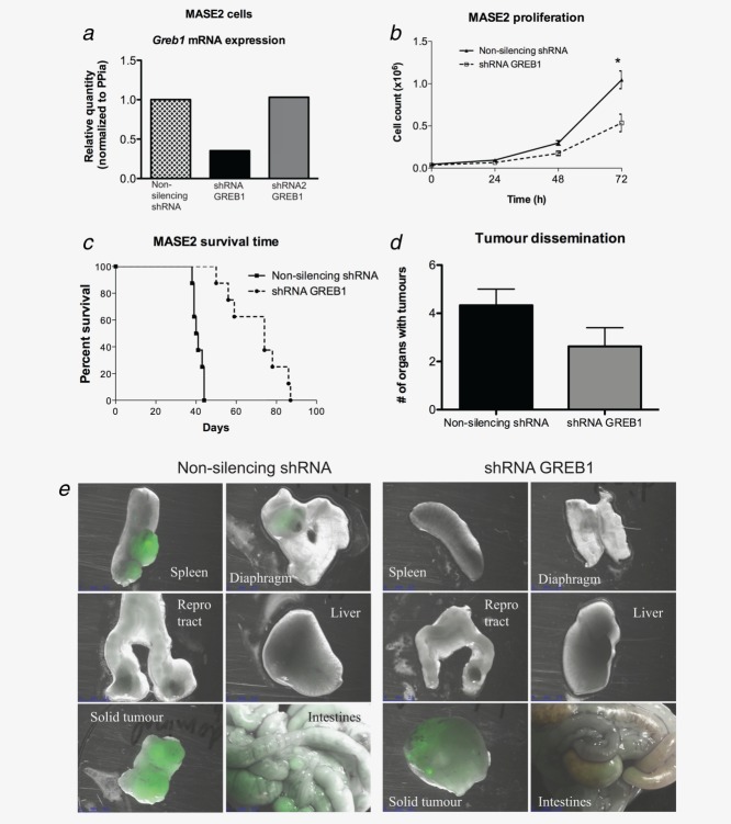
Exogenous 17β-estradiol (E2) accelerates the progression of ovarian cancer in the transgenic tgCAG-LS-TAg mouse model of the disease. We hypothesized that E2 has direct effects on ovarian cancer cells and this study was designed to determine the molecular mechanisms by which E2 accelerates ovarian tumor progression. Mouse ovarian cancer ascites (MAS) cell lines were derived from tgCAG-LS-TAg mice. Following intraperitoneal engraftment of two MAS cell lines, MASC1 and MASE2, into SCID mice, exogenous E2 significantly decreased the survival time and increased the tumor burden. Microarray analysis performed on MASE2-derived tumors treated with E2 or placebo showed that E2 treatment caused the upregulation of 197 genes and the downregulation of 55 genes. The expression of gene regulated by estrogen in breast cancer 1 (Greb1) was upregulated in mouse tumors treated with E2 and was overexpressed in human ovarian cancers relative to human ovarian surface epithelium, suggesting a role for GREB1 in human ovarian tumor progression. RNA interference-mediated knockdown of GREB1 in MASE2 cells decreased their proliferation rate in vitro and increased survival time in mice engrafted with the cells. These results emphasize the importance of E2 in ovarian tumor progression and identify Greb1 as a novel gene target for therapeutic intervention.

Keywords: GREB1; estrogen; microarray; mouse model; ovarian cancer.


Figures

Figure 1
Figure 1
E2 treatment did not affect in vitro proliferation or substrate-independent growth of mouse ovarian cancer cells. (a) The MAS cell lines proliferated at comparable rates and treatment with 500 nM of E2 did not alter the rate of proliferation over 48 hr (compared to those treated with vehicle). (b) E2 (250 nM) did not affect the ability of MASC1 and MASE2 cells to form colonies in soft agar (n = 3; inset is a close up of the mOSE, MASC1 and MASE2 data). (c) Q-PCR analysis revealed that MASE2 cells had higher endogenous Esr1 mRNA levels than MASC1 cells (n = 3, p < 0.05, One-Way ANOVA and Tukey post hoc test), but the abundance of Esr1 transcripts was not altered by treatment with E2 (500 nM). Esr2 transcript levels were very low in the MAS cell lines and were not induced by E2 treatment. (d) ESR1 protein expression (in duplicate) was higher in the MASE2 cells compared to the mouse ovary (control) and to the very low levels seen in the MASC1 cells.
Figure 2
Figure 2
Exogenous E2 treatment decreased the survival time of immunodeficient mice grafted IP with mouse ovarian cancer cell lines MASC1 and MASE2. (a) MASC1 or MASE2 cells were injected IP into SCID mice and 5 days later the mice were treated with E2 or placebo (via a subcutaneous pellet). Mice treated with E2 had a significantly shorter survival time compared to those treated with placebo (MASC1 survival curve, p < 0.0001, n = 9 mice/group; MASE2 survival curve p = 0.0004, N = 7–8 mice/group, Log-rank test). Mice grafted with MASC1 cells and treated with E2 had significantly elevated volumes of ascites compared to placebo controls (*p = 0.0070, t test, N = 8–9). The tumor burden of mice grafted with the MASE2 cells and treated with E2 was significantly more than the placebo controls (*p < 0.0001 t test, N = 5–8). (b) The tumors from MASC1 and MASE2 cells grafted into SCID mice were highly mitotic tumors with serous papillary structures. Low magnification scale bars are 500 μm and the high magnification scale bars are 100 μm. (c) E2 treatment did not affect the proliferation (Ki67 immunoreactivity) of ovarian cancer cells in vivo (One-way ANOVA, N = 4–5 tumors/group). (d) E2 treatment in vivo induced GREB1 expression in both MASC2 and MASE2 tumors.
Figure 3
Figure 3
Q-PCR validation of four microarray targets in MASE2-derived tumors (n = 4–5 tumors/group) treated with and without E2. Greb1 (*p = 0.02, t test) and Cyp11a1 (*p = 0.02, t test) gene expression was significantly increased in tumors from mice treated with E2 compared to controls. There was a strong trend toward increased mRNA expression of Stc2 (p = 0.07, t test) and Pgr (p = 0.09, t test) in E2-treated tumors compared to placebo controls. Whole mouse ovary was used as a positive control.
Figure 4
Figure 4
Greb1 mRNA was expressed in human and mouse ovarian cancers. (a) GREB1 was highly expressed in four epithelial ovarian carcinoma subtypes, including high grade serous, low grade serous, endometrioid and clear cell (N = 4–5 tumors/subtype), but was almost absent in four independent early-passage cultures of human OSE cells. MCF7 breast cancer cells were used as a positive control for GREB1 mRNA expression. (b) GREB1 was expressed in the human ovarian cancer cell lines HEY, ES2 and A2780cp, but was not induced by E2 treatment in vitro. (c) ESR1 mRNA levels were very low in ES2 cells and were undetectable by Q-PCR in HEY and A2780cp cells. (d) GREB1 levels were upregulated by E2 in two ESR1-expressing ovarian cancer cell lines, OVCA 432 (p = 0.051) and OVCAR-3 cells (*p = 0.016). (e) Greb1 mRNA was present in MASC1 and MASE2 cells and was significantly increased in the MASE2 cells following treatment with 500 nM E2 for 48 hr in vitro (*p < 0.05). (f) GREB1 protein level was increased in MASE2 cells by in vitro treatment with E2 (10–500 nM). GAPDH was used as a loading control.
Figure 5
Figure 5
GREB1 knockdown slows the proliferation of MASE2 cells and prolongs survival in an orthotopic model of ovarian cancer. (a) Q-PCR demonstrating decreased mRNA expression of Greb1 in MASE2 cells following shRNA knockdown with construct 1 (shRNA GREB1) but no knockdown with construct 2 (shRNA2 GREB1). (b) GREB1 knockdown in the MASE2 cells significantly slowed cellular proliferation (*p = 0.021, linear regression). (c) Knockdown of GREB1 (shRNA GREB1) significantly prolonged survival (median survival 74 days) after grafting to SCID mice (non-silencing shRNA controls, median survival time 40.5 days, p < 0.0001, Log-rank test). (d) GREB1 knockdown decreased the dissemination of tumors throughout the peritoneal cavity (non-silencing shRNA, N = 6; shRNA GREB1, N = 8). (e) The reduction in tumor dissemination was visualized by immunofluorescent microscopy (green immunofluorescence indicates the presence of cancer cells). Repro, reproductive

Similar articles

Cited by

References

    1. Siegel R, Ma J, Zou Z, Jemal A. Cancer statistics, 2014. CA Cancer J Clin. 2014;64:9–29. - PubMed
    1. Naora H, Montell DJ. Ovarian cancer metastasis: integrating insights from disparate model organisms. Nat Rev Cancer. 2005;5:355–66. - PubMed
    1. Lacey JV, Jr, Mink PJ, Lubin JH, et al. Menopausal hormone replacement therapy and risk of ovarian cancer. JAMA. 2002;288:334–41. - PubMed
    1. Glud E, Kjaer SK, Thomsen BL, et al. Hormone therapy and the impact of estrogen intake on the risk of ovarian cancer. Arch Intern Med. 2004;164:2253–9. - PubMed
    1. Beral V, Bull D, Green J, et al. Ovarian cancer and hormone replacement therapy in the Million Women Study. Lancet. 2007;369:1703–10. - PubMed

Publication types

MeSH terms