. 2014 Feb 11;111(6):2146-51.
doi: 10.1073/pnas.1314138111. Epub 2014 Jan 27.

Septin assemblies form by diffusion-driven annealing on membranes

Affiliations

Septin assemblies form by diffusion-driven annealing on membranes

Andrew A Bridges et al. Proc Natl Acad Sci U S A. .

Abstract

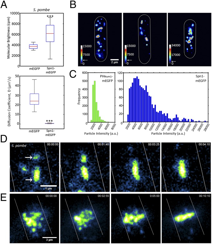
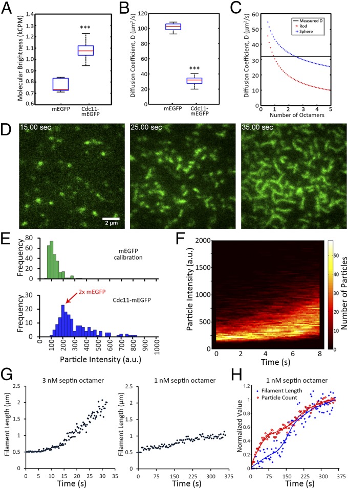
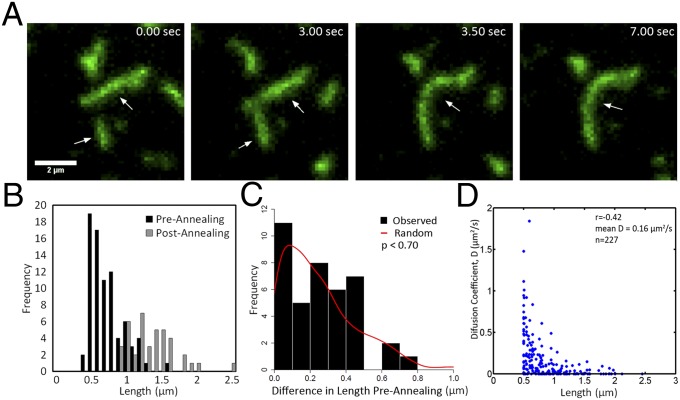
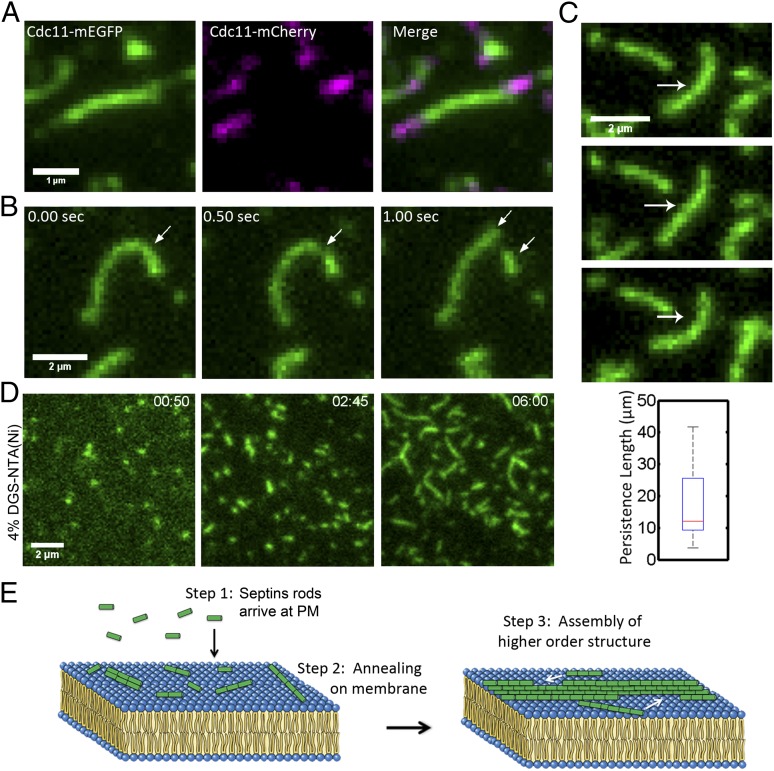
Septins assemble into filaments and higher-order structures that act as scaffolds for diverse cell functions including cytokinesis, cell polarity, and membrane remodeling. Despite their conserved role in cell organization, little is known about how septin filaments elongate and are knitted together into higher-order assemblies. Using fluorescence correlation spectroscopy, we determined that cytosolic septins are in small complexes, suggesting that septin filaments are not formed in the cytosol. When the plasma membrane of live cells is monitored by total internal reflection fluorescence microscopy, we see that septin complexes of variable size diffuse in two dimensions. Diffusing septin complexes collide and make end-on associations to form elongated filaments and higher-order structures, an assembly process we call annealing. Septin assembly by annealing can be reconstituted in vitro on supported lipid bilayers with purified septin complexes. Using the reconstitution assay, we show that septin filaments are highly flexible, grow only from free filament ends, and do not exchange subunits in the middle of filaments. This work shows that annealing is a previously unidentified intrinsic property of septins in the presence of membranes and demonstrates that cells exploit this mechanism to build large septin assemblies.

Keywords: biophysics; cytoskeleton.


Conflict of interest statement

The authors declare no conflict of interest.

Figures

Fig. 1.
Fig. 1.
Septins grow by annealing on the plasma membrane of S. pombe. (A) Molecular brightness and diffusion coefficient of cytoplasmic Spn1-mEGFP (AGY093) determined by FCS compared with those of cells expressing cytoplasmic mEGFP (AGY109) alone in S. pombe. Box plots display median, lower, and upper quartiles and whiskers show range n > 20 cells, ***P < 0.001 by Kruskal–Wallace one-way analysis of variance. (B) Spn1-mEGFP particles and short filaments localized on the cortex of S. pombe monitored using TIRF microscopy. Heat maps represent relative particle intensities. Arrows on the intensity scale indicate the estimated intensity of a complex containing two Cdc11-mEGFP molecules, ∼4,000 a.u., based on calibration. Cell outlines are shown in white. (C) Distribution of PHNum1-GFP and Spn1-mEGFP particle intensities on the plasma membrane measured from images collected using the same laser intensity and exposure time. (D) Annealing event between two diffusing short septin filaments on the cell cortex of S. pombe. Arrows indicate particles involved in annealing. Cell outines are shown in white. (E) Formation of Spn1-mEGFP septin ring by annealing of intermediate-size particles at the plane of division in S. pombe. Cell outines are shown in white. With the exception of FCS (A), all images were acquired using TIRF microscopy.
Fig. 2.
Fig. 2.
Reconstituted septin assembly on supported lipid bilayers. (A) Molecular brightness of pure mEGFP compared with that of copurified, recombinant septins containing Cdc11-mEGFP in solution, as measured by FCS. Box plots display median, lower, and upper quartiles and whiskers show range. ***P < 0.001 by Kruskal–Wallace one-way analysis of variance. (B) Diffusion coefficients of pure mEGFP compared with Cdc11-mEGFP containing septin complexes, estimated by fitting FCS results to a single-component anamolous diffusion model. (C) Diffusion coefficients were predicted for complexes of different numbers of septin octamers, using previously measured dimensions (i.e., length = 32 nm, diameter = 4 nm, molecular mass = 443 kDa) modeled as either rod-like or spherical in organization for a given molecular mass. Horizontal black line is measured diffusion constant, red asterisks are predicted diffusion constants for septin complexes in a rod-like shape, and blue asterisks are diffusion constants of complexes arranged in a sphere. (D) Cdc11-mEGFP signal accumulates and forms filaments at the lipid bilayer over time (3 nM octamer concentration), monitored by TIRF microscopy. (E) Quantification of fluorescence intensity (background subtracted) of single molecules of mEGFP (green histogram) and Cdc11-mEGFP complexes in the first moment of arrival at the bilayer (3 nM, blue histograms). (F) TIRF microscopy was used to measure septin particle brightness in each frame during the first 8 s of assembly (3 nM) in conditions where single molecules of mEGFP fluoresce to 100 a.u. The number of particles of a given intensity is represented by the color code beside the plot (n > 10,000 particles). (G) Filament length was measured over time and the mean length of elongated particles is plotted for each time point in a 3-nM and a 1-nM mixture of septin complex. (H) Particle count and filament length over time in a 1-nM assembly (same data as in G). Values were normalized to maximum values of length or particle count and minimum values were set to zero. These values were fitted and plotted normalized to the best-fit curve.
Fig. 3.
Fig. 3.
Septin filaments grow by annealing on supported lipid bilayers. (A) Representative TIRF image series of filament growth by annealing. Arrows indicate short filaments that merge. (B) Measured filament lengths before annealing and after annealing are plotted (n = 40 annealing events). (C) Histogram shows the length difference between pairs of filaments before annealing. These filaments that participated in annealing events were then paired at random, and differences were calculated from the random pairs and plotted as a red line. The simulated random distribution of differences is not distinguishable from the actual data (insignificant two-sample Kolmogorov–Smirnov test). (D) Filament length and diffusion coefficients were measured and plotted for a population of septin particles at the bilayer.
Fig. 4.
Fig. 4.
Septin filaments elongate at ends, are flexible, and can be formed on a membrane without phosphatidylinositide. (A) Cdc11-mEGFP septin filaments were assembled on a bilayer, unbound complexes were washed out, and Cdc11-mCherry complexes were added and monitored in TIRF. Addition of new complexes occurred at both filament ends. (B) Cdc11-mEGFP filaments fragment on a supported lipid bilayer. Arrows indicate site of fragmentation event and new short filaments arising from the break. (C) Example of filament-bending image series used for determining persistence length of septin filaments. Arrow indicates bending filament. Box plot shows a median persistence length of 12 μm. Filaments used ranged between 1.2 µm and 2.1 µm, with a median of 1.8 µm (n = 10). Box plots display median, lower, and upper quartiles and whiskers show range. (D) Cdc11-mEGFP containing septin filaments assembled on supported lipid bilayers containing 96% (mol%) phosphatidylcholine and 4% (mol%) DGS-NTA(Ni). (E) Model. Step 1: Septins arrive at plasma membranes from cytoplasm as short rods. Step 2: Rods then assemble into short filaments on the plasma membrane through annealing. Step 3: Short filaments then build higher-order structures.

Comment in

References

    1. Dobbelaere J, Barral Y. Spatial coordination of cytokinetic events by compartmentalization of the cell cortex. Science. 2004;305(5682):393–396. - PubMed
    1. Gilden JK, Peck S, Chen YC, Krummel MF. The septin cytoskeleton facilitates membrane retraction during motility and blebbing. J Cell Biol. 2012;196(1):103–114. - PMC - PubMed
    1. Byers B, Goetsch L. A highly ordered ring of membrane-associated filaments in budding yeast. J Cell Biol. 1976;69(3):717–721. - PMC - PubMed
    1. Oh Y, Bi E. Septin structure and function in yeast and beyond. Trends Cell Biol. 2011;21(3):141–148. - PMC - PubMed
    1. Culotti J, Hartwell LH. Genetic control of the cell division cycle in yeast. 3. Seven genes controlling nuclear division. Exp Cell Res. 1971;67(2):389–401. - PubMed

Publication types