. 2014 Feb 11;111(6):2337-42.
doi: 10.1073/pnas.1316294111. Epub 2014 Jan 27.

TBC1D24 regulates neuronal migration and maturation through modulation of the ARF6-dependent pathway

Affiliations

TBC1D24 regulates neuronal migration and maturation through modulation of the ARF6-dependent pathway

Antonio Falace et al. Proc Natl Acad Sci U S A. .

Abstract

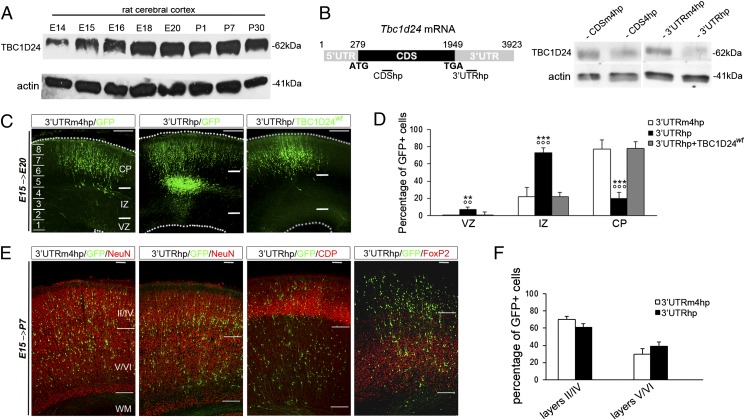
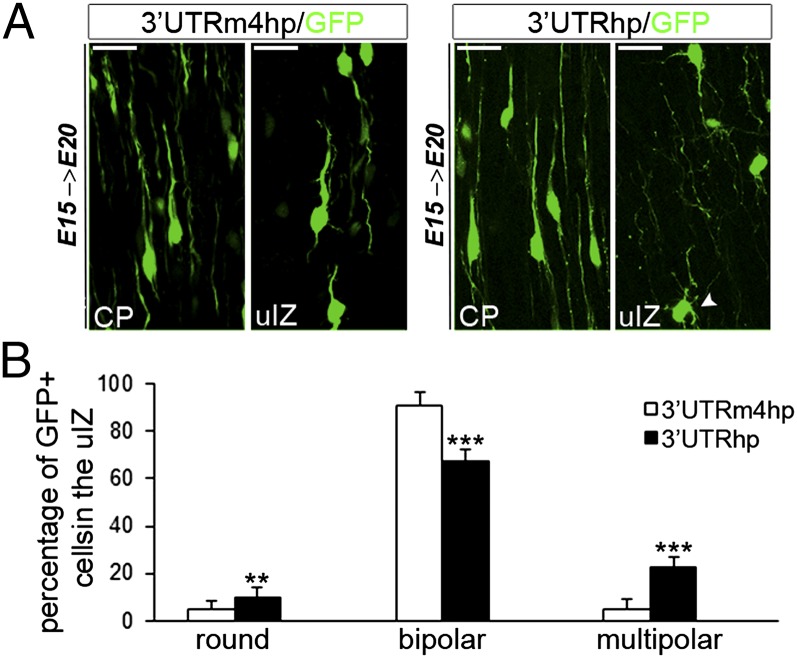
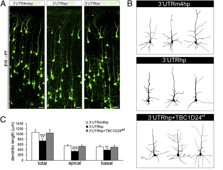
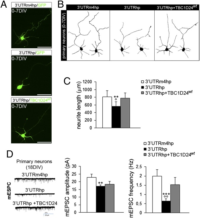
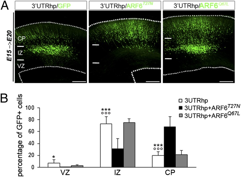
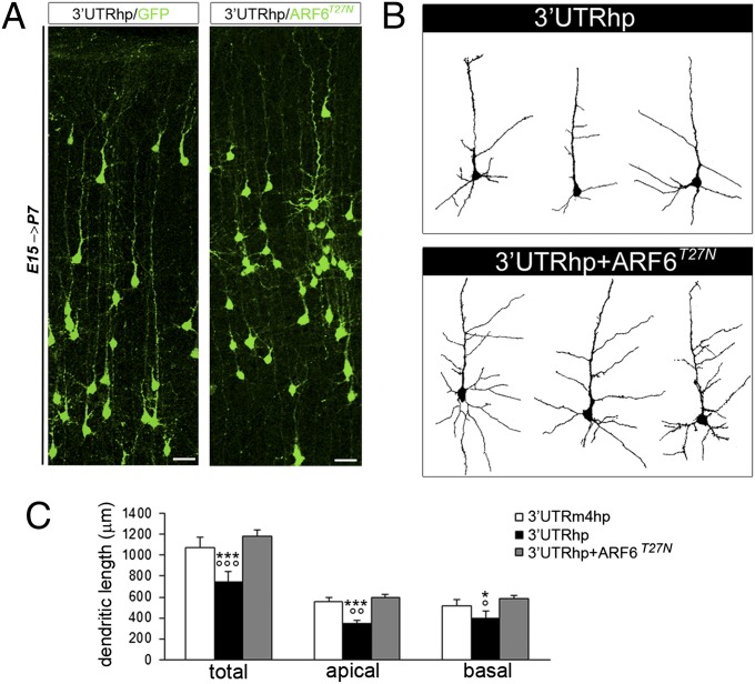
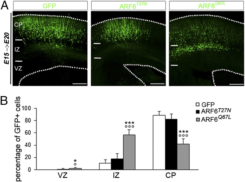
Alterations in the formation of brain networks are associated with several neurodevelopmental disorders. Mutations in TBC1 domain family member 24 (TBC1D24) are responsible for syndromes that combine cortical malformations, intellectual disability, and epilepsy, but the function of TBC1D24 in the brain remains unknown. We report here that in utero TBC1D24 knockdown in the rat developing neocortex affects the multipolar-bipolar transition of neurons leading to delayed radial migration. Furthermore, we find that TBC1D24-knockdown neurons display an abnormal maturation and retain immature morphofunctional properties. TBC1D24 interacts with ADP ribosylation factor (ARF)6, a small GTPase crucial for membrane trafficking. We show that in vivo, overexpression of the dominant-negative form of ARF6 rescues the neuronal migration and dendritic outgrowth defects induced by TBC1D24 knockdown, suggesting that TBC1D24 prevents ARF6 activation. Overall, our findings demonstrate an essential role of TBC1D24 in neuronal migration and maturation and highlight the physiological relevance of the ARF6-dependent membrane-trafficking pathway in brain development.

Keywords: RNA interference; dendritogenesis; epileptic encephalopathies; gene; synaptogenesis.


Conflict of interest statement

The authors declare no conflict of interest.

Figures

Fig. 1.
Fig. 1.
Neuronal migration following embryonic TBC1D24 knockdown. (A) WB analysis of TBC1D24 protein expression from rat cortical lysates during development. (B, Left) Diagram showing the position of shRNAs targeting the coding sequence (CDShp) and the 3′UTR (3′UTRhp) of rat Tbc1d24 mRNA. (B, Right) TBC1D24 protein expression in rat primary neurons transfected at the day of plating with the indicated plasmids and analyzed after 12 d by WB. (C) Representative neocortical coronal sections showing migration of transfected cells 5 d after electroporation at E15 with pCAGGs-IRES-EGFP (GFP) construct combined with the indicated plasmids (3′UTRm4hp, n = 10; 3′UTRhp, n = 12; 3′UTRhp + TBC1D24wt, n = 9). (Scale bar: 200 μm.) (D) Quantification of GFP-positive cell distribution 5 d after transfection at E15 expressed as a percentage of total transfected cells. Data, expressed as means ± SEM, were compared via one-way ANOVA for repeated measures, followed by the Bonferroni’s multiple comparison test. ***P < 0.0001 and **P < 0.005 vs. 3′UTRm4hp; °°°P < 0.0001 and °°P < 0.005 vs. 3′UTRhp + TBC1D24wt. (E) Representative P7 neocortical sections showing the laminar position of GFP cells in rats electroporated at E15 with the indicated plasmids and immunostained with the neuronal marker NeuN, cortical II–IV layers marker CDP and the V–VI layers marker FoxP2. (Scale bar: 500 μm.) (F) Quantification of GFP cells in cortical upper layers (II–IV) and the cortical deep layers (V–VI) and expressed as a percentage of the total number of transfected cells (n = 7 for each condition).
Fig. 2.
Fig. 2.
TBC1D24 knockdown impairs multipolar/bipolar transition in uIZ. (A) E20 rat cortices electroporated at E15 with 3′UTRhp or 3′UTRm4hp in the indicated regions. (Scale bar: 20 μm.) In the TBC1D24-knockdown model, a high percentage of migrating neurons persisted at the multipolar stage (arrowhead). (B) Histograms showing the average percentages (means ± SEM) of the polarity in the uIZ from similar sections as in A (3′UTRm4hp, n = 7; 3′UTRhp, n = 9). One-way ANOVA for repeated measures: ***P < 0.0001; **P < 0.001.
Fig. 3.
Fig. 3.
TBC1D24 is required for dendritic arborization in vivo. (A) Representative coronal sections of P7 rat brains transfected with the indicated constructs by IUE at E15. (Scale bar, 50 μm.) (B) Representative reconstructed layers II/III pyramidal neurons showing dendritic arborization patterns from the same experimental conditions shown in A. (C) Quantitative analysis (means ± SEM) of total dendritic length revealed that TBC1D24 knockdown significantly impairs neuronal morphological maturation affecting both apical and basal dendrites (n = 130–140 in each group). Two-tailed t test: ***P < 0.0005 and *P < 0.05 vs. 3′UTRm4hp; °°°P < 0.0005, °°P < 0.005, and °P < 0.05 vs. 3′UTRhp + TBC1D24wt.
Fig. 4.
Fig. 4.
TBC1D24 regulates neuronal morphogenesis and maturation in vitro. (A) Representative images of 7 DIV cultured cortical neurons transfected at the time of plating with the indicated plasmids. (Scale bar: 50 μm.) (B) Representative traces of 7 DIV cortical neurons treated as in A. (C) Histogram showing quantitative analysis of total neurite length (three independent preparations; 90–100 cells for each group). Data are means ± SEM and were analyzed by paired two-tailed t test. **P < 0.001 vs. 3′UTRm4hp; °P < 0.05 vs. 3′UTRhp + TBC1D24wt. (D, Left) Representative traces of mEPSCs recorded at −70mV in 18 DIV cortical neurons transfected at 13 DIV with the indicated constructs. (D, Right) Histograms showing average amplitude and frequency of mEPSCs. Data are expressed as means ± SEM from 30 to 35 neurons per experimental condition obtained from three independent preparations. Two-tailed t test: **P < 0.01 and ***P < 0.0005 vs. 3′UTRm4hp; °°P < 0.01 vs. 3′UTRhp + TBC1D24wt.
Fig. 5.
Fig. 5.
TBC1D24 regulates neuronal migration through ARF6. (A) Representative coronal sections of E20 rat brains 5 d after electroporation with indicated plasmids. (Scale bar: 200 μm.) (B) Quantification of GFP-positive cell distribution in the cortex expressed as a percentage of total transfected cells (3′UTRhp, n = 12; 3′UTRhp + ARF6T27N, n = 7; 3′UTRhp + ARF6Q67L, n = 4). Data, expressed as means ± SEM, were compared using one-way ANOVA for repeated measure, followed by the Bonferroni’s multiple comparison test. ***P < 0.0001 and *P < 0.05 vs. 3′UTRhp + ARF6T27N; °°°P < 0.0001 vs. 3′UTRhp + ARF6Q67L.
Fig. 6.
Fig. 6.
In vivo down-regulation of ARF6 restores dendritic outgrowth in TBC1D24-knockdown model. (A) Representative images of coronal slices of P7 rat brains transfected with indicated constructs by IUE at E15. (Scale bar: 50 μm.) (B) Representative dendritic arborization patterns in cortical neurons from the same experimental conditions shown in A. (C) Concomitant expression of ARF6T27N in TBC1D24-knockdown neurons restores both apical and basal dendritic outgrowth. Data are expressed as means ± SEM and analyzed by two-tailed t test. ***P < 0.0001 and *P < 0.05 vs. 3′UTRm4hp; °°°P < 0.0001, °°P < 0.0005, and °P < 0.005 vs. 3′UTRhp + ARF6T27N (3′UTRm4hp, n = 130 from six rats; 3′UTRhp, n = 140 from six rats; 3′UTRhp + ARF6T27N, n = 81 from four rats).
Fig. 7.
Fig. 7.
Dysregulation of ARF6 activity impairs neuronal migration. (A) Representative coronal sections of E20 rat brains 5 d after electroporation with indicated plasmids. (Scale bar: 200 μm.) (B) Quantification of GFP-positive cell distribution in the cortex expressed as a percentage of the total (GFP, n = 10; ARF6T27N, n = 6; ARF6Q67L, n = 7). Data, expressed as means ± SEM, were compared via one-way ANOVA for repeated measure, followed by the Bonferroni’s multiple comparison test. ***P < 0.0001 and *P < 0.05 vs. GFP; °°°P < 0.0001 and °P < 0.05 vs. ARF6T27N.

References

    1. McConnell SK. Strategies for the generation of neuronal diversity in the developing central nervous system. J Neurosci. 1995;15(11):6987–6998. - PMC - PubMed
    1. Kriegstein A, Alvarez-Buylla A. The glial nature of embryonic and adult neural stem cells. Annu Rev Neurosci. 2009;32:149–184. - PMC - PubMed
    1. Rakic P. Evolution of the neocortex: A perspective from developmental biology. Nat Rev Neurosci. 2009;10(10):724–735. - PMC - PubMed
    1. Ayala R, Shu T, Tsai LH. Trekking across the brain: The journey of neuronal migration. Cell. 2007;128(1):29–43. - PubMed
    1. Rakic P, Ayoub AE, Breunig JJ, Dominguez MH. Decision by division: Making cortical maps. Trends Neurosci. 2009;32(5):291–301. - PMC - PubMed

Publication types

LinkOut - more resources