. 2014 Feb 11;111(6):2265-70.
doi: 10.1073/pnas.1317431111. Epub 2014 Jan 27.

Tumor-specific IL-9-producing CD8+ Tc9 cells are superior effector than type-I cytotoxic Tc1 cells for adoptive immunotherapy of cancers

Affiliations

Tumor-specific IL-9-producing CD8+ Tc9 cells are superior effector than type-I cytotoxic Tc1 cells for adoptive immunotherapy of cancers

Yong Lu et al. Proc Natl Acad Sci U S A. .

Abstract

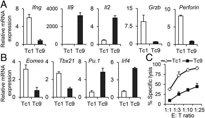
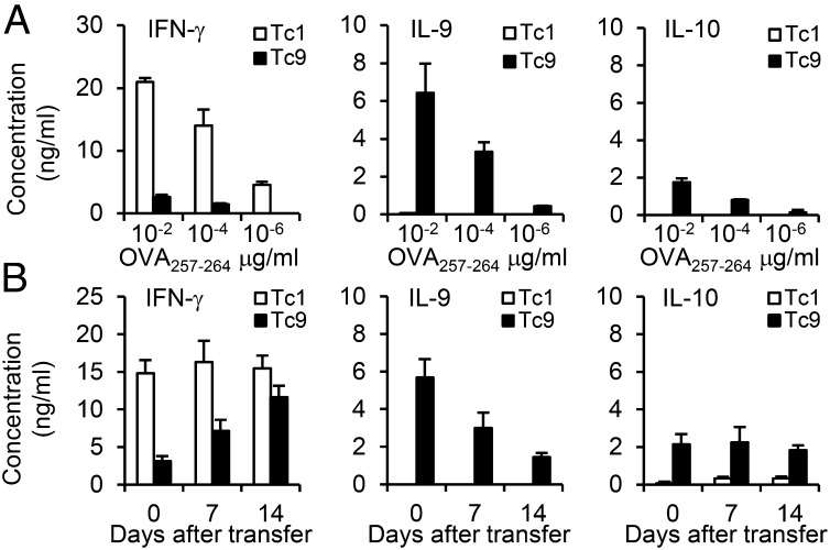
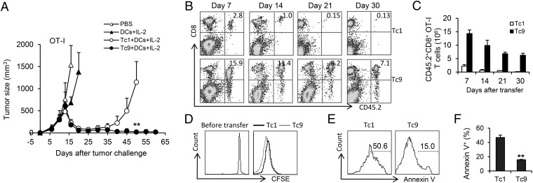
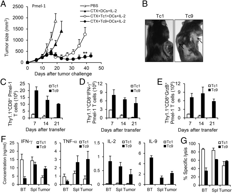
Because cytokine-priming signals direct CD8(+) T cells to acquire unique profiles that affect their ability to mediate specific immune responses, here we generated IL-9-skewed CD8(+) T (Tc9) cells by priming with Th9-polarized condition. Compared with type-I CD8(+) cytotoxic T (Tc1) cells, Tc9 secreted different cytokines and were less cytolytic in vitro but surprisingly elicited greater antitumor responses against advanced tumors in OT-I/B16-OVA and Pmel-1/B16 melanoma models. After adoptive transfer, Tc9 cells persisted longer and differentiated into IFN-γ- and granzyme-B (GrzB)-producing cytolytic Tc1-like effector cells. Phenotypic analysis revealed that adoptively transferred Tc9 cells secreted IL-2 and were KLRG-1(low) and IL-7Rα(high), suggesting that they acquired a signature of "younger" phenotype or became long-term lived cells with capacity of self-renewal. Our results also revealed that Tc9-mediated therapeutic effect critically depended on IL-9 production in vivo. These findings have clinical implications for the improvement of CD8(+) T-cell-based adoptive immunotherapy of cancers.

Keywords: T-cell lineage plasticity; adoptive cell therapy; less-exhausted T cells.


Conflict of interest statement

The authors declare no conflict of interest.

Figures

Fig. 1.
Fig. 1.
Tc9 cells produced IL-9 and were diverted from cytolytic differentiation. (A and B) Real-time PCR analysis of relative mRNA expression of cytokines and cytolytic-related molecules (A) or transcription factors (B) in OT-I Tc1 and Tc9 cells. Expression relative to Gapdh is displayed. (C) T cells were added at the indicated ratios to CFSEhi B16-OVA target cells or CFSElo B16 nontarget cells in duplicate. Percent of specific lysis was determined after 8 h. Representative results from one of two performed experiments are shown.
Fig. 2.
Fig. 2.
Cytokine expression profile of Tc9 cells. (A) OT-I Tc1 or Tc9 cells were primed in polarized conditions and expanded with IL-2. The cells were then restimulated with splenocytes pulsed with OVA257–264 at indicated concentrations for 24 h. Production of indicated cytokines was determined by ELISA. (B) OT-I Tc1 or Tc9 cells (2 × 10⁶) were adoptively transferred into CD45.1-transgenic mice, followed by i.v. injection of 5 × 10⁵ OVA257–264-pulsed DCs and i.p. injection of four doses of exogenous IL-2. CD45.2+ transferred cells were sorted from splenocytes at days 7 and 14. Day 0 represents T cells before transfer. The cells were then restimulated with splenocytes pulsed with 0.01 µg/mL OVA257–264 in triplicate for 24 h. Production of indicated cytokines was determined by ELISA. Representative results from one of two repeated experiments are shown.
Fig. 3.
Fig. 3.
OT-I Tc9 cells mediated enhanced antitumor response and displayed greater persistence. (A–C) Tc1 or Tc9 cells (2 × 10⁶) were adoptively transferred into CD45.1-transgenic mice bearing 10-d large established B16-OVA melanoma. DC vaccination and IL-2 were administered to some group of mice as indicated. (A) Tumor responses (n = 5) to adoptive transfer of Tc1 or Tc9 were shown. (B and C) Persistence of transferred Tc1 or Tc9 cells in the spleens of treated tumor-bearing mice was analyzed by FACS. Numbers in histograms (B) represent the percentage of CD45.2+CD8+ OT-I T cells in splenocytes. (C) Total number of CD45.2+CD8+ OT-I T cells was calculated from B. The spleens of three mice per condition were examined at each time point. (D) CFSE-labeled Tc1 or Tc9 cells were transferred into tumor-bearing mice. Shown is CFSE dilution of gated CD45.2+CD8+ splenocytes 4 d after transfer. (E and F) Annexin V expression was measured in Tc1 and Tc9 cells 4 d after transfer. (E) Numbers in histograms represent the percentage of Annexin V+ apoptotic Tc1 or Tc9 cells in splenocytes. Summarized (n = 3) percentages of apoptotic transferred cells were shown in F. Representative results from one of two repeated experiments are shown. **P < 0.01.
Fig. 4.
Fig. 4.
Tc9 cells display less exhausted phenotype and switch to Tc1-like cells in tumor-bearing mice. Tumor-bearing mice (n = 3) were transferred with OT-I Tc1 or Tc9 cells and treated the same as described in Fig. 1. Splenocytes were harvested and analyzed. (A) Expression of IL-7Rα by transferred cells 7 d after transfer. (B) Expression of indicated exhaustion markers by transferred cells 7 d after transfer. (C) FACS determination of intracellular cytokine production by Tc1 or Tc9 cells before and after transfer. (D) Total number of IFN-γ–producing Tc1 or Tc9 cells after transfer was calculated from FACS analysis. (E) FACS determination of GrzB-producing Tc1 or Tc9 cells after transfer. (F) Total number of GrzB-producing Tc1 or Tc9 cells after transfer was calculated from FACS analysis. Representative results from one of two performed experiments are shown. *P < 0.05; **P < 0.01.
Fig. 5.
Fig. 5.
CTX synergizes with Pmel-1 Tc9 cells to control large established B16 melanoma in vivo. Pmel-1 Tc1 or Tc9 cells were primed in polarized conditions and expanded with IL-2. Tc1 or Tc9 cells (2 × 10⁶) were adoptively transferred into C57BL/6 mice bearing 10-d large established B16 melanoma. One dose of CTX was given 1 d before T-cell transfer. DC vaccination and IL-2 were administered to some group of mice after T-cell transfer. (A) Tumor responses (n = 5) to adoptive transfer of Tc1 or Tc9 were shown. (B) Representative autoimmune vitiligo of tumor-bearing mice 25 d after T-cell transfer. Arrows indicated the presence of vitiligo. (C) Persistence of transferred Tc1 or Tc9 cells in the spleens of treated tumor-bearing mice was analyzed by FACS. (D and E) Total numbers of IFN-γ–producing (D) or GrzB-producing (E) Thy1.1+CD8+ cells after transfer were calculated from FACS analysis. (F) Transferred Tc1 or Tc9 cells were sorted from the spleens or tumor tissues at day 14 after transfer. The cells were then restimulated with splenocytes pulsed with 0.01 µg/mL hgp10025–33 peptide in triplicate for 24 h. Production of indicated cytokines was determined by ELISA. BT represents cells before transfer. (G). Transferred Tc1 or Tc9 cells were sorted from the spleens or tumor tissues at day 14 after transfer. Cytolytic function of T cells was tested by in vitro cytotoxicity assay at 10 to 1 effector to target ratios with CFSEhi B16 target cells and CFSElo MC38 nontarget cells in duplicate. Percentage of specific lysis was determined overnight. Representative results from one of two performed experiments are shown. **P < 0.01.
Fig. 6.
Fig. 6.
IL-9 contributes to Tc9 cell-mediated tumor rejection. Pmel-1 Tc9 cells (2 × 10⁶) were adoptively transferred into C57BL/6 mice bearing 10-d large established MC38-gp100 tumor. One dose of CTX was given 1 d before T-cell transfer. DC vaccination and IL-2 were administered to the mice that received T-cell transfer. mAbs neutralizing IL-9 or IFN-γ or control IgG were i.p. injected to mice as indicated. (A) Tumor responses (n = 5) to adoptive transfer of Tc9 and antibody treatment were shown. (B) FACS determination of the percentage of tumor-infiltrating, adoptively transferred Thy1.1+CD8+ T cells, IFN-γ–producing or GrzB-producing tumor-infiltrating, adoptively transferred Thy1.1+CD8+ T cells in the leukocyte fraction. Tumor tissues were harvested 3 wk after transfer. (C) Total number of tumor-infiltrating, IFN-γ–producing or GrzB-producing Thy1.1+CD8+ T cells 3 wk after transfer was calculated from FACS analysis. Cell number was normalized to 500-mg tumor tissues. Representative results from one of two performed experiments are shown. *P < 0.05; **P < 0.01.

Similar articles

Cited by

References

    1. Restifo NP, Dudley ME, Rosenberg SA. Adoptive immunotherapy for cancer: Harnessing the T cell response. Nat Rev Immunol. 2012;12(4):269–281. - PMC - PubMed
    1. Rosenberg SA, Dudley ME. Adoptive cell therapy for the treatment of patients with metastatic melanoma. Curr Opin Immunol. 2009;21(2):233–240. - PMC - PubMed
    1. Rosenberg SA, et al. Durable complete responses in heavily pretreated patients with metastatic melanoma using T-cell transfer immunotherapy. Clin Cancer Res. 2011;17(13):4550–4557. - PMC - PubMed
    1. Hinrichs CS, et al. Adoptively transferred effector cells derived from naive rather than central memory CD8+ T cells mediate superior antitumor immunity. Proc Natl Acad Sci USA. 2009;106(41):17469–17474. - PMC - PubMed
    1. Hinrichs CS, et al. Human effector CD8+ T cells derived from naive rather than memory subsets possess superior traits for adoptive immunotherapy. Blood. 2011;117(3):808–814. - PMC - PubMed

Publication types

MeSH terms