. 2014 Feb 11;111(6):2200-5.
doi: 10.1073/pnas.1305609111. Epub 2014 Jan 27.

Maternal tract factors contribute to paternal seminal fluid impact on metabolic phenotype in offspring

Affiliations

Maternal tract factors contribute to paternal seminal fluid impact on metabolic phenotype in offspring

John J Bromfield et al. Proc Natl Acad Sci U S A. .

Abstract

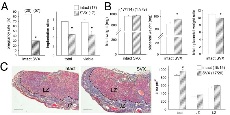
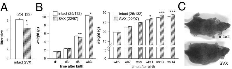
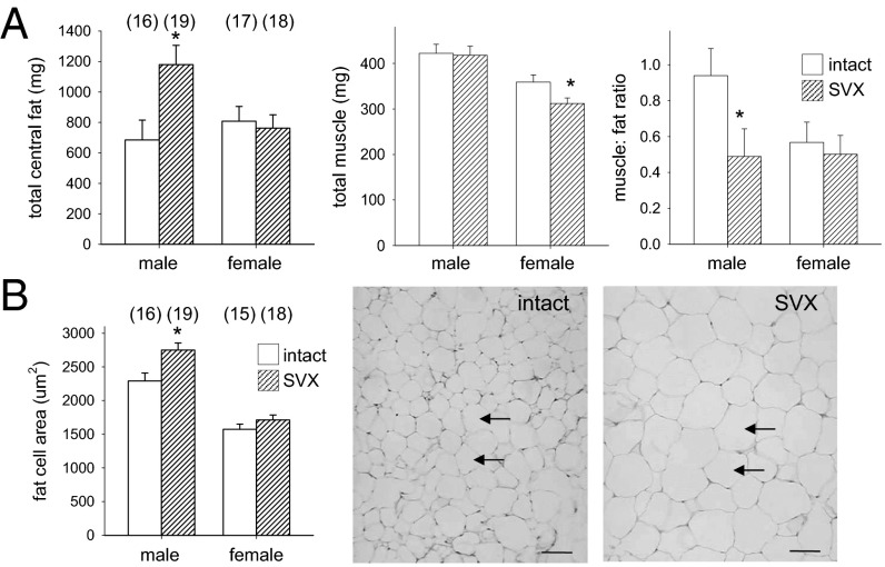
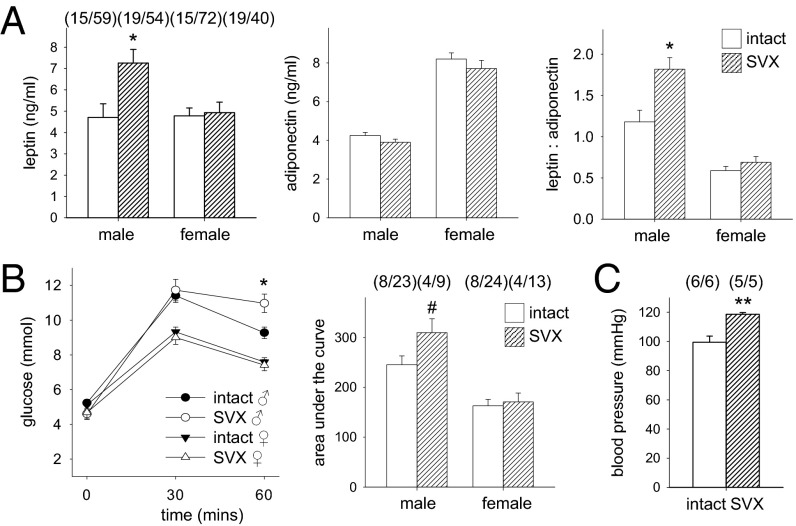
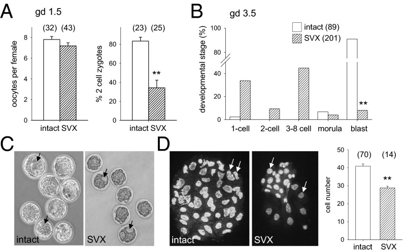
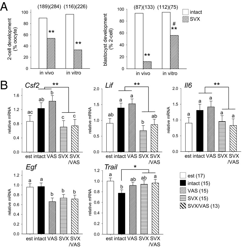
Paternal characteristics and exposures influence physiology and disease risks in progeny, but the mechanisms are mostly unknown. Seminal fluid, which affects female reproductive tract gene expression as well as sperm survival and integrity, provides one potential pathway. We evaluated in mice the consequences for offspring of ablating the plasma fraction of seminal fluid by surgical excision of the seminal vesicle gland. Conception was substantially impaired and, when pregnancy did occur, placental hypertrophy was evident in late gestation. After birth, the growth trajectory and metabolic parameters of progeny were altered, most profoundly in males, which exhibited obesity, distorted metabolic hormones, reduced glucose tolerance, and hypertension. Altered offspring phenotype was partly attributable to sperm damage and partly to an effect of seminal fluid deficiency on the female tract, because increased adiposity was also evident in adult male progeny when normal two-cell embryos were transferred to females mated with seminal vesicle-excised males. Moreover, embryos developed in female tracts not exposed to seminal plasma were abnormal from the early cleavage stages, but culture in vitro partly alleviated this. Absence of seminal plasma was accompanied by down-regulation of the embryotrophic factors Lif, Csf2, Il6, and Egf and up-regulation of the apoptosis-inducing factor Trail in the oviduct. These findings show that paternal seminal fluid composition affects the growth and health of male offspring, and reveal that its impact on the periconception environment involves not only sperm protection but also indirect effects on preimplantation embryos via oviduct expression of embryotrophic cytokines.

Keywords: embryo development; fertility; growth factors; metabolic disorder; programming.


Conflict of interest statement

The authors declare no conflict of interest.

Figures

Fig. 1.
Fig. 1.
Seminal vesicle excision reduces fertility and fecundity and affects placental size. (A) Female mice mated with SVX males achieved pregnancy infrequently, compared with controls mated with intact males. In pregnancies sired by SVX males, total and viable implantation sites visible in the uterus were fewer than in controls on gd 17.5. Numbers of mated and pregnant females are in parentheses. (B) In pregnancies sired by SVX males, fetal weight was not changed but placental weight was larger, and fetal-to-placental weight ratio was smaller, than in controls. (C) Total cross-sectional area of midsagittal sections of placentas in pregnancies sired by SVX males, stained with Masson’s trichrome, was increased compared with controls, with an increase in both the labyrinthine zone (LZ) and junctional zone (JZ). See Fig. S4 for an enlargement. (Scale bars, 450 μm.) The dotted line is the JZ/LZ boundary. Pregnancy rate is analyzed by χ2 test; other data are the estimated marginal mean ± SEM and were compared by mixed-model ANOVA, with mother as subject and litter size as covariate (*P < 0.05). Numbers of mated dams and implantation sites are in parentheses.
Fig. 2.
Fig. 2.
Seminal vesicle excision alters postnatal growth and weight of adult offspring. (A) Litters sired by SVX males contained fewer pups at birth, compared with control litters sired by intact males. (B) Postnatal weight of pups sired by SVX males was unchanged on days 1 and 3 after birth but reduced at days 8 and 21, compared with control pups. Postweaning growth trajectory of progeny sired by SVX males was accelerated compared with control progeny. Data are the estimated marginal mean ± SEM, and the effect of seminal fluid was determined by mixed-model ANOVA, with litter size and sex as covariates (*P < 0.05; **P < 0.01; ***P < 0.001). See Fig. S1 for offspring growth data according to sex. Numbers of mated dams and progeny are in parentheses. (C) Representative male progeny at 14 wk.
Fig. 3.
Fig. 3.
Seminal vesicle excision increases central fat in adult offspring. (A) In adult progeny of SVX males at 14 wk, the total central fat mass was increased in males, the total muscle mass was reduced in females, and the muscle-to-fat ratio was reduced in males, compared with control adult progeny sired by intact males. (B) Average adipocyte area in retroperitoneal fat was increased in male progeny of SVX males, compared with controls. (Scale bars, 50 µm.) Arrows indicate adipocytes. See Fig. S4 for an enlargement. Data are the estimated marginal mean ± SEM, and the effect of seminal fluid was evaluated for each sex by mixed-model ANOVA, with litter size as covariate (*P < 0.05). Numbers of mated dams and progeny are in parentheses.
Fig. 4.
Fig. 4.
Seminal vesicle excision alters metabolic parameters in adult offspring. (A) In adult male progeny of SVX males at 14 wk, plasma leptin and leptin-to-adiponectin ratio were elevated, compared with control male progeny sired by intact males. (B) Glucose clearance assessed at 60 min after challenge and as the area under the curve was slowed compared with control males at 14 wk. (C) Resting systolic blood pressure of male progeny was elevated, compared with control males at 14 wk. Data are the estimated marginal mean ± SEM, and the effect of seminal fluid was evaluated by mixed-model ANOVA (*P < 0.05; **P < 0.01; #P = 0.062). Numbers of mated dams and progeny are in parentheses.
Fig. 5.
Fig. 5.
Seminal vesicle excision impairs preimplantation embryo development. (A) Embryos flushed on gestational day (gd) 1.5 from the oviducts of females mated with SVX males were comparable in number but the rate of zygote cleavage to two-cell stage was reduced, compared with control embryos sired by intact males. (B) Embryos flushed from the uterus on gd 3.5 showed impaired development to blastocyst stage when sired by SVX males, compared with controls. (C and D) Blastocysts sired by SVX males showed frequent irregularities in blastocoel cavity formation (arrows) (C) and contained fewer blastomeres compared with control blastocysts (D). Embryo developmental stage was analyzed by χ2 test; other data are mean ± SEM and were compared by one-way ANOVA (**P < 0.01). Numbers of mated dams and embryos are in parentheses.
Fig. 6.
Fig. 6.
Seminal vesicle excision imparts maternal tract-mediated inhibitory effects and alters oviduct expression of embryotrophic cytokines. (A) The rate of cleavage to two-cell stage in zygotes from females mated with SVX males was less compared with control zygotes sired by intact males when zygotes were developed in vivo or flushed from the oviducts on gd 0.5 and developed in vitro (**P < 0.01). In contrast, development to blastocyst in two-cell embryos sired by SVX males was increased when embryos were developed in vitro, compared with in vivo (#P < 0.01), but less than in controls (**P < 0.01). (B) Oviduct expression of the cytokines Csf2, Lif, Il6, Egf, and Trail on gd 0.5 after mating with intact, VAS, SVX, SVX/VAS, or estrous females (est). Data are the estimated marginal mean ± SEM, and the effect of seminal fluid was evaluated by mixed-model ANOVA. Different superscripts represent statistical difference between groups. *P < 0.05, **P < 0.01 compared with females exposed to seminal plasma. Numbers of embryos and mated females are in parentheses.

References

    1. Poiani A. Complexity of seminal fluid: A review. Behav Ecol Sociobiol. 2006;60(3):289–310.
    1. Avila FW, Sirot LK, LaFlamme BA, Rubinstein CD, Wolfner MF. Insect seminal fluid proteins: Identification and function. Annu Rev Entomol. 2011;56:21–40. - PMC - PubMed
    1. Robertson SA. Seminal plasma and male factor signalling in the female reproductive tract. Cell Tissue Res. 2005;322(1):43–52. - PubMed
    1. Sharkey DJ, Tremellen KP, Jasper MJ, Gemzell-Danielsson K, Robertson SA. Seminal fluid induces leukocyte recruitment and cytokine and chemokine mRNA expression in the human cervix after coitus. J Immunol. 2012;188(5):2445–2454. - PubMed
    1. Tremellen KP, Seamark RF, Robertson SA. Seminal TGFbeta1 stimulates GM-CSF production and inflammatory cell recruitment in the murine uterus. Biol Reprod. 1998;58(5):1217–1225. - PubMed