. 2014 Feb 11;111(6):2116-21.
doi: 10.1073/pnas.1321022111. Epub 2014 Jan 27.

A vertebrate myosin-I structure reveals unique insights into myosin mechanochemical tuning

Affiliations

A vertebrate myosin-I structure reveals unique insights into myosin mechanochemical tuning

Henry Shuman et al. Proc Natl Acad Sci U S A. .

Abstract

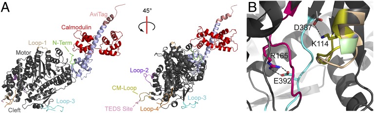
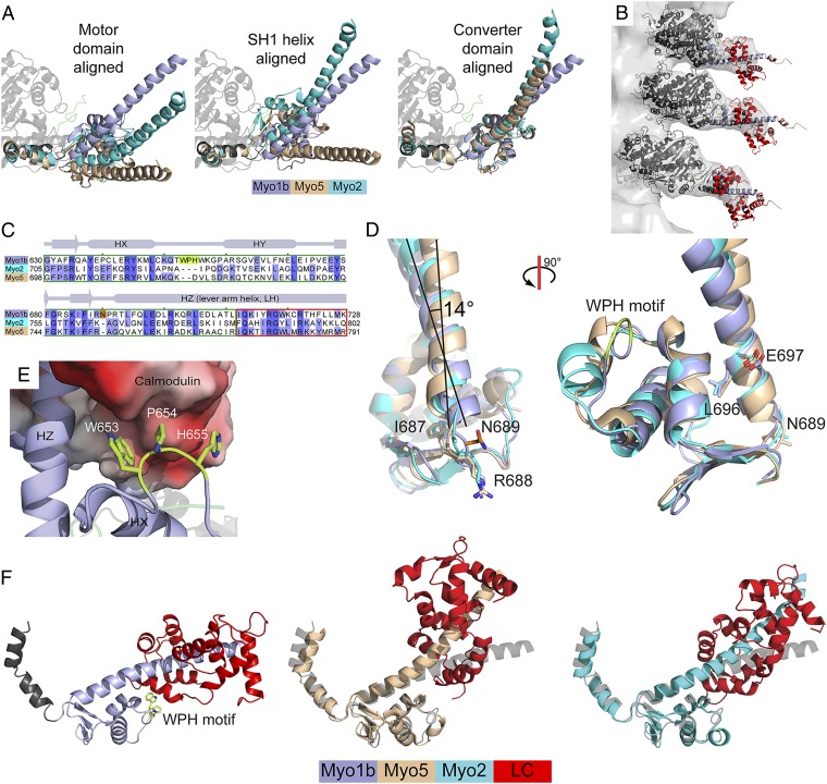
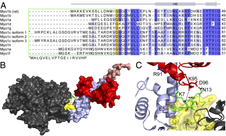
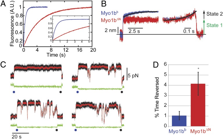
Myosins are molecular motors that power diverse cellular processes, such as rapid organelle transport, muscle contraction, and tension-sensitive anchoring. The structural adaptations in the motor that allow for this functional diversity are not known, due, in part, to the lack of high-resolution structures of highly tension-sensitive myosins. We determined a 2.3-Å resolution structure of apo-myosin-Ib (Myo1b), which is the most tension-sensitive myosin characterized. We identified a striking unique orientation of structural elements that position the motor's lever arm. This orientation results in a cavity between the motor and lever arm that holds a 10-residue stretch of N-terminal amino acids, a region that is divergent among myosins. Single-molecule and biochemical analyses show that the N terminus plays an important role in stabilizing the post power-stroke conformation of Myo1b and in tuning the rate of the force-sensitive transition. We propose that this region plays a general role in tuning the mechanochemical properties of myosins.

Keywords: cryo-EM; mechanochemistry; optical tweezers; structural biology.


Conflict of interest statement

The authors declare no conflict of interest.

Figures

Fig. 1.
Fig. 1.
Structure of apo-Myo1bIQ. (A) Overall structure of Myo1bIQ shows key structural elements and the AviTag. (B) Structural motifs from the active site of Myo1b (red box from Fig. S1B) are colored: P-loop, yellow; SW-1, blue; SW-2, red. Conserved interactions are highlighted. A113 is colored green, and G112, which is partially occluded by A113, is colored blue. The P-loop of Myo5 (PDB ID code 1OE9) is shown in tan.
Fig. 2.
Fig. 2.
(A) Comparison of the LAH of Myo1bIQ, Myo5, and Myo2. The structures are aligned using the core motor domain (Myo1bIQ residues 26–607, Left), the SH1 helix (residues 621–629, Center), and the converter domain (residues 634–700, Right). (B) Helical reconstruction of cryoelectron micrographs shows that the long axis of the LAH of Myo1bIQ in the nucleotide-free state is nearly perpendicular to the long axis of the actin filament. Three duplicate models of the Myo1bIQ structure are docked into the EM map. The low signal-to-noise ratio of the density from the N-terminal lobe of the calmodulin likely resulted from conformational flexibility and the greater disorder that occurs at the higher radius of the decorated filament. The flexibility of the calmodulin N-terminal lobe is consistent with the higher b-factors for this region in the crystal structure. (C) Structure-based alignment of the converter domains and IQ motifs of Myo1bIQ, Myo5 (PDB ID code 1OE9), and Myo2 (PDB ID code 2MYS). The two myosin-I–specific insertions are highlighted in lime and orange. (D) N689 insertion shifts the base of the LAH between conserved residues immediately N-terminal (R688) and C-terminal (L696), which are structurally equivalent Myo1b, Myo5, and Myo2. The insertion buckles the loop in which it resides, causing a 3.9-Å shift in the N terminus of the LAH. (E) Myo1b WPH motif interacts with a hydrophobic and acidic surface on the C-terminal lobe of the calmodulin bound to the IQ motif. (F) Distinct LAH positions of Myo1bIQ, Myo5, and Myo2 result in distinct positions of the bound light chain (LC). Structures were aligned using converter domains (residues 634–700 in Myo1bIQ). Light chains are shown in red, and Myo1bIQ is shown in gray.
Fig. 3.
Fig. 3.
Myo1bIQ NTR is located between the motor domain and LAH. (A) Sequence alignment of the N termini of human myosin-I isoforms and the rat Myo1b isoform used in this study. The green box shows the divergent NTR. High sequence conservation is present after the invariant glycine residue (orange box). For clarity, the first 18 residues of Myo1c isoform-1 are shown below the alignment. (B) NTR (green) is sandwiched between the motor (gray) and LAH (blue). A hydrophobic core (yellow), composed of residues from the motor, NTR, and LAH, is at the base of the LAH. (C) Magnification of the base of the LAH shows the concentration of hydrophobic residues. Interactions between K7 of the NTR and the C-terminal lobe of calmodulin are shown. The molecule is rotated ∼180° about the y axis, compared with B.
Fig. 4.
Fig. 4.
Transient kinetic and optical trapping measurements of Myo1bb (blue) and Myo1bΔN (red). (A) Rate of ADP release, as measured using stopped-flow transient kinetics (Materials and Methods), is slowed ∼10-fold by deletion of the NTR. Shown are the normalized fluorescence transients as a function of time. (Inset) Transient is shown at a shorter time scale. Fluorescence is plotted in arbitrary units (A.U.). (B) Ensemble averages of the Myo1bb (n = 561) and Myo1bΔN (n = 230) working stroke were constructed as described in Materials and Methods. The time-forward ensemble averages with a single-exponential fit to the Myo1bb data (Left) and the time-reversed averages with a single-exponential fit to the Myo1bΔN data (Right) are shown. Both constructs show a two-step working stroke with similarly sized substeps. The measured values for the step and substep sizes can be found in Table 2. (C) Sample data collected with the Myo1bΔN construct under a load imposed by the isometric optical clamp. Each panel is a single actomyosin-binding event that begins when the covariance of the two trapped beads, shown in green, becomes low (blue asterisk) and ends when the covariance becomes high (black asterisk). Raw data are shown in black, and the Savitzky–Golay-filtered data are shown in red. The first actomyosin-binding event shows no reversals, whereas the other binding events show transient decreases in force without actomyosin detachment (as seen by the constant covariance over the course of the binding event). (D) Quantification of the percentage of time spent in the reversed state as described in Materials and Methods. Myo1bΔN spends fourfold the time reversed compared with Myo1bb.
Scheme 1.
Scheme 1.
Actomyosin ATPase Cycle.

Similar articles

Cited by

References

    1. De La Cruz EM, Ostap EM. Relating biochemistry and function in the myosin superfamily. Curr Opin Cell Biol. 2004;16(1):61–67. - PubMed
    1. Greenberg MJ, Ostap EM. Regulation and control of myosin-I by the motor and light chain-binding domains. Trends Cell Biol. 2013;23(2):81–89. - PMC - PubMed
    1. Veigel C, Schmitz S, Wang F, Sellers JR. Load-dependent kinetics of myosin-V can explain its high processivity. Nat Cell Biol. 2005;7(9):861–869. - PubMed
    1. Takagi Y, Homsher EE, Goldman YE, Shuman H. Force generation in single conventional actomyosin complexes under high dynamic load. Biophys J. 2006;90(4):1295–1307. - PMC - PubMed
    1. Capitanio M, et al. Ultrafast force-clamp spectroscopy of single molecules reveals load dependence of myosin working stroke. Nat Methods. 2012;9(10):1013–1019. - PubMed

Publication types

Associated data

LinkOut - more resources